Quadriceps Resections
Jacob Bickels
Yair Gortzak
Amir Sternheim
Martin M. Malawer
BACKGROUND
The quadriceps muscle group is the most common site for extremity soft tissue sarcomas.
The most common sarcomas at this site are liposarcomas, undifferentiated pleomorphic sarcomas, and leiomyosarcomas.
Although tumors of the anterior compartment of the thigh can be extremely large at presentation, it is possible to perform limb-sparing resections in most patients. By using induction chemotherapy and either preoperative or postoperative adjuvant radiation therapy to eradicate possible residual microscopic disease, resections of the anterior compartment of the thigh are often feasible and safe.
In addition, when resection necessitates en bloc removal of a considerable amount of muscle tissue, reconstruction of the extensor mechanism with the sartorius muscle, the hamstring muscles, or both produces good functional results.
The most common indications for amputation (ie, hip disarticulation or hemipelvectomy) are very large tumors with extracompartmental extension into the adductor and hamstring musculature, tumors with intrapelvic extension through the femoral triangle and inguinal ligament, large fungating tumors, and massive tumor contamination, with or without infection.
ANATOMY
The thigh consists of three distinct anatomic compartments, separated by thick fascial layers: the anterior compartment (quadriceps and sartorius muscle), the medial compartment (thigh adductor muscles), and the posterior compartment (the hamstring muscles).
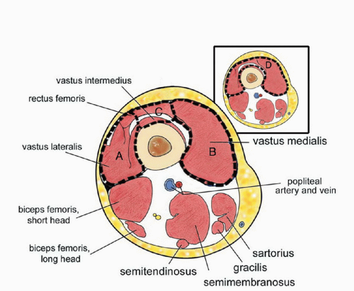
The quadriceps muscle group consists of the vastus medialis, vastus lateralis, rectus femoris, and vastus intermedius muscles. The vastus medialis and lateralis arise from the proximal femur and intermuscular septum. The vastus intermedius arises from the surface of the femur and the linea aspera and covers the entire femoral shaft. The rectus femoris arises from the supra-acetabular tubercle at the superior part of the acetabulum. All four heads merge distally into the quadriceps tendon, which inserts onto the patella.
By covering the anterior aspect of the femur, the vastus intermedius protects the underlying femur from direct tumor extension by tumors of the other components of quadriceps muscle.
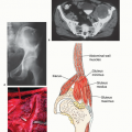
The fact that soft tissue sarcomas often remain localized to one muscle belly permits partial muscle group resection for many quadriceps sarcomas (FIG 1).
The medial and lateral intermuscular septum of the thigh separates the anterior thigh muscles from the medial and posterior compartments, respectively. However, the medial intermuscular septum “runs out” proximally, and quadriceps tumors may therefore extend into the posterior and medial compartments and complicate and sometimes obviate a limb-sparing resection. Likewise, tumors arising from the medial and posterior compartments of the thigh may extend into the quadriceps group.
The femoral triangle is the key to resection of the quadriceps muscle group. It is formed by the adductor longus medially, the sartorius muscle laterally, and the inguinal ligament proximally. The pectineus muscle forms the floor of the triangle. A thick fascia covers the roof.
The superficial femoral artery and vein pass from below the inguinal ligament through the femoral triangle and into the sartorial canal at the apex. The femoral nerve enters the canal laterally and quickly branches to innervate the quadriceps muscle components. The superficial femoral artery and vein pass along the medial wall of the sartorial canal throughout the length of the thigh and are separated from the anterior group (vastus medialis) by a thick fascia, which often permits a safe resection.
This fascia forms a good border for quadriceps resections.
INDICATIONS
Almost all low-grade soft tissue sarcomas of the anterior thigh may be safely resected by a partial muscle group resection. The large majority of high-grade soft tissue sarcomas can be resected by partial or total compartmental removal. The contraindications to limb-sparing resection are as follows:
Groin involvement: Tumors arising or involving the groin and femoral triangle often cannot be reliably resected and may require amputation.
Extracompartmental extension: In general, resection of a single muscle group permits a viable extremity. If two muscle groups have to be completely removed, the extremity may not be functionally salvageable. Large tumors of the anterior thigh may involve the adductor group as well as the posterior muscle group by passing through the linea aspera or the intermuscular septum. In this situation, amputation might be necessary.
Intrapelvic extension: On rare occasions, large tumors of the proximal thigh and groin extend below the inguinal ligament into the retroperitoneal space, necessitating amputation.
Recurrent tumors of the quadriceps, infection, extensive tumor hemorrhage, or extensive tumor contamination from previous surgical procedures may require amputation.
Neurovascular involvement of the tumor does not necessarily obviate limb salvage resections. Most tumors of the quadriceps muscle will displace but not invade the superficial femoral or common femoral arteries. If the surgical margins are positive for tumor cells or extremely close, resection of the involved artery and replacement with a vascular graft often allows limb salvage.
En bloc resection of the femoral nerve is also not a contraindication for limb-sparing resection; reconstruction techniques often provide patellar stabilization and allow limited knee extension, even when the entire quadriceps muscle is resected or paralyzed secondary to femoral nerve resection (Tables 1 and 2).
Table 1 Histopathologic Diagnoses of 15 Patients Treated with Soft Tissue Tumor Resection from the Anterior Compartment of the Thigh and Extensor Mechanism Reconstruction | ||||||||||||||||||||
|---|---|---|---|---|---|---|---|---|---|---|---|---|---|---|---|---|---|---|---|---|
|
Table 2 Grading System for Quadriceps Muscle Strength | ||||||||||||||||||||||||
|---|---|---|---|---|---|---|---|---|---|---|---|---|---|---|---|---|---|---|---|---|---|---|---|---|
| ||||||||||||||||||||||||
Unique Anatomic Considerations
An important criterion for the success of a muscle flap transfer (FIG 2A-D) is maintenance of a pattern of circulation that is consistent in location and resistant to the effect of radiation therapy and superficial trauma (FIG 2E).
The surgical manipulation of the muscle flap must not interrupt its circulation; therefore, a precise knowledge of the location and pattern of the vascular pedicles is required. The sartorius muscle is supplied by the superficial femoral artery and has a segmental vascular pattern (type IV vascular pattern according to Mathes and Nahai6). Each pedicle provides circulation to a portion of the muscle, and division of more than three pedicles during the elevation of the flap may result in distal muscle necrosis. The hamstring muscles are supplied by branches of the profunda femoris artery and have proximal dominant vascular pedicles and distal minor pedicles (type II vascular pattern). Complete elevation of the muscles is possible when the dominant proximal vascular pedicles are preserved.
IMAGING AND OTHER STAGING STUDIES
Computed Tomography and Magnetic Resonance Imaging
Magnetic resonance imaging (MRI) and computed axial tomography (CAT) cross-sectional imaging are essential for determining the location and extent of the lesion and its relations to the femur and the neuromuscular bundle. Large tumors of the quadriceps muscle often displace the superficial and deep femoral vessels (FIG 3). It is important to determine the anatomic relations of these vessels to the tumor before resection. Large tumors of the proximal thigh may require ligation of the profundus femoris artery and vein; therefore, knowing before surgery whether the superficial artery is patent is essential. This is particularly true in the older patient in whom the superficial femoral artery may be occluded secondary to peripheral vascular disease. Displacement of the superficial femoral artery usually does not indicate direct tumor extension; however, if the surgical margins are positive, the artery should be resected and replaced with a saphenous or artificial graft.
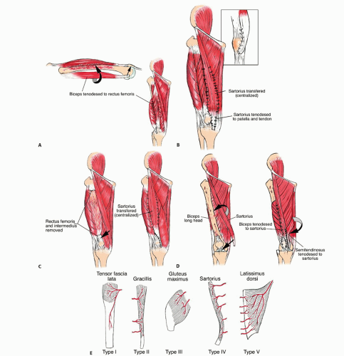
FIG 2 • A. Muscle transfers for type A resection (vastus lateralis with or without vastus intermedius). The long head of the biceps femoris is transferred anteriorly and sutured to the patella, the quadriceps tendon, and the rectus femoris muscle. B. Muscle transfers for type B resection (vastus medialis with or without vastus intermedius). The sartorius muscle is transferred anteriorly but not detached from its distal insertion and is sutured to the patellar tendon, patella, quadriceps tendon, and rectus femoris muscle. C. Muscle transfers for type C resection (rectus femoris and vastus intermedius). The sartorius muscle is mobilized anteriorly and sutured to the patella and the remains of the quadriceps tendon. D. Muscle transfers for type D resection (subtotal resection). The biceps femoris laterally and the sartorius and semitendinosus medially are transferred anteriorly, tenodesed to each other, and sutured to the patella. E. Vascular anatomy of muscles. There are five patterns of vascular supply to muscles based on the distribution of major and minor vascular pedicles. The sartorius muscle has a type II vascular pattern, and the hamstring muscles have a type IV vascular pattern (represented by the gracilis muscle in the schematic).
Stay updated, free articles. Join our Telegram channel

Full access? Get Clinical Tree

 Get Clinical Tree app for offline access

