Pulmonary Disease in the Elderly


Pulmonary Disease in the Elderly


Eleanor M. Summerhill


CLINICAL PEARLS


  • Although the physiologic reserve of the respiratory system declines with age, dyspnea with usual activity is abnormal and should trigger an investigation for underlying disease.


  • The perception of dyspnea appears to be blunted in the elderly and may delay seeking medical attention.


  • An alveolar-to-arterial oxygen gradient of 20 to 25 mm Hg is the upper limit of normal in any population, and a higher gradient in the elderly patient warrants further evaluation.


  • The most common causes of cough include postnasal drip, cough-variant asthma, chronic bronchitis, and gastroesophageal reflux disease. Therapy should be directed at the underlying mechanism following diagnostic evaluation.


  • The differential diagnosis of asthma in the elderly is broad and often overlooked.


  • Asthma may present at any age.


  • The negative predictive value of methacholine challenge testing is higher than the positive predictive value, so that it is more useful in ruling out a diagnosis of asthma than establishing one.


  • Special treatment considerations in the elderly with asthma and chronic obstructive pulmonary disease include a higher risk of adverse effects of medication and exacerbation of coexistent disease, medical noncompliance, and precipitation of bronchospasm by commonly used medications for comorbid illness.


  • Although a number of medications are indicated for symptomatic relief in chronic obstructive pulmonary disease, smoking cessation and supplemental oxygen therapy are the only therapies shown to favorably impact outcome.


  • Elderly patients with pneumonia often present with nonspecific symptoms and may not manifest the classic constellation of fever, dyspnea, and cough. This may lead to delayed diagnosis or misdiagnosis and a greater mortality risk.


  • Initiation of appropriate antibiotic therapy for pneumonia within the first 8 hours of presentation has been shown to significantly reduce mortality.


  • Prevention may be the best way to reduce the morbidity and mortality associated with pneumonia in the elderly. Recommended immunizations include the 23-valent pneumococcal polysaccharide and influenza vaccines.


  • Because of the waning of cell-mediated immunity, the two-step purified protein derivative skin test is recommended in those elderly with an initial nonreactive test result.


  • The risk of isoniazid-induced hepatitis is low. Therefore, isoniazid-based treatment regimens are recommended for the treatment of both latent and active tuberculosis in the elderly.


  • Sleep patterns change with normative aging, and sleep disturbances such as insomnia, nocturnal awakening, and sleep-disordered breathing appear to be more common in the elderly population. Their clinical significance is unclear.


INTRODUCTION

With normal aging come changes in pulmonary function that reduce the physiologic reserve of the respiratory system. This progressive decline that occurs with advancing age in various organ systems is called homeostenosis. However, it is important to remember that despite this age-related decline, it is never normal for an individual to have the need to curtail normal activities because of pulmonary limitation. Dyspnea with usual activity is always abnormal and is indicative of underlying disease.1

As in many areas of medicine, there are few pulmonary diseases entirely unique to the geriatric population. However, the prevalence, presenting manifestations, diagnosis, and treatment may differ from those in younger adult populations. This chapter contains an overview of presenting signs and symptoms of pulmonary disease in the elderly, the normal physiology of the aging lung, and some specific disease processes particularly relevant to ambulatory geriatric clinical practice.


PULMONARY SIGNS AND SYMPTOMS

Pulmonary disease may be associated with a variety of signs and symptoms. The most common symptoms pointing to pulmonary pathology include dyspnea, cough, pleuritic chest pain, and hemoptysis.

Dyspnea is defined as an abnormally uncomfortable awareness of breathing2 and is most often described as breathlessness or shortness of breath. Recently, dyspnea was defined by the American Thoracic Society (ATS) as “a subjective experience of breathing discomfort that consists of qualitatively distinct sensations that vary in intensity.”3 Dyspnea is a very common presenting symptom of pulmonary disease. However, as delineated in Table 30.1, dyspnea may also be indicative of cardiac disease, hematologic disease, neuromuscular disease, metabolic disease, deconditioning, and/or obesity, or it may be psychosomatic
in origin. A thorough history including acuity of onset, exacerbating factors such as exertion and positional change, and associated symptoms are vital in determining the etiology of dyspnea. Abrupt onset points to relatively acute processes such as pulmonary infection, pulmonary embolism (PE), and congestive heart failure (CHF). Subacute or chronic presentations of dyspnea are more likely to be indicative of underlying chronic bronchitis, emphysema, interstitial lung disease (ILD), or chronic CHF. Dyspnea may occur at rest or may only be apparent on exertion. In making an assessment of the severity of chronic dyspnea, it is important that the practitioner ask the patient “what are your usual daily activities?” and “what activities have you had to discontinue recently?” because patients often reduce activities to minimize discomfort.4


TABLE 30.1 COMMON CAUSES OF DYSPNEA


Cardiovascular


Left ventricular failure


Valvular dysfunction


Pericardial effusion


Right-to-left shunt


Pulmonary


Obstructive airways disease


Parenchymal lung disease


Pulmonary vascular disease


Pleural disease


Respiratory muscle weakness


Neuromuscular disorders


Phrenic nerve paralysis


Metabolic and systemic illness


Mechanical


Upper airway obstruction


Chest wall and thoracic spine abnormalities


Exogenous obesity


Massive ascites


Hematologic


Anemia


Psychosomatic


Anxiety


Deconditioning


Evidence suggests that the perception of dyspnea may be blunted in the elderly. This may be due to a number of complex processes involving the mechanical properties of the lung and chest wall, central and peripheral chemoreceptors, neural input and output, and deconditioning. For example, there is a lower response to hypercapnic hypoxemia in the elderly, largely because of a decrease in carbon dioxide sensitivity.5 And, despite similar levels of decline in forced expiratory volume in 1 second (FEV1), older subjects given methacholine bronchoprovocation testing noted less subjective discomfort compared to younger individuals. This may be due either to reduced number or activity of stretch receptors or to decreased perception of resistive respiratory loads.6

A history of positional change eliciting or exacerbating dyspnea may be helpful in elucidating the underlying etiology. Orthopnea, or dyspnea in the supine position, is characteristic of CHF. It is usually related to gravitational elevation of pulmonary and venous capillary pressures and a reduction of vital capacity (VC). However, it may also be indicative of bilateral diaphragmatic paralysis and may occur in obstructive lung disease. Paroxysmal nocturnal dyspnea, like orthopnea, is usually indicative of CHF. It may also be present in chronic bronchitis because of pooling of secretions or in asthma because of circadian changes in airway obstruction. Platypnea, which occurs in the upright position, is most often associated with orthodeoxia, or oxygen desaturation when upright. Platypnea and orthodeoxia may occur because of changes in ventilation-perfusion (V/Q) matching or may be indicative of intracardiac shunt.

The underlying etiology of dyspnea in the elderly may usually be elucidated by means of a thorough history (including tobacco and occupational exposures), physical examination, and simple diagnostic testing. Initial testing should include a chest radiograph (CXR), electrocardiogram (ECG), and complete blood count (CBC). If a pulmonary cause for dyspnea is suspected, further investigation is indicated. This should include pulse oximetry or arterial blood gas (ABG) analysis and pulmonary function tests (PFTs). PFTs include spirometry, inspiratory and expiratory flow loops, lung volumes, and diffusing capacity for carbon monoxide (DLCO). If respiratory muscle weakness is suspected, maximal inspiratory and expiratory pressures should be obtained. If the cause of dyspnea remains unclear, echocardiography and, finally, cardiopulmonary exercise testing may help in differentiating between cardiac or pulmonary limitation and deconditioning.1,4

Cough is a common symptom referable to the respiratory system. It is a physiologic mechanism for clearing and protecting the airway. It may be stimulated by a number of different processes, including airway irritation or inflammation, parenchymal disease, CHF, or medications. Cough is generally categorized as either acute or chronic on the basis of a duration of < or >3 weeks. In the overwhelming majority of cases, acute cough is related to either a viral or a bacterial upper respiratory tract infection. However, particularly in the elderly, acute cough may be a presenting manifestation of more serious illness such as pneumonia, aspiration, CHF, or PE.7

Studies have shown that chronic cough is usually due to one or a combination of four processes: Postnasal drip, cough-variant asthma, chronic bronchitis, or gastroesophageal reflux disease (GERD).8,9 Other causes to consider in the elderly include bronchiectasis, CHF, ILD, bronchogenic lung cancer, postviral airway hyperreactivity, and recurrent aspiration.7 An additional etiology that should be recognized in this population is angiotensinconverting enzyme (ACE) inhibitors. Approximately 5% to 20% of patients placed on ACE inhibitors will develop a dry cough. This is a class effect of these antihypertensive medications and is most likely related to the accumulation of the inflammatory mediators bradykinin or substance P, both of which are degraded by ACE. The development of cough is idiosyncratic and is not dose related. The time course may be quite variable, ranging from several hours to even months after initiation of ACE inhibitors. After discontinuing the medication, the cough generally resolves within 4 days.10

Evaluation of chronic cough should begin with a complete history and physical examination, as well as a CXR. If the CXR is normal, and nothing in the history and physical points to a diagnosis, the next most useful step is a methacholine challenge test to assess for bronchial hyperreactivity (BHR).8 GERD may be silent. Therefore, if another cause of chronic cough cannot be found and treated, prolonged esophageal pH monitoring should be considered.11 Some patients will be found to have more than one diagnosis. Therapy should be directed toward the underlying pathophysiologic mechanism of the cough. Specific therapy has been shown to be effective in patients compliant with the therapeutic regimen in 97% to 98% of cases.8,9 Patients with chronic bronchitis, asthma, and postviral airway hyperreactivity respond to inhaled bronchodilators and corticosteroids; those with postnasal drip respond to decongestants and nasal steroid preparations and those with GERD to
histamine-2 (H2) blockers and proton pump inhibitors. Symptomatic treatment with antitussive agents such as dextromethorphan, and narcotics such as codeine is useful to supplement specific therapy, but is ineffective alone.

Pleuritic chest pain is most often sharp but may also be described as dull, achy, or a “catching” sensation. Characteristically, it worsens with deep inspiration, cough, or positional change. Pleuritic pain originates in the parietal pleura, which has extensive pain fiber innervation; the visceral pleura does not. Pain is usually relatively well localized over the area of involvement. However, if the diaphragmatic parietal pleura is affected, pain may be referred to the shoulder.12 Inflammation in the peripheral lung parenchyma may spread to the visceral and then parietal pleura, causing pleuritic pain. Some of the most common causes of pleuritic chest pain include pneumonia, PE, pneumothorax, pleuritis due to acute viral infection or collagen vascular disease, pericarditis, and radiation pneumonitis.

Hemoptysis may be noted by the patient as ranging from blood-streaked sputum to the expectoration of gross blood. If massive (defined as >200 to 600 mL in a 24-hour period, depending on the author), it represents a medical emergency requiring hospitalization and immediate evaluation by a pulmonologist and/or thoracic surgeon.13 Studies performed in the 1940s to 1960s found that the most frequent causes of hemoptysis included bronchiectasis, bronchogenic carcinoma, and tuberculosis (TB). More recently, bronchitis has become one of the most frequent etiologies of hemoptysis in developed countries. In contrast, TB is much less common.14,15 Table 30.2 lists some of the more common etiologies of hemoptysis.

Evaluation should begin with a detailed history and physical, ruling out other sources of bleeding such as the upper respiratory or gastrointestinal tracts. Duration and amount of hemoptysis, smoking history, and features suggestive of the presence of bronchitis or a pulmonary parenchymal infection should be elicited. The most important diagnostic test is the CXR. Additional testing to consider includes a CBC, coagulation profile, renal function studies, and urinalysis. If the CXR is abnormal, it may be helpful in elucidating the underlying etiology. If normal or without localizing findings, studies have shown that independent risk factors for occult carcinoma include age >40 years, smoking history of >40 pack-years, and duration of hemoptysis of >1 week.15, 16, 17

If hemoptysis is not massive, <1 week in duration, occurs in the setting of acute bronchitis, and if CXR is normal, further workup is not usually necessary, provided there is resolution with appropriate antibiotic therapy. However, if there is an abnormal finding on CXR, hemoptysis is massive, persists for >1 week, occurs in a setting inconsistent with acute bronchitis, or two or more of the above patient risk factors for carcinoma are present, pulmonary or thoracic surgery consultation is advised for further evaluation.15, 16, 17 Fiberoptic bronchoscopy and/or high-resolution computed tomography scanning are the next steps in evaluation, depending on the clinical setting, and may provide complementary information.18,19 Further evaluation is particularly important in the geriatric population, which is at higher risk for carcinoma, especially those individuals with a significant history of smoking or second-hand exposure.


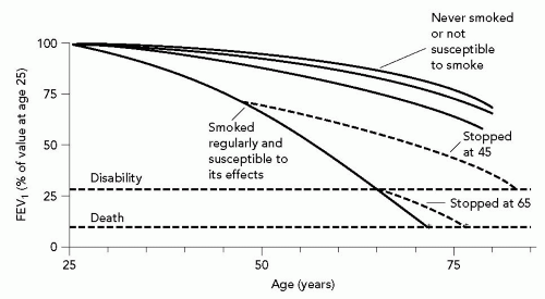
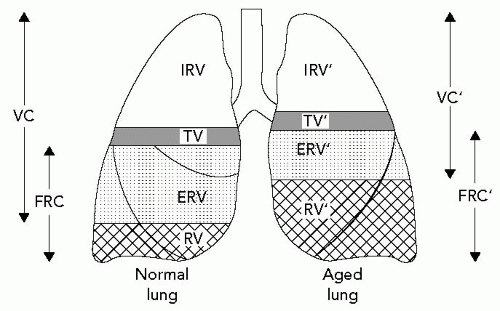
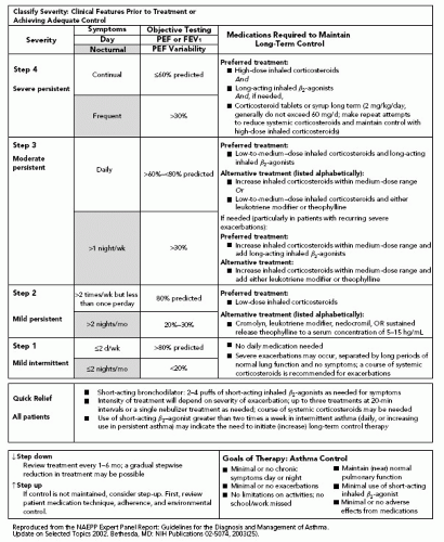

TABLE 30.2 MAJOR CAUSES OF HEMOPTYSIS


Airway diseases—most common


Bronchitis


Bronchogenic carcinoma


Bronchiectasis


Pulmonary parenchymal diseases


Lung abscess


Mycetoma


Necrotizing pneumonia


Parasitic infection


Fungal infection


Tuberculosis


Vascular


Pulmonary embolism


Pulmonary hypertension


Arterial-venous malformation


Systemic diseases


Goodpasture syndrome


Wegener granulomatosis


Systemic lupus erythematosus


Other systemic vasculitides


Cardiac


Mitral stenosis


Iatrogenic


Bronchoscopy


Swan-Ganz catheter-induced infarction


Pulmonary artery rupture


Transtracheal aspiration


Hematologic—rare


Coagulopathy


NORMAL AGE-RELATED CHANGES IN THE RESPIRATORY SYSTEM

Pulmonary function deteriorates with age, even in the healthy elderly population.5,20 The anatomic and structural changes that occur with normal aging and result in diminished pulmonary function are discussed in the subsequent text.


Changes in the Lung

Changes in the lung associated with aging include smaller airway size, changes in the morphology of the alveolar ducts and sacs, and possibly, alterations in the composition and/or thickness of the alveolar basement membrane.5,20
Mean bronchiolar diameter, the main determinant of airways resistance, has been found to decrease significantly after age 40.21 This change is thought to be the result of alterations in the collagenous matrix and elastic components of the underlying connective tissue, which support the airways, tethering them open.20 With age, the alveolar ducts become enlarged, and the alveolar sacs shallower, resulting in a decrease in the airspace surface area-to-lung volume ratio.22 In addition, there is some evidence that changes in the alveolar basement membrane composition and, thickness may also occur with aging.5,20 Taken together, these changes contribute to a decline in the measured DLCO. This decline occurs later in women than in men, perhaps secondary to estrogen effects.5,20


Figure 30.1 Decline in lung function with aging. As depicted in this graph, forced expiratory volume in 1 second (FEV1) decreases over time with normative aging. In susceptible smokers, the rate of decline in FEV1 has a much steeper slope. After smoking cessation, the rate of decline reverts to that of a nonsmoker. (From Fletcher C, Peto R. The natural history of airflow obstruction. Br Med J. 1977;1:1645-1648. Graph reproduced with permission from the BMJ Publishing Group).


Changes in the Chest Wall and Respiratory Muscles

The thoracic cage and respiratory muscles also undergo significant alterations. The chest wall becomes less compliant because of calcification of the intercostal cartilages, arthritis of the costovertebral joints, and in some individuals, osteoporosis and kyphoscoliosis of the spine. There is also an age-related decrease in diaphragm and intercostal muscle strength. The exact mechanism is unclear, and there is wide variability within the normal range.5,20


Mechanics

Taken together, the structural changes described in the preceding text are responsible for the changes in lung mechanics noted with normal aging. These include decreased elastic recoil, and therefore, increased lung compliance; increased airways resistance; premature airway closure; and decreased gas exchange capacity. These changes are similar to those encountered in emphysema, but without the same clinical consequences. In addition, there is increased chest wall stiffness leading to decreased compliance.


Changes in Pulmonary Function

Because of the loss of elastic recoil of the lung, increased chest wall stiffness, and decreased force generated by the respiratory muscles, there is a progressive decrease in VC. It has been estimated that forced vital capacity (FVC) declines about 14 to 30 mL per year in nonsmoking men, and 15 to 24 mL per year in nonsmoking women, starting at approximately 30 to 40 years of age. Rates for decline of FEV1 are similar.20 Of note, this rate of decline is markedly increased in susceptible tobacco smokers. Within a short period of smoking cessation, the rate of decline in FVC reverts to resemble a nonsmoker’s decay curve, as shown in Figure 30.1.23 With the reduction in FVC, there is a symmetric increase in residual volume (RV) so that total lung capacity (TLC) remains relatively constant (see Fig. 30.2). An exception is the patient with kyphoscoliosis.
In these individuals, there is often a significant drop in TLC and a resultant restrictive defect. Just as RV increases with aging, so does functional residual capacity. Therefore, the small airways (terminal bronchioles) close early, especially in the dependent parts of the lung, leading to V/Q mismatching and increased dead space ventilation. These contribute to the increase in the alveolar-to-arterial oxygen gradient (A-a gradient).5,20


Figure 30.2 Changes in lung volume associated with aging. As depicted above, vital capacity (VC) decreases with aging. Correspondingly, residual volume (RV) and functional residual capacity (FRC) increase so that total lung capacity (TLC) does not significantly change. TV, tidal volume; IRV, inspiratory reserve volume; ERV, expiratory reserve volume. (From Chan ED, Welsh CH. Geriatric respiratory medicine. Chest. 1998;114:1704-1733. Reproduced with permission from Chest).


Arterial Oxygen Tension

In addition to increased dead space ventilation, there is also an increase in shunt fraction that occurs with aging. In combination with decreased gas exchange capacity, these processes result in a decrease in arterial oxygen tension (PaO2) with aging. Recently, it was shown that this increase in alveolar to arterial (A-a) gradient is not linear after age 74. At sea level, PaO2 in the healthy elderly patient remains approximately 83 mm Hg. Therefore, it must be emphasized that a maximum A-a gradient of 20 to 25 mm Hg is the upper limit of normal in any population. A gradient in excess of this is indicative of a pathologic process.20,24


Loss of Respiratory Reserve

The decrease in lung function associated with age results in diminished respiratory reserve. Although this loss of pulmonary reserve is not apparent during normal activities, it may be unmasked under conditions such as acute illness, surgery, or strenuous exercise. With aging, ventilatory performance becomes less efficient. This results in a greater utilization of oxygen ([V with dot above]O2) to achieve the same minute ventilation as in youth. In addition, the [V with dot above]O2 max, a measure of pulmonary, cardiac, and metabolic performance, declines steadily after age 25 to 30, resulting in a decrement in maximum work capacity over time.20


ASTHMA IN THE ELDERLY

As defined by the National Asthma Education and Prevention Program (NAEPP) Expert Panel 1 in 1997, asthma is “a chronic inflammatory disease of the airways in which many cells, including mast cells, eosinophils, neutrophils, T-lymphocytes, and epithelial cells play a role. In susceptible individuals, this inflammation causes recurrent episodes of wheezing, breathlessness, chest tightness, and cough, especially at night or in the early morning. These symptoms are usually associated with widespread but variable airflow limitation that is at least partially reversible either spontaneously or with treatment. This inflammation also causes an associated increase in airway hyperresponsiveness to a variety of stimuli.”25


Epidemiology

Asthma is considered a young person’s disease, and therefore is a frequently overlooked diagnosis in the elderly. However, onset may be at any age. Given the presence of confounding factors such as tobacco smoking, second-hand smoke exposure, and occupational exposures, it is often impossible to distinguish asthma from chronic bronchitis in the older population. Asthma prevalence peaks in childhood at approximately 8% to 10% of the population and declines in young adulthood to a rate of 5% to 6%. Subsequently, there is another peak after age 70, with a prevalence rate of approximately 7% to 9%.26 Elderly patients with asthma represent two distinct populations. One includes those with long-standing asthma or recrudescence of childhood or early adulthood asthma, and the other includes those with new- or recent-onset disease. Estimates of the incidence of new-onset asthma in the elderly have been highly variable. In one study, approximately 50% of asthmatics over the age of 70 had developed the disease after age 65. Those with early onset asthma had a greater likelihood of previous atopic disease and of chronic persistent airway obstruction, as occurs in chronic obstructive pulmonary disease (COPD).27


Diagnosis

The approach to making a diagnosis of asthma in the geriatric patient is similar to that in younger individuals. It is based on a compatible clinical history, physical examination, and objective assessment of reversible airflow limitation and/or bronchial hyperresponsiveness (BHR). The most common symptoms include episodic wheezing, dyspnea, and cough. Exertional dyspnea and paroxysmal nocturnal dyspnea are less frequently encountered. The differential diagnosis in this population is broad. Diseases that may present with similar symptoms include chronic bronchitis, emphysema, bronchiectasis, occupational asthma, recurrent aspiration, sarcoidosis, constrictive bronchiolitis, CHF, GERD, PE, laryngeal dysfunction, endobronchial tumors, and ACE inhibitor-induced cough.20,25,26 The physical examination may be normal, or there may be evidence of hyperinflation and/or end-expiratory wheezing on examination of the chest. Other findings supporting the presence of atopic disease are helpful. These include nasal mucosal swelling and polyps, as well as eczema. Objective testing must show either a significantly reversible obstructive ventilatory defect or evidence of BHR. An obstructive ventilatory defect is demonstrated by spirometry showing a reduction in FEV1/FVC ≤70%. Significant reversibility is defined as ≥15% as well as ≥200 mL improvement in FEV1 following administration of bronchodilator.28 On occasion, full PFTs with a DLCO will be necessary to rule out emphysema or restrictive lung disease. If spirometry is normal, or there is an irreversible obstructive defect, methacholine challenge testing is helpful. BHR is present if there is a ≥20% decrement in FEV1. The negative predictive value of the methacholine challenge test is higher than the positive predictive value, making it more useful in excluding a diagnosis than establishing one. If the baseline FEV1 is ≤65% of the predicted value, methacholine of the predicted value, methacholine challenge testing is potentially harmful and therefore not recommended.25,29


Treatment Considerations in the Elderly

Treatment is generally similar to that for younger individuals and should follow the stepped care approach outlined in Table 30.3 (Evidence Levels A and B), adapted from the NAEPP Expert Panel Report 2 update in 2002.30 Using this approach, patients are assigned to the step that includes the most severe feature of their disease. The aim is to gain control as quickly as possible, with a short course of corticosteroids, if necessary, and then step down to the least medication needed to maintain control. Overreliance on short-acting β-agonists is one of the indicators of poor asthma control. In addition to pharmacotherapy, patients should be educated about environmental controls. Referral to an allergist or pulmonologist should be considered for patients who fit the criteria for steps 3 and 4.

Only gold members can continue reading. Log In or Register to continue

Stay updated, free articles. Join our Telegram channel

Jul 21, 2016 | Posted by in GERIATRICS | Comments Off on Pulmonary Disease in the Elderly

Full access? Get Clinical Tree

Get Clinical Tree app for offline access