Figure 7.1
USA epidemiology
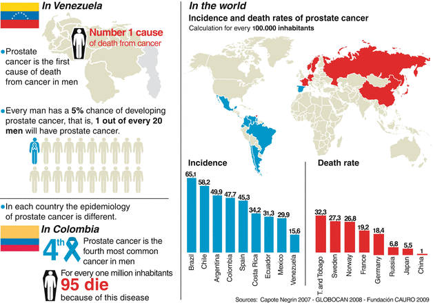
Figure 7.2
World epidemiology
Epidemiology
Prostate cancer is the most common cancer diagnosed in North American men, excluding skin cancers. Prostate cancer is now the second leading cause of cancer death in men, exceeded only by lung cancer. It accounts for 29 % of all male cancers and 9 % of male cancer-related deaths.
Predisposition
Before 50 years of age, it’s rare that men develop prostate cancer, so efforts to diagnose it are usually intensified after that age. Although it is not known with precision what triggers prostate cancer, several factors that might be related have been described, including:
Age
This is one of the determinants. On the average, the diagnosis is made beginning at 71 years. Fewer than 1 % of the cases of prostate cancer are detected in men under 50 years of age, and only 16 % in men between 50 and 64 years of age. However, with the development of new technologies, the diagnosis in young men has increased.
Hormonal factors
Hormonal action is without doubt another trigger of the disease. It has been observed that the men suffering from a failure of the pituitary gland, those who have had their testicles removed, or those suffering from a hormonal problem that decreases androgen levels are less prone to develop this type of cancer. Similarly, the appearance of this disease in young men has been related to the use of testosterone.
Hereditary factors
It has also been shown that a man whose father or brother has developed prostate cancer has twice the risk of occurrence of the disease compared to that of the average man. For this reason it is recommended that if there is any family history of prostate cancer, examinations such as the digital rectal examination and the prostate-specific antigen should be carried out at early ages, starting at 40, and a urologist should be seen at least every 6 months after the age of 50. Between 11 and 15 % of the patients with prostate cancer have a family history of this disease (Fig. 7.3).
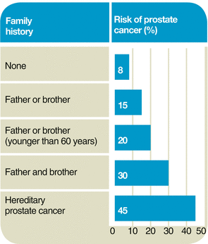
Figure 7.3
Family history
Genetic susceptibility
Studies of the human genome and more recently of the proteins that form part of the human DNA (that is, all of the genetic information of the species) have described several mutations or changes in certain chromosomes that generate cellular alterations, thereby accelerating the appearance of cancer. In the very near future, new markers that have been studied will be the gateway to new perspectives in the prevention and treatment of prostate cancer.
Diet
This also plays an important role. It is known that people who are overweight or have problems with fat metabolism tend to develop more prostate cancer than those who follow a balanced diet. Several research studies have been conducted to study the effect of selenium, vitamin E, as well as the isoflavonoids found in various food products on prostate cancer prevention. However, at this time there is not enough conclusive evidence that any of these agents can effectively prevent prostate cancer.
Geographic location
It appears that men who live in countries with less exposure to the sun are more likely to suffer from prostate cancer because of their low levels of vitamin D.
Tobacco use
Important, though not conclusive, studies indicate that smokers may be at greater risk of developing prostate cancer. It appears that nicotine, which is highly toxic, might generate some cellular and hormonal change that favors the disease.
Exposure to cadmium
Cadmium is a mineral found in cigarettes as well as in alkaline batteries. It interferes with zinc, a chemical element vital to many activities of the human cells, including some of those processes that occur in the prostate. Patients with prostate cancer have been found to have low levels of zinc in the body.
Vasectomy
This common procedure used for birth control in men (comparable to tubal ligation in women), may play a role in the predisposition to prostate cancer; however, there is no evidence suggesting a direct relation between prostate cancer and vasectomy (Fig. 7.4).
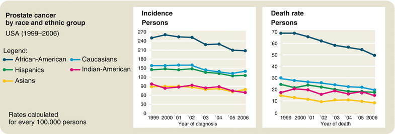
Figure 7.4
Race and ethnic group
Prevention
Screening
One of the great controversies in preventative medicine is whether the prostate-specific antigen test should or should not be part of the periodic health examination. To answer this question it should be taken into account that prostate cancer is the most frequent of the lethal tumors and the leading cause of death by cancer in men.
The use of the prostate-specific antigen has contributed to increased detection of prostate cancer. Now more than ever prostate cancer is diagnosed. At the same time, the death rate has decreased. Men with localized prostate cancer have a 20–60 % chance of dying from this cause if they do not receive treatment. For example, at present in North America about 60 % of the tumors detected at the initial PSA screening are localized, making it possible for the urologist to offer the patient a treatment that will cure the disease.
In the past 10 years, thanks to screening programs, the diagnosis of prostate cancer in advanced stages has declined significantly.
Fact
Screening, according to the World Health Organization, is the application of a simple test in a healthy population to identify those individuals who have some related pathology but who do not as yet show symptoms. In the case of prostate cancer, the screening is directed toward men 50 years of age or older and those 40 years and older if they are of African descent or have a first-degree relative who has been affected.
Primary prevention
Current information about the risk factors for prostate cancer suggests that some cases can be prevented. One possible risk factor that can be changed is the diet. The risk of prostate cancer can be reduced with a diet low in fats and high in vegetables, fruits, and cereals.
The American Cancer Society recommends eating a variety of health foods, with emphasis on those of vegetable origin and limiting the consumption of red meat. It also suggests the daily consumption of fruit and products with fiber, such as bread, cereals, and rice. These nutritional suggestions also help reduce the risk of other types of cancer.
Tomatoes, citric fruits, and watermelon are rich in lycopenes. These substances, similar to vitamins, are antioxidants, which can help prevent mutations of the DNA and, therefore, reduce the risk of prostate cancer. The effects of vitamin E and selenium as preventatives have also been studied.
Several studies associate daily use of nonsteroidal, anti-inflammatory drugs, such as Aspirin®, with a lower incidence of prostate cancer in men 60 or more years old.
It should be taken into account that the exact cause of prostate cancer is unknown and that there are risk factors such as age, race, or family history that cannot be controlled.
Secondary prevention
Early treatment through early detection reduces morbidity and mortality associated with the disease. In the United States and Europe, studies have been carried out in the past few years to clarify whether screening with the prostate-specific antigen reduces mortality from prostate cancer or not. Up until now, screening in healthy men has proved to be useful, in spite of criticism of some of the studies. What is found in medical literature suggests that it is a useful tool and it reduces mortality.
One of the most recognized studies is that of Tyrol, an Austrian city where screening with the prostate-specific antigen began in the 90s. The research showed that there was a reduction in mortality from prostate cancer in the period analyzed (between 1999 and 2003) in comparison with other cities where the antigen tests were not performed.
The effect of the screening programs depends on the sensitivity and the specificity of the detection method, but also on the effectiveness of the therapy applied in the cases detected. In Tyrol, the majority of the patients underwent surgical removal of the prostate and seminal vesicles (radical prostatectomy) with good results.
In the United States, the most important study of screening for the detection of prostate cancer showed that the diagnosis has increased drastically since 1988, but then demonstrated controversy about the benefits of the program. One study compared two groups of men, one in which the prostate-specific antigen was tested and the other group in which it was not. After 10 years of follow-up, a very low death rate was found, without any significant difference between the two groups. The European study, mentioned above, showed a mortality reduction of 20 % in patients subjected to the screening 8 years after starting the program.
A recent and important study carried out in Sweden compared two groups of 10,000 men between 50 and 65 years old. What was found was a higher incidence in the group of men who underwent PSA testing, as there was a diagnosis of prostate cancer in 12.7 % of these men. In the second group (without PSA), the diagnosis was reduced to 8.2 % of the participants. This means there were 1.6 times more diagnoses of prostate cancer in those who underwent the test regularly. They concluded that screening with PSA reduces mortality from prostate cancer by half.
Symptoms
Prostate cancer rarely causes symptoms, and when it does, most probably it is a tumor in an advanced stage. This is because the majority of the tumors grow in the peripheral zone of the prostate, that is, far from the urethra, so they can continue growing without causing any discomfort. When the tumor grows near the urethra or the neck of the bladder, then it can cause symptoms of irritation or obstruction (see Chap. 2), which do not arouse much suspicion of cancer because they also occur in other prostate diseases. Prostate cancer with local invasion can grow and affect the bladder floor and, thus, occlude the emptying of urine from both kidneys, leading to total obstruction and kidney failure.
Other symptoms, such as pain in the bones and anemia associated with renal failure, suggest metastatic prostate cancer (Fig. 7.5).
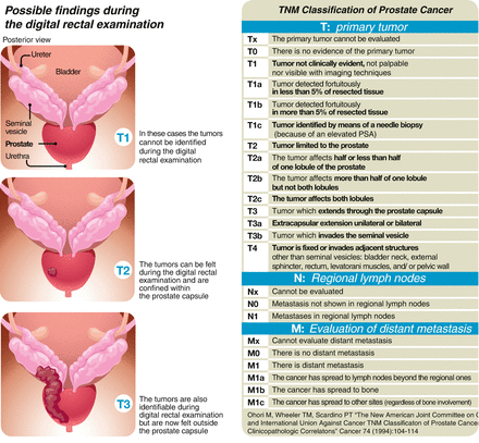
Figure 7.5
Classification
Surgical Treatments
Radical prostatectomy
This is the name given to surgery used to remove the prostate in patients with cancer. It involves the removal of the entire prostate gland, together with the seminal vesicles, and the bladder is then reconnected with the urethra, the channel through which the urine flows from the bladder through the penis to the exterior. In special cases, the surgeon might also remove the regional lymph nodes to evaluate them and determine if the cancer has spread beyond the prostate (Fig. 7.6).
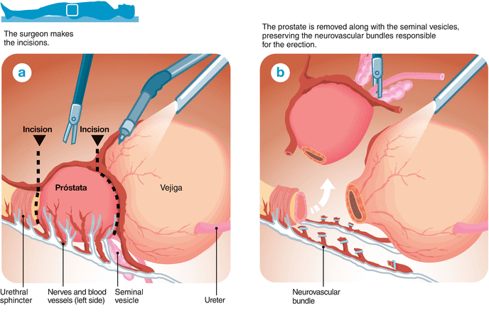
Figure 7.6
(a, b) Radical prostatectomy
An important consideration is that after surgery, the patient will no longer ejaculate due to the removal of the prostate and the seminal vesicles, which store the seminal fluid; although this happens, the patient will conserve exactly the same sensation of pleasure during orgasm. In the case where the patient wishes to have children after the procedure, some assisted reproductive techniques to achieve fatherhood will be needed, such as those that involve the extraction of sperm from the testicles with subsequent injection into the female reproductive cells (as such is the case with in vitro fertilization) (Fig. 7.7).
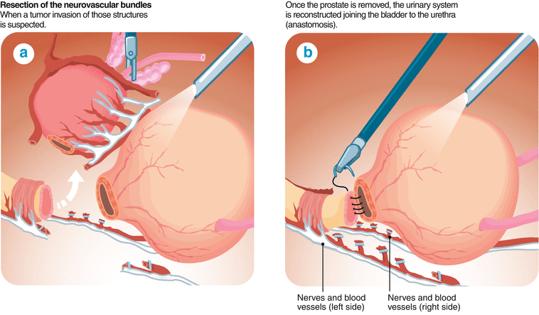
Figure 7.7
(a, b) Radical prostatectomy
Indications
In general, a radical prostatectomy is recommended only for men who have clinical, biochemical, and/or radiological evidence of the cancerous disease located only in the prostate, as well as being in good health in general, with a life expectancy of 10 years or more. An exception to this are young men with suspicion of a localized advanced disease, who could benefit more from a combined treatment including radical prostatectomy, radiotherapy, and hormonal blocking (as opposed to just receiving radiotherapy and hormonal blocking). The urologist can make use of certain normograms, like those shown in the table, to estimate the risk of extraprostatic disease and evaluate the pros and cons of surgery.
Diagnostic studies currently available do not make it possible to precisely evaluate whether the prostate tumor has extended beyond the prostate. This can only be known after surgery, once the pathological examination has been completed. Furthermore, on rare occasions (less than 3 % of patients), it happens that even when the results of the pathological study indicated that the disease was limited to the prostate, the prostate-specific antigen begins to rise after surgery, revealing that possibly cancerous cells still remain at the prostate bed or has entered the bloodstream and disseminated in the body. For this reason, patients who have undergone this operation, even when according to the biopsy the disease was contained, must continue annual control during at least 10 years after surgery.
Lymphadenectomy
This is the removal of lymph node chains near the prostate (iliac and obturator nodes) to determine the presence of cancer cells in them. It is indicated for patients who are considered medium to high risk, as shown in the table (Fig. 7.8).
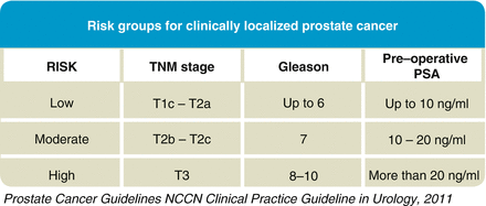
Figure 7.8
Risk groups
Types of radical prostatectomy
The purpose of surgery is to eradicate the prostate cancer, attempting to protect the nervous and vascular structures responsible for erection without compromising the cancer control goal, and delicately separating the area of the urinary sphincter to re-establish urinary continence.
The neurovascular structures responsible for erections are located along the sides of the prostate and in close contact with it. Therefore, on occasion, if it is suspected that the cancer may be on the edge of the prostate, the total or partial removal of these structures is indicated in order to increase the possibility of the complete extraction of the cancer.
At present, the use of certain statistical analyses of the results of patients operated on under similar conditions makes it possible to create tables of probabilities. When the results of a specific patient are situated within these tables, it is possible to know the risk of the disease extending beyond the prostate, and even the possibility that the regional lymph nodes are affected. The analysis of these probabilities, the results of the biopsies, and the digital rectal examination (sometimes a nuclear magnetic resonance is added), along with the intra-operative findings, condition the surgeon’s decision to remove or preserve the nerves linked to erection.
The operation can be carried out in different ways, with various methods of incision. The preservation of the healthy nerves is possible in any of the techniques, and success depends more on the expertise of the surgeon than on the method used.
Minimally invasive laparoscopic techniques, with or without robot, offer the advantage of very small incisions with more rapid recovery (and return of the patient to normal activities), as well as lower risk of complications from surgical wounds, such as infections or hernias, since only five incisions, between 5 and 12 mm in length, are made. However, it should be taken into account that robotic surgery in the hands of non-experts is not better than open surgery in expert hands.
Some publications report that, in addition to these advantages, robotic surgery enables a faster return to urinary continence and erectile function. Furthermore, there are surgeons, experts in open surgery, who have decided to start operating with the robot and have reported more than 2,000 cases in which the results with positive margins (indicating that tumor tissue was left) are fewer with the use of the robot (Vanderbilt University). However, another series of studies reports similar results in the hands of experts, independent of the technique used.
There are different approaches to a prostatectomy. Some can be carried out with regional anesthesia, others require general anesthesia. On the average, the surgery lasts about 3 h.
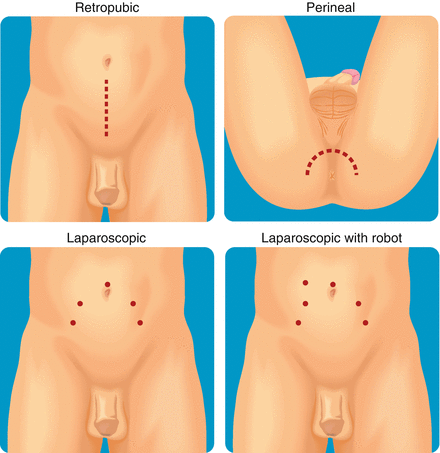
Radical retropubic prostatectomy: This is the most common approach. Regional anesthesia can be used and it is a conventional open surgery. The incision in the skin extends from the umbilicus to the pubis. Bleeding is a known complication to this procedure. The surgeon will remove the prostate gland, taking care to preserve the nerves that control erections on either side of the prostate. The surgeon may also remove the lymph nodes nearby, if advanced disease is suspected.
Radical perineal prostatectomy: In this procedure, the surgeon makes an incision in the skin between the anus and the base of the scrotum (perineum). Due to the location of the incision, it is difficult to remove the regional lymph glands. The perineal surgery has less risk of hemorrhage and normally takes less time to complete than the retropubic procedure.
Radical laparoscopic prostatectomy: This procedure is carried out in specialized medical centers. This type of technique requires general anesthesia, and in place of a single large incision, the surgeon makes various small incisions of between 5 and 12 mm each in the abdominal wall. Through one of them, gas (carbon dioxide CO2) is fed into the abdominal cavity, creating a working space between the wall and the abdominal viscera. Then a special tube with its own light source and video camera (laparoscope) is introduced; this helps observe inside the abdomen, with the possibility of amplifying the view of the operating field with optimal quality in high definition (HD). Into the other incisions (up to four) other working instruments are introduced, such as forceps and scissors, used for performing the surgery. One of the difficulties in carrying out this procedure is that the surgeon must learn and have adequate training in a series of maneuvers that will allow for an adequate angle for cutting or suturing while using these instruments. The air in the abdominal cavity compresses the veins and thus reduces the bleeding. With this technique it is also possible to preserve the nerves around the prostate and remove the lymph nodes. Clearly the recovery of the patient and his reincorporation into his daily life is much faster than in any of the open surgery techniques.
Robotic laparoscopic radical prostatectomy: Today, this is the most frequently employed prostate cancer surgery in the United States, and is used in very specialized centers. It follows the same oncologic principles as the previous techniques and can preserve the nerves for erection as well as obtain lymph nodes, when necessary. The air in the cavity and the points of access to the abdomen are obtained in the same way as in the laparoscopic technique, but through them the robot arms of the Da Vinci® system are introduced. The robot arms give the surgeon finer and more precise control for certain tasks, such as preservation of nerves. Visual amplification like that in laparoscopy is used (Fig. 7.9).
Figure 7.9
Prostatectomy incisions
Another difference between laparoscopy and robotics is that the forceps used in laparoscopy are manipulated directly by the hands of the surgeon, where as in robotics the surgeon is seated at a control console from which the movements of his hands are transmitted to the hands of the robot, which are inside the patient’s abdomen.
In laparoscopy, the surgeon has the indirect sensation of touch between the organs and his forceps, a sensation not experienced by the surgeon who uses the robot. Also, the robotic camera has one lens for each eye, which when fused give the surgeon a three-dimensional view similar to normal human vision, in contrast to the laparoscopic technique. The robotic procedure just like the laparoscopic technique requires general anesthesia and the time in surgery is a bit longer than with open techniques, but just like laparoscopy the recovery and the reincorporation of the patient into his daily activities are faster.
One important difference is that for surgeons the learning curve to use the robot is faster than with conventional laparoscopy, since three-dimensional vision and the movements of the ends of the instruments are similar to those of the human hand. Both procedures, laparoscopy with or without the robot, require a learning process estimated at over 250 cases to achieve the best oncologic results.
In the United States today there are more than a 1,000 robotic systems employed in surgery, and statistics show that robotic surgery is more frequent than traditional open surgery. Of course, one drawback to the use of these complex technologies is the cost: besides the price of the robot, approximately three million dollars, there is the cost of maintenance and replacement of the instruments, since each pair of forceps can be used in only ten procedures.
The advance of these technological platforms is evolving. At present, the most modern robot has two consoles; at one is the surgeon, and at the other the expert professor who guides him in order to accelerate the learning curve. Additionally, a series of simulators are available where the surgeon can practice. Moreover, a robot is being developed that will enter through a single orifice and will deploy a series of forceps internally, reducing further still the number of punctures needed for the surgery.
Preparation Prior to Surgery and Aftercare
Ten days before surgery: All treatment with Aspirin® or other anticoagulants should be suspended (Warfarin, Plavix®, etc.). All of the preoperative examinations should be done: laboratory exams, chest X-ray, and cardiovascular evaluation. If diabetic, stop taking tablets of the oral hypoglycemic type (Glibenclamide, Metformin, etc.) 48 h before surgery.
The day before surgery: Have a light breakfast at 9 am. Starting at noon, take one-half of a bottle (50 ml) of undiluted oral laxative (Fleet Fosfosoda®) followed by several glasses of sweetened juice, which will help relieve the salty and unpleasant taste of the laxative. Three hours after taking the first dose, take the remaining half of this laxative, also followed by the sweetened drinks. This oral laxative will cause an increase in bowel movements in order to clean the intestine for the day of the operation.
For supper, have clear liquids such as water, juice, or tea; consuming liquid is acceptable, if thirsty, until midnight. Anti-hypertensive drugs should not be suspended nor should the times they are habitually taken be altered. Even on the morning of the day of the surgery, this medication should be taken with a small quantity of water (just a sip).
Finally, pack luggage for the hospital stay: take comfortable pants with an elastic waist, a shirt with buttons (which can be opened in front), and a bathrobe.
The day of surgery: A full fast all day is required. If necessary, take only the anti-hypertensive medication with a sip of water.
The first hours after surgery: In the first hours the patient might feel cold, as an effect of the anesthesia, as well as some pain at the incision sites. This is easily controlled with painkillers.
A very common feeling is the discomfort produced by the presence of the catheter inside the urethra of the patient; generally this is perceived as an irritation or an intermittent desire to urinate without being able to, but the sensation is mild and transitory. This is a normal effect due to the surgery and catheter in the urethra and bladder. The feeling disappears completely after a few hours. Family members and persons accompanying the patient play an important role in the first hours after the operation to help control anxiety. If more intense discomfort occurs, the medical personnel should be notified.
Fact
One of the principal advantages of laparoscopic surgery is that usually there is less bleeding. Only 3 % of the patients require a transfusion of 1 or 2 units of blood. At 6 and 24 h after surgery, blood tests are performed to determine hemoglobin levels.
Aftercare—getting up progressively: After surgery, remain quietly in bed for 2–3 h without talking. Gradually begin to move in bed, then raise the bed to a sitting position. It is recommended that you try to get to your feet during the first 6–8 h after operation. Obviously this should be a slow process: progressively the head of the bed should be raised. If you tolerate this without feeling dizzy, after 10–15 min you can sit up. Having achieved this, wait about the same time and finally try to stand beside the bed. Then you should try to take a few steps in the room. It is very important that this be done after the doctor’s evaluation.
The possibility of being able to get up soon afterward is one of the principal benefits of laparoscopic and robotic surgery. This will prevent complications such as pneumonia, intestinal paralysis, and thromboembolic disorders.
Early initiation of the diet: After 4–6 h, try a few sips of water. If this is tolerated adequately (no nausea or vomiting), try some juice (non-citric) or jell-O, and begin a complete diet in the following hours. Likewise, this is done as per the recommendations of the doctor.
Care of the urinary catheter: The catheter performs a fundamental role during the postoperative period. It will remain in place for a period of 7–10 days, as decided by the doctor. Consequently, it is very important that it not be tugged or pulled at. The presence of the catheter can cause a burning feeling and the desire to urinate without being able to do so, but this is normal and will disappear spontaneously. The catheter has an internal system that prevents it from coming out of place. However, if this should occur, the medical staff should be notified immediately. Also, it might happen that from time to time a little urine escapes outside the catheter; this is normal if the quantity is small. The bag for collecting the urine, which is connected to the catheter, should always be below the level of the abdomen. Therefore, when in bed, should be hung in side and not rely on the mattress (Fig. 7.10).
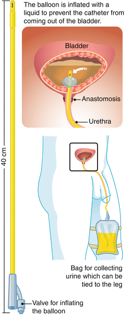
Figure 7.10
Catheter
Similarly, when you walk, you can carry it in your hand, with your arms extended downward. There is no need to worry about the fact that the urine has a reddish color, since it is normal for it to be mixed with a little blood during the first days after the operation. Mainly it turns reddish when walking or having a bowel movement. All of this subsides spontaneously during the first days after the operation and the urine assumes a normal color more quickly if you drink plenty of clear fluid (water, crystalline drinks, etc.) On occasion when passing stool, a few drops of blood might appear on the penis, around the catheter. This is usually a non-alarming sign. Also it is important to verify that urine is coming out adequately thorough the catheter. For this it is necessary to empty the urine-collecting bag every 2 h, thereby being able to confirm that it fills up regularly.
Usually a cream is prescribed to apply at the tip of the penis around the catheter, to avoid inflammation of the skin.
At home, after surgery:
The great majority of prostate patients who have undergone surgery by laparoscopy or robot are released from the hospital the day after the operation and are sent home in good condition to continue their convalescence. The following is indicated for them:
Take antibiotics: If instructed by the doctor, take antibiotics after leaving the hospital. This treatment must be completed as indicated in order to avoid infections.
Take painkillers: The doctor will indicate the most adequate painkiller to avoid and alleviate the pain. Generally, a few days after the operation the requirements for painkillers are low, and these should be taken according to how the patient feels and the guidelines from the doctor.
Rest: Remain at home, resting. During the first 5–7 days it is acceptable to go to the bathroom, take a shower, eat while seated at a table, and carry out normal activities that do not require much physical effort. It is advisable to sit on soft surfaces or on a pillow while the catheter is still in place. After 2 weeks, begin to do more activities, such as driving an automatic car for short distances, carrying out light office work, etc. as long as your are not having pain the require the use of narcotic pain killers, but do not return to demanding jobs such as masonry, gardening, or plumbing; do not engage in any physical exercises or sports. After 30 days it is acceptable to fully return to work, sports, and other daily routines. Sexual activity can also be renewed a month after the operation, either with natural erections or with the treatment recommended to improve them, which can be through intracavernous injection or oral medication (see a more detailed discussion later in this chapter). However, until 3 months after the operation, do not ride a bicycle, motorcycle, or horse.
Clean the wounds: It is only necessary to wash the wounds with soap and water during a daily bath. The wounds can be left uncovered; it is not necessary to cover them with bandages or dressings.
The drain is a small rubber hose placed in the abdomen of the patient, extending to the exterior through one of the holes used in the laparoscopy. It facilitates the drainage of any liquid that may accumulate inside the abdominal cavity (blood, urine, serous fluid, etc.)—a routine occurrence after surgeries.
Care of the drain
Generally, the doctor removes the drain between the second and forth day after the operation, but in some cases it may be necessary to maintain it in place a few days longer. In these situations, the patient should clean it just like the other wounds and arrive punctually at the appointment made by the doctor to remove it. For this appointment it is recommended to measure the quantity of liquid collected in the reservoir during the past 24 h, to help the doctor with the decision to remove the drain (Fig. 7.11).
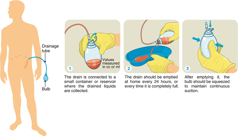
Figure 7.11
Drainage
Medical visit for catheter removal
Approximately between 7 and 10 days after the operation, you should see the doctor for evaluation and removal of the catheter. Typically, this appointment will be made by the doctor. There will be a certain degree of incontinence when the catheter is removed, something which is normal and will disappear in time. Therefore, you should take an adult diaper to the appointment. The duration and severity of the incontinence is variable and depends on many factors, so it is necessary to be patient and understand that it is part of normal convalescence. Statistically, more than 50 % of the patients have complete continence when the catheter is removed; after 3 months, that figure goes up to more than 85 %; and at 6 months, almost all (95 %) recover urinary continence.
Diet at home
Upon returning home from the hospital, eat a regular, healthy diet. Drink abundant clear fluids (eight to ten glasses a day) to keep the urine clear. Also, it is good to eat easily digestible food like vegetables, salads, fruits, greens, and broth. The diet should be rich in fiber to ease bowel movements and include food like cereals, wheat germ, wheat bran, bran, vegetables, prunes, prune yogurt, etc.
The overall diet should also have low-fat content, and give preference to meats like chicken or fish (as opposed to red meat), either grilled or baked. Those patients who previously suffered from constipation and who were taking laxatives (Sennosides, Plantago Psyllium, etc.) should start taking them again.
Whole milk is not recommended nor are citric fruits, fried eggs, or alcoholic drinks. This diet is oriented toward controlling constipation, since it is normal for patients to not evacuate up to 4 or 5 days after the operation. This is basically because in the first days there is hardly any intestinal content, a result of the cleansing done by the enema prior to surgery, and also because surgery produces a certain degree of slowing of the intestinal movements, which reduces the frequency of the bowel movements.
Resumption of previous treatments
Patients who suffer from any type of illness, which requires daily medicine, can restart their treatments after the operation. In cases of diabetes mellitus, oral hypoglycemic agents should be resumed once the patient resumes his regular diet and the functions are back to normal. Treatments with anticoagulants or Aspirin® should be suspended until the catheter is removed.
Sleep
Undergoing surgery causes certain discomfort and generates some anxiety in some patients; thus, there may be some difficulty in sleeping. During the first and second weeks after the operation the patient is in a period of convalescence, which requires a greater expenditure of energy. For this reason he should take a nap during the day to restore his strength.
Pathological Interpretation of the Prostate Removed During Surgery
The specimen obtained from surgery consists of the prostate and seminal vesicles. When advanced disease is suspected, the lymph nodes are also included. The specimen is studied to identify certain parameters that will determine whether cancer treatments are necessary after surgery. These parameters are established by the Gleason grade, the histological features of the surgical margins, which show if the disease has extended outside the prostate, whether it has reached seminal vesicles, and if there is metastasis to the regional lymph nodes.
The information obtained from this analysis is used to predict the evolution of the illness, measure the probability of biochemical recurrence, and calculate long-term survival. It is also useful to indicate or select the adjuvant therapy, radiotherapy, or hormone therapy.
The three dimensions of the prostate are measured and it is weighed without the seminal vesicles. It should be immediately immersed in a solution of 10 % formalin to prevent self-destruction of the tissues. This process must be completed in full, prior to proceeding to its study. The specimen should be immersed in a volume at least 20 times greater. The time depends on the size. One hour per millimeter of thickness of the tissue is estimated. Usually it takes at least 24 h. Then the outer surface is stained with different colors for topographical identification of the tumor and precise determination of the surgical margins. It is cut into thin slices in order to begin the microscopic examination.
The number of histological slides of each specimen varies, with an average of 20 per case. In selected cases the entire piece is included, which means up to 60 slides for study, which explains why it takes several days to get the final result of the biopsy.
Histological examination of the apex, which corresponds to the tip and the base of the prostate, is essential for the evaluation, in addition to the margins of resection, which are considered positive if the tumor is in contact with the stained surface.
Biopsy Results
Positive margins of resection
The positive surgical margin is defined as the extension of the tumor to the outer surface of the specimen, which is manifested by the contact of the tumor cells with the painted surface of the prostate.
The average rate of positive margins in radical prostatectomy specimens is 28 %, with values that can range between 0 and 53 %. The possibility of positive margins appearing is related to various factors, including the value of the pre-operative prostate-specific antigen (PSA), the clinical stage, the tumor volume, the percent of cancer in the biopsy and its Gleason number (Chap. 4), the pathological processing of the specimen, and even the surgeon’s experience.
Today the incidence in positive margins is progressively diminishing because the majority of prostate cancers are diagnosed and operated on in an early stage. Moreover, more surgical experience has been acquired and new techniques continue to be perfected (Fig. 7.12).
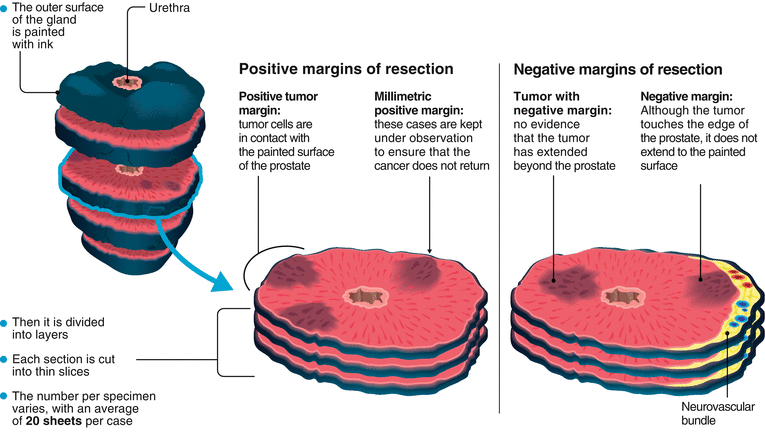
Figure 7.12
Positive margins
The treatment for positive margins is different in each patient. It depends basically on the quantity of tissue affected. In patients with only one positive millimetric margin identified, the choice can be to conduct observation and close monitoring according to the value of the prostate-specific antigen (PSA); if this tends to rise, complementary treatment will be started, such as radio therapy or hormone blockade. In the case of patients who show an extensive zone or multiple zones with positive margins, the urologist may decide to start early adjuvant treatment in conjunction with the radiation oncologist and medical oncologist to prevent the spread of the disease.
It should be understood that every patient is different and will receive the most adequate therapies in a personalized way, since not only is the number of millimeters of tumor in contact with the surface important but the Gleason histological grade present is also taken into account, which is what determines the aggressiveness of the tumor.
Negative resection margins
A negative surgical margin means there is no evidence of contact between the tumor and the surface of the cutting edge of the surgery, suggesting that it has not extended beyond the prostate; therefore, there is a better prognosis.
Prognosis
The surgery should eliminate all cancerous cells. Even so, small tumor cells may have escaped before surgery without being identified. Therefore, the doctor will keep close watch on the patient for some time to be sure the cancer does not reappear. The patient will have to undergo regular checkups, including blood tests for the prostate-specific antigen and urine cultures. Generally the followup will be every 3 months during the first 2 years, then every 6 months for three more years, then annually.
Complications of Radical Prostatectomy
Traditionally, this has been a greatly feared operation because of the complications and consequences associated; however, thanks to research in recent years, it has been possible to describe the anatomical details and exact location of the blood vessels and nerves (neurovascular bundles) that are responsible for both urinary continence and for the erection. Now that the anatomy of the region is known perfectly, the technique has evolved and surgery can be performed while respecting each of these important structures and minimizing the associated risks. These risks are inherent in the general condition of the patient and the surgical procedure itself. They can be classified into general and specific risks.
1.
General: These are the same complications as for any other surgery, and include the following:
Allergic reactions: Allergies may occur to any drug, including anesthetics, as well as to operating room materials such as gloves, sheets, etc.
Infection: Of specific concern is the surgical wound but infection may also present itself in the lungs (pneumonia), in the abdominal cavity, in the bladder, or in the kidneys.
Pulmonary problems: Atelectasis (lung volume reduction), pneumonia, pulmonary thromboembolism.
Hemorrhage: Bleeding, at the time of surgery or afterwards, that warrants transfusion or reoperation.
Cardiovascular problems: Heart attack or stroke during or after surgery.
2.


Specific: These are the complications related to the specific surgery used in the treatment of prostate cancer.
Intestinal injury: This is the most dangerous of all; it consists of fecal material leaking into the abdominal cavity due to the opening of the colon or rectum during surgery. This complication is very rare, but in very specific cases, such as inflammation of the prostate, history of multiple biopsies of the prostate, locally advanced disease, and a salvaged surgery after failure of brachytherapy or radiotherapy, it becomes difficult to separate the rectum from the prostate, thereby increasing the risk of injury. If it occurs, the important thing is to diagnose it. Generally, when repaired by suture, there is no additional complication. If not diagnosed or if the repair fails, a rectovesical fistula may occur.
Rectovesical fistula: This is a communication between the rectum and the location where the bladder was joined to the urethra (anastomosis site). This condition is manifested by the outflow of fecal matter through the urinary catheter, and/or the outflow of urine through the rectum. This complication is rare and is reported in the United States in 1 % of patients who undergo open retropubic prostatectomy.
Urinary incontinence: This is the involuntary loss of urine, requiring the use of diapers. Usually it is transient. The patient progressively notes his recovery. First, at night, he is aware that he can get up and go to the bathroom. Then, during the day, he will perceive that the loss between urinations decreases and that he requires fewer diapers. Then he substitutes the diapers for sanitary napkins or towels for protection, and so on until he loses only a few drops when exerting himself, doing exercises, or drinking alcohol.
Related posts:

Stay updated, free articles. Join our Telegram channel
Full access? Get Clinical Tree
