Primary and Metastatic Tumors of the Spine: Total En Bloc Spondylectomy
Primary and Metastatic Tumors of the Spine: Total En Bloc Spondylectomy
Hideki Murakami
Norio Kawahara
Katsuro Tomita
BACKGROUND
Conventionally, curettage or piecemeal excision has been the usual approach to vertebral tumors.
These approaches have clear disadvantages, however, including high risk of tumor cell contamination to the surrounding structures and residual tumor tissue at the site due to difficulty in demarcating tumor from healthy tissue.
These contribute to incomplete resection of the tumor as well as high local recurrence rates of the spinal malignant tumor.
To reduce local recurrence and to increase survival, we have developed total en bloc spondylectomy (TES).
10,
11,
14
In this method, the entire vertebra or vertebrae containing the malignant tumor are resected, together with en bloc laminectomy, en bloc corpectomy, and bilateral pediculotomy using a T-saw through the posterior approach.
9
Using this technique, we are able to excise the tumor mass together with a wide or marginal margin.
ANATOMY
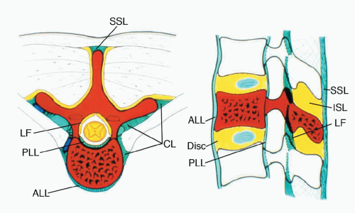
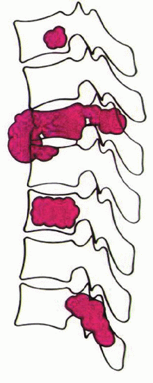
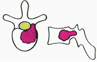
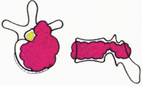
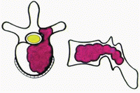
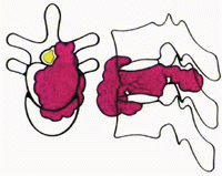
The following tissues serve as barriers to spinal tumor progression: the anterior longitudinal ligament (ALL), the posterior longitudinal ligament (PLL), the periosteum abutting the spinal canal, the ligamentum flavum (LF), the periosteum of the lamina and spinous process, the interspinous ligament (ISL), the supraspinous ligament (SSL), the cartilaginous endplate, and the cartilaginous annulus fibrosus. However, both the PLL and the periosteum on the lateral side of the vertebral body are “weak” anatomic barriers. In contrast, the ALL, cartilaginous endplate, and annulus fibrosus are “strong” barriers. In the spine, one vertebra could be regarded as a single oncologic compartment and the surrounding tissues as barriers to tumor spread
(FIG 1).
5


