Primary POF
Genetic aberrations
X-linked (monosomy, trisomy, deletions, translocations, fragile X)
Autosomal dominant (FSH receptor gene polymorphism, inhibin B mutation, etc.)
Enzyme deficiency
Metabolic
Autoimmune disease
Secondary POF
Surgical
Bilateral oophorectomy
Hysterectomy without oophorectomy/uterine artery
embolization
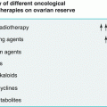
Chemotherapy or radiotherapy
Infections
Various terms have been suggested to define deviation from healthy ovarian function, including premature menopause (PM) or premature ovarian insufficiency (POI). Many authors regard ovarian insufficiency as more accurate than ovarian failure, because ovarian insufficiency can be used to described a wide range of impaired ovarian function. The term also suggests that ovarian follicular activity might intermittently recover, even years after diagnosis and lead to pregnancy in about 5–10 % of these women [2].
Follicle depletion or dysfunction in adolescent or young women may be caused by many different factors. POF might also result from genetic defects, chemotherapy, radiotherapy, or surgery. A common cause of POF in adolescent is gonadal dysgenesis, with or without Turner syndrome. When ovarian failure presents as a primary amenorrhea and no associated comorbidities, approximately 50 % are found to have abnormal karyotypes. However, most causes of spontaneous POF present as secondary amenorrhea and among younger women aged 30 years or younger with secondary amenorrhea, 13 % also have been noted to have an abnormal karyotype. Thus, in most cases the diagnosis will be 46, XX spontaneous POF, meaning the karyotype is normal [2]. In 90 % of cases an unknown mechanism leads to premature exhaustion of the resting pool of primordial follicles and no etiology for spontaneous POF will be identified even after a thorough evaluation. Approximately 4 % of women with 46, XX spontaneous POF will have steroidogenic cell autoimmunity as the mechanism of POF [3]. Approximately 6 % of women with 46, XX spontaneous POF will have premutations in the FMR1 gene. This is the gene responsible for fragile X syndrome, the most common cause of familial mental retardation. The risk of having an FMR1 premutation is higher if there is a family history of POF, therefore, in these patients it is crucial to take a family history, and women who have relatives with spontaneous POF should be referred for genetic counseling. Moreover, approximately 6 % of women with familial POF will have a premutation in the FMR1 gene as compared with 2 % of women with isolated POF. Women found to have a premutation in the FMR1 gene are at risk of having a child with mental retardation, should they be one of the 5–10 % who conceive [4]. There are other rare genetic causes of familial POF for which routine genetic testing in sporadic cases is now not clinically indicated, such as mutations involving FSHR (FSH receptor), GALT (galactose-1-phosphate uridylyltransferase associated with galactosemia), FOXL2 (a forkhead transcription factor associated with the blepharophimosis/ptosis/epicanthus inversus syndrome), INHA (inhibin alpha gene), EIF2B (a family of genes associated with central nervous system leukodystrophy and ovarian failure), BMP15 (bone morphogenetic protein 15), and AIRE (autoimmune regulator gene associated with the autoimmune polyendocrinopathy-candidiasis-ectodermal dystrophy syndrome) [5] (Table 12.2).
Table 12.2
Genes implicated in Premature Ovarian Failure (POF)
Categories | Chromosomes | Gene |
|---|---|---|
Identified mutations | X | FMR1, 2, BMP15 |
Autosomal genes | FOXL2, FSHR, LHR, FSHβ, LHβ, inhibin A, GALT, AIRE, NOGGIN, POLG | |
Unidentified mutations | X | AT2, c-kit, sox 3 |
Autosomal genes | MIS | |
Candidate genes | X | DIAPH2, DFFRX, XPNPEP2, 2FX, FSHPRH1, XIST |
Autosomal genes | WT1, ATM |
POF can be explained by different autoimmune mechanisms including a general immune dysregulation such as polyglandular syndrome (i.e., hypothyroidism, adrenal insufficiency, and hypoparathyroidism 14–27 %), dry-eye syndrome, myasthenia gravis (2 %), rheumatoid arthritis, diabetes mellitus (2 %), or systemic lupus erythematosus. It might also arise from an inflammatory autoimmune process against ovarian-specific germ line antigens or regulatory factors, making the disorder more complex than initially thought. The prevalence of these diseases in women with POF is higher than in the general population, suggesting that there may be an autoimmune component in these women that is not well understood. Autoimmunity in ovarian failure can be divided into two categories: ovarian failure associated with autoimmune adrenal insufficiency and ovarian failure associated with other autoimmune disease. It is only when POI is associated with adrenal autoimmunity or insufficiency that true autoimmune oophoritis can be demonstrated. Approximately 2–10 % of POF is associated with adrenal failure or autoimmunity and would thus be expected to result from autoimmune oophoritis. Circulating steroidogenic cell antibodies have been recorded in these autoimmune cases, therefore, steroid cell and/or enzyme antibody markers are present in 60–87 % of women with secondary amenorrhea and adrenal autoimmunity and/or Addison’s disease; therefore, it has been suggested that antibodies discriminate best between autoimmune POF and POF of other etiologies. However, ovarian antibody assays have a poor specificity and testing is therefore not recommended in women with primary ovarian insufficiency [6]. Although POF can happen as a result of ovarian surgery, oophorectomy, or exposure to viral or environmental toxic agents such as smoking, the most common cause of acquired disorder is chemotherapy or radiotherapy for cancer treatment. Chemotherapy-induced ovarian damage can arise through impairment of follicular maturation or primary follicle depletion, or both. Moreover, oocytes are very sensitive to radiation and age of the patient, and extent, type, and schedule of irradiation are key prognostic factors for development of primary ovarian failure. One dose is more destructive to the oocytes than are fractionated doses, and pelvic and abdominal irradiation has the highest risk, although scatter radiation can cause substantial damage even when the ovaries are not within the radiation field [7].
In the absence of symptoms, a change in the regular menstrual bleeding pattern is the main presenting symptom with POF. The absence of the menses in a 15-year-old girl (primary amenorrhea) or the cessation of menses for 4 months or more (secondary amenorrhea) can point towards the diagnosis; however, there is no consensus on criteria to identify primary ovarian failure in adolescence and delay in diagnosis is common. Some adolescent females report hot flushes and night sweats or vaginal symptoms like dryness or dyspareunia, low libido, low energy levels, sleep disturbance, lack of concentration, stiffness, skin/hair changes, and mood swings, however the most frequent symptoms of POF is primary or secondary amenorrhea. Among patients with amenorrhea, the incidence of POF ranges from 2 to 10 %. Abnormal bleeding patterns also include oligomenorrhea (bleeding that occurs less frequently than every 35 days), non-structural causes of abnormal uterine bleeding (ovulatory dysfunction, iatrogenic, or not classified), or polymenorrhea (bleeding that occurs more often than every 21 years). Because irregular menstrual cycles are both common during early adolescence and an initial symptom of early POF, diagnosis can be difficult in this population. Although less than 10 % of women presenting abnormal menses will ultimately be found to have POF, the condition has such detrimental consequences on bone health that early diagnosis of this condition is important [8]. Therefore in young female it is important to evaluate amenorrhea or change from regular to irregular menses for three or more consecutive months in the absence of hormonal preparations such as oral contraceptives (OCs). Differential diagnosis is based on the exclusion for all potential causes of primary and secondary amenorrhea including pregnancy, polycystic ovary syndrome, hypothalamic amenorrhea, thyroid abnormalities, and hyperprolactinemia. In a woman aged less than 40 years, the diagnosis of POF is usually confirmed by the combination of the triad of oligo/amenorrhea for at least 4–6 months, sex steroids deficiency, and two recording of serum concentrations of follicle-stimulating hormone (FSH) of more than 30–40 IU/L at least 1 month apart. If the result indicates that FSH is elevated, a diagnosis of POF can be established. Estradiol levels of less than 50 pg/ml indicate hypoestrogenism. However FSH is not an ideal diagnostic tool because it rises only in the later stages of follicle depletion, has marked cycle- to-cycle variability, and it is poor at predicting reproductive status. In this view, there has been interest in more direct markers of ovarian reserve such as anti-Mullerian hormone (AMH), which closely follows the reduction in follicle number over time in healthy women and falls to very low levels prior to menopause. In assessment of amenorrhea, AMH or transvaginal ultrasound scan will exclude polycystic ovarian syndrome as a cause. In POF, the antral follicle count is very low, and seeing this as a related marker of ovarian function may help to understand the diagnosis. However, even in POF, the intermittent ovarian function means that follicular activity is seen in the majority of women [9, 10].
In the absence of an obvious cause of POF (oophorectomy, chemotherapy, or pelvic radiotherapy), young women should be offered investigation in etiology, however, in the majority of cases no cause is found. Even if causation is established, the management, including fertility options, remains unchanged. E genetic etiology will be identified in approximately 5 % and autoimmune in up to 30 % [10]. Genetic counseling is nowadays recommended for several reason when a genetic form of POF is suspected or identified and a karyotype should be offered to women with POF in the onset of amenorrhea or oligomenorrhea before age 25. A karyotype is also indicated in women of any age in whom Turner’s syndrome mosaicism is suspected, as well as e FMR1 (fragile x) permutation testing. Overall the permutation is found in 4–5 % of women with POF, among those with a family history of POF, 14 % have a positive result, in this view a full family history is important as many cases of spontaneous POF appear to be inherited, estimates vary from 4 to 31 %. Thirty percent of the cases of POF are estimated to be owing to autoimmunity. Presumed autoimmune etiology may be inferred from a family history of autoimmune disease or a positive anti-thyroid antibody result, which is found in approximately 24 % of women affected by POF. If a POF patient appears to be a sporadic and not hereditary case, the risk of other female relatives developing POF will probably be equal to the risk in the general population [11].
Related posts:


Stay updated, free articles. Join our Telegram channel

Full access? Get Clinical Tree


