Figure 5.1
The Semmes-Weinstein monofilament used to check foot skin sensation. The open circles indicate areas tested
The biothesiometer (or neurothesiometer) is a simple handheld device that gives semi-quantitative assessment of vibration perception threshold (VPT) (Fig. 5.2). A VPT > 25 V is abnormal and has been shown to be strongly predictive of subsequent foot ulceration. A large multicenter study showed a significant increase in risk with each volt increase of VPT over 25 V [1].
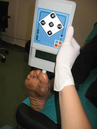
Figure 5.2
The measurement of vibration perception threshold on the toe of a diabetic patient
The development of a neuropathic ulcer is complex, involving repetitive injuries of which a patient may be unaware on account of loss of sensation. The propensity to injury is also associated with limited joint mobility and structural foot deformities, and hence, high plantar pressures over certain areas that are regarded as pre-ulcerative areas. Raised dynamic plantar pressures significantly increase the risk of foot ulceration in patients with PN who have lost protective sensation.
Clinically, findings such as callus, sub-keratosis haemorrhages, blisters, macerated skin, limited hallux dorsiflexion of less than 30°, prominence of metatarsal heads or other types of plantar prominences such as rocker-bottom deformity seen in the Charcot foot, suggest high-pressure areas in the plantar surface of the foot. Patients with a history of foot ulcers or surgery to the metatarsal head may also have high plantar pressures and therefore should be considered as high risk of developing foot ulcers.
Pressure may be measured using platform devices for interface measurement between the foot and the floor as well as in-shoe systems to measure pressure between the sole of the foot and the shoe. No accepted threshold for the onset of ulceration currently exists since it is difficult to directly compare values obtained using different plantar pressure measurement devices. A threshold of 700 KPa has been suggested for predicting foot ulceration using an EMED® platform [2].
Peripheral Arterial Disease
PAD is important in the aetiology of diabetic foot ulcers. It is essential to detect PAD in the pre-ulcerous diabetic subject in order to prevent skin breakdown. Clinical signs of PAD include loss of hair on the dorsum of the foot, thin skin and fragile and deformed nails. A history of intermittent claudication suggests the presence of PAD and, although some patients may not experience pain due to neuropathy, walking is usually limited by fatigue.
Rest pain signifies severe PAD. Pallor of the foot on elevation and rubor on dependency suggest the presence of severe ischaemia, but the absence of these signs does not permit its exclusion. The first step in the assessment is palpation of the posterior tibial and dorsalis pedis arteries, which supply the foot. Palpating for pulses is not always easy. Absent pulses usually indicate the presence of PAD, but this does not quantify the perfusion deficit. Objective measurements of the macrocirculation are achieved by measuring the ankle brachial pressure index (ABPI) using Doppler ultrasound, and duplex ultrasound imaging for haemodynamic assessment.
ABPI is measured using a blood pressure cuff and a hand-held Doppler probe. It should be measured in the supine patient, with the patient rested (for 5 min). Ankle systolic blood pressure should be measured over the posterior tibial and dorsalis pedis arteries; brachial systolic pressure on the same side should be recorded. If possible the pressure in the peroneal artery should also be recorded as in patients with diabetes this vessel is often preserved and becomes an important blood supply to the foot. The ratio of the highest ankle pressure to the brachial pressure is the ABPI.
ABPI is a simple screening test, and low values are associated with the presence of peripheral vascular disease [3]. An ABPI <0.5 signifies the presence of significant PAD, while values between 0.85 and 1.2 permit the exclusion of significant arterial disease. In the diabetic subject, ABPI may be falsely high due to Mönckeberg sclerosis (calcification of the tunica media), resulting in rigid arteries. It is related to peripheral neuropathy, but does not itself result in ischaemia. The predictive variables related to the presence of calcification in a series of patients admitted with foot disease were duration of diabetes greater than 20 years, retinopathy, albuminuria, and PAD. Patients with calcification of pedal arteries underwent more amputations and re-operations in a series, but differences in outcomes were related to the association of calcification of pedal arteries with PAD [4].
Since calcification affects digital arteries less commonly, toe pressures are measured either for an absolute value or to derive the toe brachial pressure index (TBI). Toe pressures are measured using an optical probe and a small pressure cuff (in a foot acclimatized in a warm environment [5]) (Fig. 5.3). Toe pressures less than 55 mmHg or a TBI of less than 0.7 strongly suggest the presence of PAD. Pressure measurements are reliable for diagnosis, but colour flow duplex ultrasound is the recommended next step for haemodynamic data to determine clinical management.
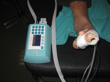
Figure 5.3
The measurement of toe blood pressure (could be used to derive toe brachial index) using an optical sensor and cuffs (not shown)
Duplex ultrasound imaging yields reliable data on both structure and function of the arteries down to the level of the dorsalis pedis artery. Beyond this the calibre of the vessels is too small to routinely image, and other imaging modalities, such as computerised tomography, are indicated.
Elevating the limb whilst insonating with a Doppler probe (the “Pole Test”) is a useful bedside test that can be performed in patients who elevated ankle pressures which seem out of keeping with their symptoms and signs. If the Doppler signal disappears as the limb is elevated an re-appears as it is lowered then the perfusion pressure of that limb is very low [6].
Once an Ulcer Appears
Where an ulcer is present, the aim is to heal the ulcer. However, the most difficult decision is to determine which ulcers will heal without intervention and which will only heal with revascularisation. Most commonly simple observation of the wound is used to assess wound-healing potential. The logic of this is sound, and the percentage change in the area of the foot ulcer after 4 weeks of observation is a robust predictor of healing at 12 weeks. Therefore, patients in whom ulcer size fails to reduce by 50 % over the first 4 weeks of treatment are unlikely to achieve wound healing over a reasonable period [7]. However, this is a slow and potentially dangerous technique, which delays revascularisation in those patients who ultimately need it.
Related posts:
 Foot Deformity and Pressure Management in the Diabetic Foot
 Screening and Treatment of Early Complications in the Diabetic Foot
The Role of the Multidisciplinary Team in the Management of Diabetic Foot Complications
The Role of the Orthopaedic Surgeon in Diabetic Foot Complications
Screening and Treatment of Early Complications in the Diabetic Foot
The Role of the Multidisciplinary Team in the Management of Diabetic Foot Complications
The Role of the Orthopaedic Surgeon in Diabetic Foot Complications
 Imaging in the Patient with Foot Complications
 Neuro-osteoarthropathy: The Charcot Foot – Pathology, Diagnosis, and Treatment
Stay updated, free articles. Join our Telegram channel

Full access? Get Clinical Tree


