Polycystic Ovary Syndrome


Polycystic Ovary Syndrome


It’s not how much you have that makes people look up to you, it’s who you are.

Elvis Presley, American rock “n” roll icon (1935-1977)


Introduction

Poetically described as “the thief of womanhood,”1 polycystic ovary syndrome (PCOS) was first diagnosed in 1935 by two Chicago obstetricians in seven women with a constellation of symptoms including hirsutism, amenorrhea, infertility, and large polycystic ovaries.2 PCOS remains characterized by irregular and chronic anovulation, signs and symptoms related to elevated androgens (hirsutism and acne), and polycystic ovaries. Additionally, PCOS is strongly associated with metabolic disorders and comorbidities including insulin resistance, prediabetes, diabetes, dyslipidemia, hypertension, obesity, endometrial carcinoma, depression, cardiovascular disease (CVD), and obstructive sleep apnea.3,4,5 PCOS can cause infertility, irregular menses, severe acne, and hirsutism. Women with PCOS constitute the largest group of women at risk for developing CVD and diabetes. The cardiovascular and metabolic risk factors associated with PCOS are potentially reversible. Although the patient’s immediate problems and concerns necessitate sensitive attention and prompt therapy, PCPs should view this disorder as an opportunity to practice proactive preventive medicine for this chronic, progressive disease. Early diagnosis, lifestyle interventions, and appropriate pharmacotherapy can result in a reduction in serious associated metabolic sequelae and restore fertility.


Prevalence and Pathogenesis of Polycystic Ovary Syndrome

PCOS is the most common endocrine abnormality in American reproductive-aged females resulting in menstrual irregularity and infertility.6 The prevalence of PCOS in adult women is estimated to be between 6% and 10%.6 PCOS symptomatology usually begins at the time of menarche. Environmental modifiers, such as weight gain, may delay the clinical presentation of PCOS until women reach puberty. Premature pubarche (appearance of pubic hair growth), the result of early secretion of adrenal steroids, may be a harbinger of the syndrome.7 Genetic studies support the increased frequency of PCOS in first-degree relatives of affected women.8


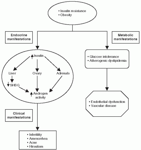
Figure 4-1 • PCOS neuroendocrine dysfunction.

Because of the heterogeneity of this disorder, multiple interrelated endocrine pathways have been implicated in the pathogenesis of PCOS (Fig. 4-1). A normally functioning hypothalamic-pituitary-ovarian axis supports the maturation of ovarian follicles via the release of luteinizing hormone (LH) from the anterior pituitary gland. Follicle-stimulating hormone (FSH) stimulates the production of the ovarian follicles and is also secreted from the anterior pituitary gland. Within the ovarian follicle, theca cells synthesize androgen. Once formed, the androgens diffuse into the ovarian granulosa cells where they are converted into estrogen.

Patients with PCOS experience increased ovarian androgen biosynthesis as a result of abnormalities occurring at all levels of the hypothalamic-pituitary-ovarian axis:


  • LH hypersecretion is a characteristic hallmark of PCOS. LH is secreted in a pulsatile manner. Women with PCOS have an increase in both the LH pulse frequency and amplitude resulting in increased 24-hour secretion. LH hypersecretion is thought to occur secondary to an increased frequency of hypothalamic gonadotropin-releasing hormone (GnRH) pulsation. Higher circulation levels of LH stimulate the production of androgens by the ovarian theca cells. (GnRH) must be accelerated in PCOS.9


  • Increasing the GnRH pulse generator favors the synthesis and release of LH over FSH.


  • When the concentration of LH increases relative to FSH, the ovaries preferentially synthesize testosterone.


  • Insulin acts synergistically with LH to increase androgen production within the theca cell.


  • Insulin also inhibits the hepatic synthesis of SHBG, which normally binds testosterone. The higher levels of unbound or “free” testosterone increase the biologic activity of the circulating hormone. The concentration of free testosterone is often elevated, whereas the total testosterone level may be only slightly increased.10


  • Testosterone further inhibits (whereas estrogen stimulates) the hepatic synthesis of SHBG.11

An increase in free and total serum testosterone levels results in androgenization, the most obvious features of which are hirsutism, acne, and diffuse alopecia. Hyperandrogenemia interferes
with the hypothalamic-pituitary axis, leading to anovulation (Fig. 4-1). The absence of a dominant follicle prevents development of nondominant follicles, resulting in the formation of multiple ovarian cysts.12 Androgen excess also affects other metabolic parameters such as lipid concentrations. A recently published study demonstrated reduced levels of glucagon-like peptide-1 (GLP-1) and glucose-dependent insulinotropic polypeptide (GIP) in patients with PCOS.13 In normal subjects, these incretin hormones are responsible for 70% of the insulin response during a meal. However, in patients with T2DM and in other insulin-resistance conditions, the incretin effect is impaired. The reduced physiologic levels of incretin hormones in patients with PCOS may have pharmacologic and therapeutic implications warranting future investigations.


Figure 4-2 • Metabolic abnormalities associated with PCOS.

Women with PCOS typically have hyperinsulinemia and insulin resistance.14 Insulin resistance is known to precede the development of T2DM. Studies have shown that 30% to 40% of women with PCOS have impaired glucose tolerance (IGT), and as many as 10% develop T2DM mellitus by the age of 40.10 Figures 4-1 and 4-2 summarize the pathogenesis and clinical outcomes associated with PCOS.


Diagnostic Criteria for Polycystic Ovary Syndrome

The 2003 Rotterdam PCOS Consensus Conference, held by the European Society of Human Reproduction and Embryology in conjunction with the American Society for Reproductive Medicine, developed the following diagnostic criteria for PCOS.15


  • A history of irregular menstrual cycles and anovulation with onset at puberty. Twenty-five percent of women with PCOS have regular menstrual cycles.


  • Elevated total and free testosterone levels.


  • The presence of polycystic ovaries and the exclusion of other hormonal disorders with similar clinical features such as adult-onset congenital adrenal hyperplasia, hyperprolactinemia, adrenal or ovarian androgen-producing adenomas, hyperthecosis, and Cushing syndrome. The term hyperthecosis refers to the presence of nests of luteinized theca cells in the ovarian stroma secondary to differentiation of the ovarian interstitial cells into steroidogenically active luteinized stromal cells. These nests or islands of luteinized theca cells are scattered throughout the stroma of the ovary, rather than being confined to areas around cystic follicles as in PCOS. The end result is greater androgen production of androgens. The clinical features of hyperthecosis are similar to those of PCOS. However, women with hyperthecosis have more hirsutism and are much more likely to be virilized.16 Hyperthecosis results in elevations of both estrogen and androgen production, increasing the risk of endometrial hyperplasia and endometrial carcinoma, especially in postmenopausal women.

The Rotterdam PCOS Consensus Panel suggested that tests of insulin resistance are not necessary to make the diagnosis of PCOS or to select the type of treatment. Obese women with PCOS should be screened for metabolic syndrome and T2DM.

The Androgen Excess Society stresses the importance of hyperandrogenism as the cardinal feature of PCOS, placing less emphasis on ovarian morphology. Thus, the cardinal diagnostic features of PCOS are based on a patient’s dysfunctional reproductive and metabolic endocrine access rather than on the physical appearance of the ovaries. The clinical manifestations of PCOS are listed in Table 4-1.


Clinical Evaluation of Polycystic Ovary Syndrome

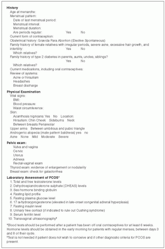
Figure 4-3 summarizes the historical and clinical features that should be evaluated in patients suspected of having PCOS. The typical history reveals a patient whose menarche begins as anticipated at the average age of 12 to 13 years. Shortly after menarche, the periods become irregular. Physicians may prescribe oral contraceptives to help regulate the menstrual cycles. Oral contraceptives lower serum androgen levels, improve menstrual cycle regularity, and reduce hirsutism and acne. Although beneficial for the treatment of PCOS, the use of oral contraceptives can result in a delay in one’s clinical diagnosis. Early, intensive lifestyle and pharmacologic management of the metabolic disorders associated with PCOS is essential for reducing one’s risk of diabetes and CVD progression.


Clinical Features Associated with Polycystic Ovary Syndrome


• Hirsutism, Alopecia, and Acne

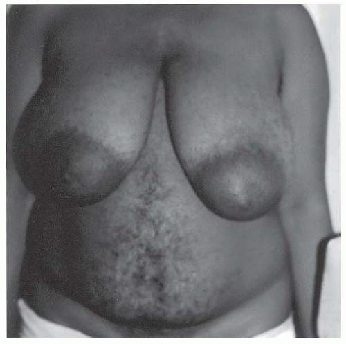
Hirsutism is the most common manifestation of androgen excess associated with PCOS. Approximately 10% of reproductive-aged women without PCOS are hirsute, versus 70% of patients with PCOS.17,18 Hair growth occurs on the chin, cheek, sideburns, neck, chest (Figs. 4-4 and 4-5) and between the breasts, on the periareolar area and upper arms, and below the umbilicus. Distribution of hair with dark hairs spreading onto the thighs may be observed in some patients. An increase in “terminal” or “androgenic” hair supersedes the appearance of short, fine, light-colored vellus hair, which is normally found throughout the body. The onset of hirsutism in PCOS follows menarche, although some girls have earlier onset of pubic hair development and some degree of hirsutism. As patients gain more weight, the hirsutism intensifies.

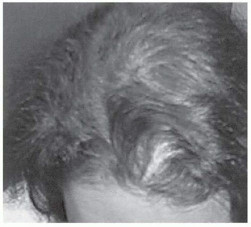
Diffuse alopecia occurs in 40% to 70% of women with PCOS. Poor nutrition (particularly related to low protein intake), anemia, genetic predisposition, and zinc deficiencies in association with androgen excess increase the risk of developing alopecia19 (Fig. 4-6).


TABLE 4-1. Clinical Manifestations of PCOS


Manifestation


Comment


Menstrual dysfunction and/or infertility


PCOS, an entity of chronic anovulation, is associated with the most frequent cause of anovulatory infertility.


Hirsutism


Hirsutism in association with menstrual dysfunction is often the most common finding in PCOS.


Acne


Biochemical evidence of hyperandrogenism may occur in the absence of hirsutism or acne.


Alopecia


Alopecia is common and often associated with other features of PCOS.


Visceral (android) fat distribution and obesity


Obesity particularly associated with a visceral fat distribution may further perpetuate the symptoms and risks for metabolic and cardiovascular risks.


Insulin resistance


Possibly related to increase in body iron stores (serum ferritin levels) which result in β-cell dysfunction. Insulin sensitizers (metformin) lower serum ferritin level in obese and overweight women with PCOS.a


Miscellaneous


Sleep apnea


Obstructive sleep apnea in PCOS is a serious symptom that must be looked for particularly in those with increased visceral fat.


Acanthosis nigricans and skin tags


The severity of skin findings may parallel the degree of insulin resistance, and a partial resolution may occur with weight reduction and treatment of the insulin-resistant state.


a Luque-Ramirez M, Alvarez-Blasco F, Botella-Carretero JI, et al. Increased body iron stores of obese women with polycystic ovary syndrome are a consequence of insulin resistance and hyperinsulinism and are not a result of reduced menstrual loss. Diabetes Care 2007;30:2309-2313.


The PCOS-associated acne often precedes the development of hirsutism. Forty percent of girls with severe acne who are resistant to oral and topical agents (including isotretinoin) have PCOS.20 Severe or moderate acne persists and even worsens in women ages 20 to 40 (Fig. 4-5). The clinical finding of acne may be the sole manifestation of androgen excess in women with PCOS.20

Generally, the androgenic-induced dermatologic anomalies observed in PCOS proceed from acne in the peripubertal period to hirsutism as a young adult to androgenic alopecia in the mature adult.


• Central Obesity

Women with PCOS may relate a sudden onset of weight gain over 6 to 18 months, further complicating symptoms associated with menstrual irregularities, infertility, acne, and hirsutism. The weight gain is often associated with a significant carbohydrate craving. Because of the exaggerated pancreatic β-cell secretion of insulin following the consumption of carbohydrate-rich meals, patients may experience postprandial symptoms suggestive of hypoglycemia such as loss of concentration, hunger, sweating, tremor, and insomnia. Weight gain is associated with an increase in visceral fat distribution, contributing to the development of IGT, atherogenic dyslipidemia, and proinflammatory markers, which constitute the components of metabolic syndrome.21 The central obesity favors progression to T2DM and increases the risk of CVD in patients with PCOS (Fig. 4-7).

Obese PCOS women are more insulin resistant than lean PCOS patients and lean controls. Although lean women with PCOS appear to secrete higher levels of basal insulin compared to age-matched controls, they do not exhibit signs of insulin resistance.22 Increased serum ferritin levels, indicating increased body iron stores, have been noted in obese and overweight women with PCOS. Insulin favors the intestinal absorption and tissue deposition of iron. β-cell dysfunction may be induced by

iron deposits in pancreatic islets similar to mechanistic models observed in hemochromatosis. The use of insulin sensitizers, such as metformin, might be useful in reversing elevated iron stores.22a


Figure 4-3 • History, physical, and laboratory workup of patients with PCOS.


Figure 4-4 • Patient with PCOS displaying diffuse hirsutism on abdomen and chest. (Photo provided courtesy of Walter Futterweit, MD.)


• Sleep Apnea

In comparison with healthy subjects, patients with PCOS have a 4- to 30-fold increased prevalence of sleep apnea.23 Major symptoms of sleep apnea include snoring, daytime somnolence, and fatigue. Sleep apnea increases the risk of developing hypertension, myocardial infarctions, stroke, and diabetes.24 The Epworth Sleepiness Scale may be used to screen patients for excessive sleepiness (Fig. 4-8).

Sleep dysfunction can be an independent risk factor toward the development of T2DM. A meta-analysis of sleep literature by Cappuccio et al.25 concluded that there is an “unambiguous increased
risk of developing T2DM at either end of the distribution of sleep duration and with qualitative disturbances of sleep.” Subjects averaging less than 5 to 6 hours per night of sleep have a 28% chance of developing T2DM, whereas those with difficulty maintaining sleep have an 84% likelihood of developing the disorder.
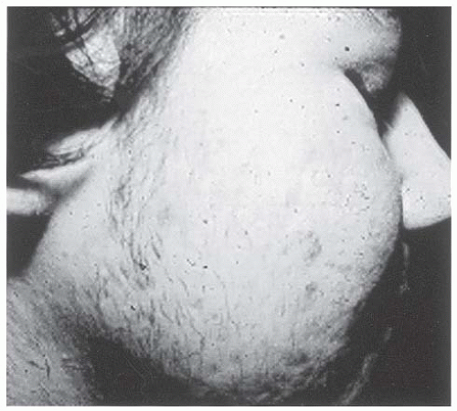


Figure 4-5 • Severe facial acne and hirsutism in a patient with PCOS. (Photo provided courtesy of Walter Futterweit, MD.)


Figure 4-6 • Alopecia associated with PCOS. (Photo provided courtesy of Walter Futterweit, MD.)

The mechanisms by which sleep apnea may potentiate insulin resistance implicate an imbalance between two hormones known to regulate food intake. Leptin is a peptide released by adipocytes
that provides information related to one’s energy expenditure to hypothalamic regulatory centers. In humans, circulating leptin levels rapidly decrease or increase in response to acute caloric shortage or surplus. Elevated plasma leptin levels decreases hunger and vice versa. Ghrelin is produced predominantly by the stomach and is also involved in energy balance regulation. However, in contrast to leptin, elevation in plasma ghrelin stimulates appetite. (See Chapter 8, Comanaging Associated Disorders of Diabetes).26,26a Individuals with insomnia, sleep fragmentation, and sleep apnea have a physiologic state that favors ghrelin secretion and leptin resistance. As the body spends more time in the “awake state,” additional nutritional intake and fat storage are needed to insure an adequate energy source for 20 hours of activity. Individuals who sleep 7 to 8 hours per night require enough energy intake and storage for only 16 to 17 hours of daily activity. The short night sleep patterns associated with insomnia, sleep fragmentation, and obstructive sleep apnea increase ghrelin secretion and favors leptin resistance. Although an individual may remain in bed for 10 to 12 hours a day, he or she is not sleeping efficiently. Ghrelin levels rise as the body strives to provide additional nutritional and energy stores to be used for during the 20 hours per day of wakefulness. The tendency for these patients is toward weight gain and insulin resistance. CPAP (continuous positive airway pressure) usage in young, obese patients with PCOS and sleep apnea has been found to improve insulin sensitivity, reduce diastolic blood pressure, and reduce sympathetic output. Patients with a BMI > 35 kg per m2 who used CPAP for 8 hours per night demonstrated the best improvement in metabolic parameters.26b
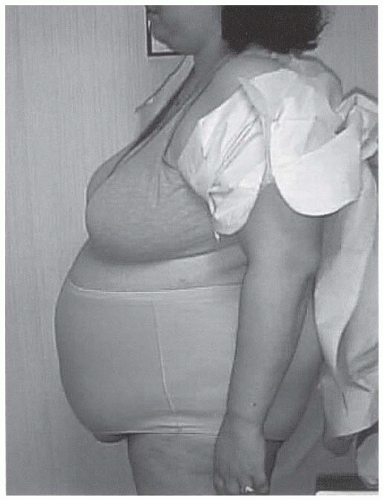


Figure 4-7 • Central obesity, a hallmark of PCOS and insulin resistance, is strongly associated with CVD risk. (Photo provided courtesy of Walter Futterweit, MD.)


Figure 4-8 • The Epworth Sleepiness Scale.


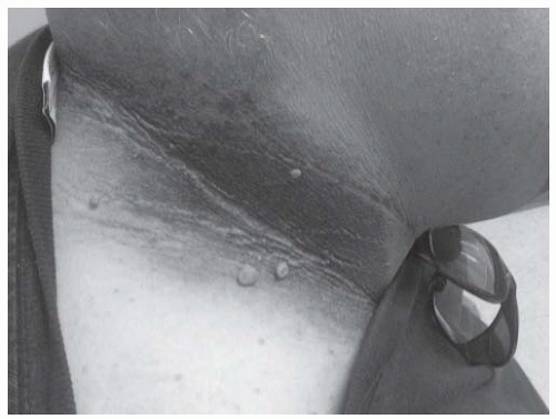
Figure 4-9 • Acanthosis nigricans and skin tags. (Photo courtesy of Jeff Unger, MD.)


• Acanthosis Nigricans and Skin Tags

The findings of acanthosis nigricans (Fig. 4-9) with or without skin tags are clinical markers of insulin resistance.27 In the absence of a paraneoplastic syndrome, acanthosis nigricans is an epiphenomenon of dermal hyperplasia seen mostly in the nape of the neck, axillae, groin, inner lips of vulvae, periumbilical and inframammary areas, and the dorsum of the fingers and knuckles. They are characterized as brownish-grey, velvety, or verrucous hyperparakeratotic pigmented areas, more commonly seen in obese women of Hispanic or African American descent, although they may be also noted in lean and white populations. Parents of children with acanthosis nigricans often express their frustration in their inability to “wash off the brown dirt with soap, water, and even bleach.” Skin tags are not often seen in women before the age of 40 years, and if present earlier, the tags are an important clinical clue to the presence of hyperinsulinism or impaired insulin sensitivity. Weight loss can improve both insulin resistance and the dark appearance of acanthosis nigricans.


Laboratory Studies and Radiographic Evaluation of Polycystic Ovary Syndrome


• Laboratory Studies

Hormonal studies are performed on patients with PCOS to determine the presence and severity of hyperandrogenism as well as whether the primary source is adrenal or ovarian. Although laboratory studies may be helpful in confirming the diagnosis of PCOS, no consensus statement has been published for the workup of these patients. Because of the circadian cyclicity of many of the hormones being evaluated, one should make note of the time the laboratory studies are obtained and when they are performed in relation to the patient’s menstrual cycle. Reference should also be made as to what medications the patient is taking at the time the tests are done, as some (such as oral contraceptives) may result in an inaccurate interpretation of the test results.

Hormonal testing should be performed in the early morning hours due to their circadian cyclicity. Patients with regular menstrual cycles should have their blood samples obtained between days 3 and 8 of the menstrual cycle. All initial blood tests on patients suspected of having PCOS should be performed at least 6 weeks after the cessation of oral contraceptives. Blood values obtained while the patient uses oral contraceptives are not useful in diagnosing PCOS because oral contraceptives lower circulating androgen levels.28

Although elevations in androgen levels are frequently observed with PCOS, some patients have normal free testosterone and total testosterone values.29 Standardized normal serum androgen levels

for adolescents and older women have not been determined. Notwithstanding these limitations, measurement of the free testosterone is thought to be a sensitive method of assessing hyperandrogenemia.30 A total testosterone value greater than 50 ng per dL is considered elevated.31 The optimal value for serum testosterone in women is unknown. Most women with a level greater than 50 ng per dL will have irregular menses and clinical symptoms related to hyperandrogenism. The level of free (unbound) testosterone is usually elevated in patients with PCOS.

Dehydroepiandrosterone sulfate (DHEAS) is a major androgen precursor secreted from the adrenal gland. Patients having elevations of both DHEAS and total testosterone should be evaluated for an androgen-secreting tumor.32 Additional tests, such as 17-ketosteroids (measuring androgen metabolites in the urine) and 17-OH progesterone, are useful in determining if the patient has an adrenocortical tumor, adrenal cancer, or adrenal hyperplasia. These tests should be obtained in patients with a DHEAS level greater than 700 ng per dL. Normal DHEAS levels range from 200 to 300 ng per dL (Table 4-2).


TABLE 4-2. Interpretation of Laboratory and Radiographic Studies in Patients Suspected of Having PCOS


Laboratory Test


Normal Rangea


Comment


Free testosterone


0-9.5 ng/mL


Hallmark feature of PCOS when elevated >50 ng/mL


The “free” testosterone is unbound to SHBG and reflects the patient’s biologically active level of testosterone.


Draw in early morning.


Repeat test to confirm abnormal values.


Dehydroepiandrosterone sulfate


35-430 µg/dL


Androgen precursor secreted from the adrenal gland


Elevated in cases of infertility, amenorrhea, hirsutism, hyperandrogenemia, adrenal tumors, and congenital adrenal hyperplasia


Elevated DHEAS requires further diagnostic testing to determine source of androgen excess.


17-ketosteroids (KS)


6-12 mg/24 h


Measures androgen metabolites in the urine


In association with elevated DHEAS may indicate adrenal tumor or adrenal hyperplasia


Spironolactone can raise levels of 17-KS.


17-OH progesterone


Day 3: 200-100 ng/dL


Midcycle: 100-250 ng/dL


Luteal phase: 100-500 ng/dL


Measures serum androgen levels. Deficiencies are noted with hyperandrongenemia.


In association with elevated DHEAS may indicate adrenal tumor or adrenal hyperplasia


Sex-hormone binding globulin (SHBG)


18-114 nmol/L


SHBG levels are low in obesity and hyperandrogenemia and with insulin resistance.


If SHBG is low, more free testosterone becomes metabolically active and induces the signs of hyperandrogenemia.


Urinary cortisol


10-100 mg/24 h


Measured in patients having cushingoid features. Levels exceeding 100 mg/24 h are suggestive of Cushing disease.


Luteinizing hormone (LH)


On day 3, the normal LH level is <7 mU/mL.


In PCOS, both the level of circulating LH and its relation to FSH levels are elevated due to an increase in amplitude and frequency of LH pulses from the pituitary gland.


On LH surge day, the LH level may increase to >20 mU/mL.


Elevated LH concentrations occur in 60% of PCOS patients. The LH/FSH ratio is >2:1 or 3:1 in up to 95% of PCOS patients.


Follicle-stimulating hormone (FSH)


3-20 mU/mL


Normally, the LH/FSH ratio is close to 1:1 A higher ratio is suggestive of PCOS.


Prolactin


<24 ng/mL


Increased prolactin levels can interfere with ovulation.


If elevated, consider further testing (MRI) to evaluated patient for pituitary adenoma.


May be elevated in PCOS


Screening tests for metabolic syndrome


Lipids


Fasting glucose 2-h post glucose challenge


Fasting plasma glucose levels of 100-126 mg/dL indicate IFG.


Plasma glucose drawn 2 h after a 75-g glucose challenge between 140-199 mg/dL is indicative of IGT.


Plasma glucose >200 mg/dL is diagnostic of diabetes.


Normal lipid values are:


Total cholesterol ≤ 200 mg/dL


HDL-C ≥ 55 mg/dL


LDL-C ≤ 130 mg/dL


Triglycerides ≤ 150 mg/dL


Ultrasound imaging


Optimally performed during the early follicular phase (days 3-6) of the menstrual cycle in women with regular menses


Anovulatory women should be scanned at random or 3-5 d after a progestin-induced withdrawal bleed.


Pelvic imaging is indicated only if the ovaries are palpable on physical examination or the total testosterone concentration is >200 ng/mL.


23% of “normal women” have characteristics of polycystic ovaries.


Polycystic ovaries are observed in many other disease states (see text).


Evaluate development of large cysts, effects of therapy on reducing cysts, and changes in the endometrium.


High frequency of fatty liver disease on ultrasonography


a Most hormone levels should be obtained on day 3 of menstrual cycle. Several determinations of plasma total and free testosterone levels are made.
HDL-C, high-density lipoprotein cholesterol; LDL-C, low-density lipoprotein cholesterol; MRI, magnetic resonance imaging.


Patients with cushingoid features should have a urine cortisol determination. Levels greater than 100 µg per 24 hours are strongly suggestive of Cushing syndrome.

If the 17-hydroxyprogesterone level at 8:00 AM (measured in the follicular phase of the menstrual cycle) is greater than 4 ng per mL, the patient probably has nonclassic adrenal hyperplasia resulting from a 21-hydroxylase deficiency. This diagnosis can be confirmed by a 60-minute ACTH stimulation test. Approximately 2% of women who present with hyperandrogenism and oligo-ovulation or anovulation have nonclassic adrenal hyperplasia resulting from a 21-hydroxylase deficiency. The prevalence of this congenital disorder varies markedly among different ethnic groups, from below 1% in Hispanic populations to as high as 5% to 8% in Ashkenazi Jewish populations.33

Only gold members can continue reading. Log In or Register to continue

Stay updated, free articles. Join our Telegram channel

May 25, 2016 | Posted by in ENDOCRINOLOGY | Comments Off on Polycystic Ovary Syndrome

Full access? Get Clinical Tree

Get Clinical Tree app for offline access