Physiology of Decompressive Stress


Physiology of Decompressive Stress


Jan Stepanek

James T. Webb


… upon the withdrawing of air … the little bubbles generated upon the absence of air in the blood juices, and soft parts of the body, may by their vast numbers, and their conspiring distension, variously streighten in some places and stretch in others, the vessels, especially the smaller ones, that convey the blood and nourishment: and so by choaking up some passages,… disturb or hinder the circulation of the blouod? Not to mention the pains that such distensions may cause in some nerves and membranous parts..

Sir Robert Boyle, 1670, Philosophical transactions

Since Robert Boyle made his astute observations in the 17th century, humans have ventured into the highest levels of the atmosphere and beyond and have encountered problems that have their basis in the physics that govern this environment, in particular the gas laws. The main problems that humans face when going at altitude are changes in the gas volume within body cavities (Boyle’s law) with changes in ambient pressure, as well as clinical phenomena secondary to formation of bubbles in body tissues (Henry’s law) secondary to significant decreases in ambient pressure. In the operational aerospace setting, these circumstances are of concern in high-altitude flight (nonpressurized aircraft >5,486 m (18,000 ft), rapid decompression at altitude, flying after diving, and in space operations in the context of extravehicular activities (EVAs). This chapter will focus on pressure changes occurring in the aerospace environment, the associated pathophysiology, pathology, and avenues for risk mitigation and treatment.

Although diving decompression illness and altitude decompression illness are evolved gas disorders, they have very distinct dynamics and clinical pictures as a result of the different gas dynamics and physics; the discussion in this chapter will focus on altitude decompression illness as opposed to diving decompression illness.

For a detailed discussion of acute hypoxia, hyperventilation, and respiratory physiology the reader is referred to Chapter 2, for details on the operational space environment and the potential problems with decompressive stress see Chapter 10, and for diving related problems the reader is encouraged to consult diving and hyperbaric medicine monographs.


THE ATMOSPHERE


Introduction

Variations in Earthbound environmental conditions place limits and requirements on our activities. Even at sea level, atmospheric environmental conditions vary considerably due to latitude, climate, and weather. Throughout the range of aerospace operations, crewmembers and their craft face even larger variations in atmospheric properties that require life support systems and personal equipment for survival and preservation of optimal function. Understanding the physical nature of our atmosphere is crucial to understanding how it can affect human physiology and what protective measures must be employed.


Constituents and Properties of the Atmosphere

The standard atmosphere of Earth at sea level pressure is expressed as 760 millimeters of mercury (mm Hg), which
is equivalent to 1,013.2 millibars [mb or hectoPascals, hPa, hundreds of Pascals (newtons per square meter)], 14.7 psi, and 29.92 in. of Hg. Constituents of the atmosphere we breathe are shown in Table 3-1 and these percentages are consistent throughout the atmosphere of interest to aerospace physiology.


TABLE 3-1


aThe Atmosphere of Earth


Gas


Percentage in Atmosphere


Partial Pressure (mm Hg)


Nitrogen


78.084


593.44


Oxygen


20.948


159.20


Argon


0.934


7.10


Carbon dioxide


0.031


0.24


Other gases


0.003


0.02


Total


100.000


760.00


a Clean, dry air at 15°C (59°F), sea level; mean of values every 15° between 15° N and 75° N; Ref: U.S. Standard Atmosphere, 1962.


Atmospheric Zones

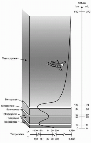
Temperature and its variation provide much of the basis for subdivisions of Earth’s atmosphere into regions defined in Figure 3-1. The lowest zone, the troposphere, is the only region of Earth’s atmosphere capable of supporting human habitation without artificial support. The troposphere starts at the Earth’s surface and extends to the tropopause, between 5 and 9 mi [8 to 14.5 kilometer (km); 26,000-48,000 ft]. At its higher levels, above 20,000 ft (3.8 mi; 6 km), at least some degree of artificial support is required in the form of supplemental oxygen. A linear decrease in temperature characterizes the troposphere from sea level (15°C) to the tropopause, typically at approximately 35,000 ft (10.7 km), where the temperature is approximately −55°C. The lapse rate, that is, the rate of decreasing temperature with increase in altitude in the troposphere, is −2°C or approximately −3.5 F per 1,000 ft. Approximately 80% of the atmospheric mass and most of the weather phenomena occur in the troposphere. Variations in temperature, pressure, and humidity in the troposphere account for extreme differences in the environmental conditions we experience as weather.

The tropopause is the division between the troposphere and stratosphere. Aircraft jet engines perform with greater efficiency at lower temperatures, which is one reason cruise is planned near the tropopause where the temperature is lowest. The stratosphere starts just above the tropopause and extends up to 50 km (31 mi). Ninety-nine percent of the mass of the air is located in the troposphere and stratosphere. The temperature throughout the lower part of stratosphere is relatively constant. Compared to the troposphere, this part of the atmosphere is dry and less dense. The temperature in this region increases gradually to −3°C due to the absorption of ultraviolet (UV) radiation. This radiation reaching the lower stratosphere from the sun is responsible for creation of ozone, the ozone layer, or ozonosphere. In the process of ozone production and in reactions with ozone, nearly all of the UV radiation is absorbed including the most hazardous form to life, UV-C (wavelengths <280 nm). Much of UV-B (wavelengths between 280 and 320 nm) is also absorbed, although the UV-B reaching the surface is sufficient to be a major cause of melanoma cancers and sunburn. Most of the UV-A (wavelengths between 320 and 400 nm) reaches the Earth’s surface, but is needed by humans for production of vitamin D. Although flight in the upper troposphere and lower stratosphere involves exposure to more UV radiation than on the surface, no health risk is currently associated with routine flying operations (see Chapter 8). Flight above the stratosphere and space flight involve risk of exposure to significant levels of radiation.

The higher regions of the atmosphere, 50,000 ft and above, are so thin that pressure suits are required to sustain life. Temperature variations result from variable absorption of the sun’s energy in several forms and thermal protection must be incorporated for any exposure in these regions. In the higher regions, flight of air-breathing aircraft becomes impossible and control surfaces are no longer effective. Further description will be left to the references and recommended reading.

The subdivision of the zones described in the preceding text relates to the ability of humans to function based on the partial pressure of oxygen available and need for artificial pressure to sustain life (Table 3-2).


Altitude

Altitude is measured in many different ways using different standards for different purposes. On low-altitude maps provided to pilots, the height of physical features of Earth, like mountains and airfields, is measured in feet above mean sea level (MSL). MSL is the average height of the surface of the sea for all stages of the tide over a 19-year period, usually determined from hourly height readings. With properly set, calibrated, and functioning altimeters, feet above MSL is the altitude viewed by the pilot in an aircraft. This is also known as pressure altitude (PA), the altitude in the Earth’s atmosphere above the standard datum plane, standard sea level pressure, measured by a pressure altimeter. Pilots are quite interested in the height of their aircraft above the ground. This altitude, above ground level (AGL), is determined by subtracting the elevation in feet above MSL of the ground below the aircraft from the elevation of the aircraft. The routine determination of a safe altitude on a route between navigational aids to avoid terrain and towers is usually viewed on low-level navigational maps as the minimum en route altitude (MEA), which is the altitude between radio fixes that assures acceptable navigational signal coverage and meets obstruction clearance requirements between those fixes. Flying at that altitude with a properly set altimeter ensures adequate separation from obstacles for the entire route segment. PA is the height in the atmosphere at which a given value of standard pressure exists. With 29.92 in. of Hg set in the Kollsman window of the
altimeter, PA is displayed in feet on the altimeters of United States Air Force (USAF) aircraft. Because hectoPascals are a standard in parts of the world, some confusion can arise when pilots “assume” that, for example, 988 means 29.98 in. of Hg when given by an air traffic controller as an altimeter setting because some controllers in the United States leave off the “2.”


FIGURE 3-1 Zones of Earth’s atmosphere.


Air Density, Pressure, and Temperature

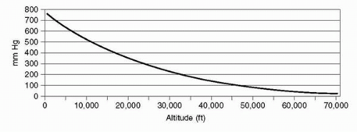
The density of air is affected by its pressure, which decreases exponentially with increasing altitude, reaching 50% of sea level density and pressure at approximately 18,000 ft (5.49 km). This relationship is affected in any specific locale by deviations from standard temperature and pressure. Figure 3-2 graphically shows how atmospheric pressure is affected by altitude. The curve depicts how each 10,000-ft increase in altitude results in less change in pressure; 0 to 10,000 ft changing by 237 mmHg, 10,000 to 20,000 ft changing by 173 mmHg, and 40,000 to 50,000 ft changing by only 54 mm Hg.

During takeoff, landing, and low-level phases of flight, aircraft altimeters are routinely set to the field altimeter setting to account for variations in local pressure. This procedure avoids significant errors in altitude of the airfield versus what is indicated on the altimeter. Temperature variations from the standard temperature of 15°C also produce errors, which affect terrain clearance. For instance, an aircraft flying at 5,000 ft in −40°C (e.g., Alaska in the winter) would be more than 1,200 ft lower than the indicated altitude after correction for local barometric pressure (PB). Local PB in the United States is based on inches of Hg. This setting would show the altitude of 0 ft at sea level on such a day. As the local pressure varies, altimeters are set to higher or lower settings to yield the correct field elevation on an aircraft altimeter at a designated point on that field. Above 18,000 ft (flight level 180; altitude in ft/100), altimeters
are routinely set to 29.92 in. of Hg to provide adequate and standardized clearance for aircraft altitude separation. Although the inches of Hg standard for altimeter settings are a pressure indication, it is not normally used in aviation for describing total atmospheric pressure at a given altitude. Elevation is typically measured in ft, meters (m), or km and pressure in psia, mm Hg, or mb.


TABLE 3-2


Physiological Divisions of the Atmosphere


Physiological Division


Altitude and Pressure Range


Problems


Solutions


Physiological zone


0-10,000 ft
0-3,048 m
760-523 mm Hg


Trapped gas expansion/contraction during changes in pressure result in middle ear or sinus blocks; shortness of breath, dizziness, headache, or nausea in unacclimatized individuals or with exercise


Acclimatization or reduced performance


Physiologically deficient zone


10,000-50,000 ft
3,048-15,240 m
523-87 mm Hg


Oxygen deficiency progresses from minor reductions in cognitive and physical capabilities at 10,000 ft to death over approximately 25,000 ft (possibly lower) without supplemental oxygen


Supplemental O2 and PBA allows good performance to approximately 35,000 ft with progressively less capability


Space equivalent zone


Above 50,000 ft
> 15,240 m
<87 mm Hg


Survival requires assisted PBAa or, above approximately 63,000 ft, a full pressure suit and delivery of 100% O2 to supply at least 140 mm Hg O2


Pressurized cabin or pressure suit with 100% O2


a PBA = positive pressure breathing for altitude. (Physiological Training, Air Force Pamphlet 160-5, 1976.)


Light and Sound

Diffusion of light in the lower atmosphere accounts for the blue color of the sky as viewed from Earth’s surface, a phenomenon which significantly dissipates as low as approximately 50,000 ft where the blackness of space begins to become apparent. The speed of sound is 761 mph(340 m/s; 1,116 ft/s) at sea level and slower, 660 mph (295 m/s) at 50,000 ft where the temperature is approximately 75°C lower. The speed of sound is a function of the square root of the temperature in °K (°C + 273).


The Gas Laws

A basic understanding of the gas laws is necessary to comprehend the physical nature of the atmosphere and how it interacts with human physiology. The gas laws define physical properties of our atmosphere and provide a basis for understanding how they affect our function during exposure to reduced atmospheric pressure.


FIGURE 3-2 Atmospheric pressure versus altitude.


Boyle’s Law

Robert Boyle (1627-1691) was an Anglo-Irish scientist noted for his work in physics and chemistry. In 1662, Boyle published the finding which states that at a constant temperature, the volume of gas is inversely proportional to its pressure. P1 and V1 are the initial pressure and volume, and P2 and V2 are the final pressure and volume. Solving this equation for the volume of a contained gas at a different pressure quantitatively describes trapped gas expansion with reduced pressure.

P1 × V1 = P2 × V2 or P1/P2 = V2/V1

Solving this equation to find the volume of a liter of dry gas taken from sea level to 20,000 ft and 40,000 ft, assuming unrestricted expansion, would result in the following:

1.0 L at sea level

(760 mmHg × 1 L)/349 mmHg = 2.2 L at 20,000 ft

(760 mmHg × 1 L)/141 mmHg = 5.4 L at 40,000 ft

The problem becomes more complicated by the inclusion of water vapor in the lungs and other spaces in the body as
described in the section Trapped Gas, Section 3. Figure 3-4 shows the volume and diameter of a wet gas sphere at various pressures and graphically shows how Boyle’s law works on trapped gases during decompression and recompression, descent.


Dalton’s Law

John Dalton (1766-1844) was an English chemist and physicist. In 1803, he observed that the total pressure of a mixture of gases is equal to the sum of the partial pressures of each gas in the mixture.

PT = P1 + P2 + P3 + … Pn

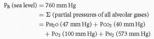
Because the standard atmosphere at sea level is 760 mm Hg, Dalton’s law indicates that the sum of partial pressures of the gases that make up the standard atmosphere must equal 760 mm Hg. The pressure of each gas in a mixture of gases is independent of the pressure of the other gases in that mixture. Multiplying the percentage of a gas in the mixture times the total pressure of the mixture yields the partial pressure of that gas.

The standard atmosphere does not include water vapor pressure, primarily due to its variation in the Earth’s atmosphere between 0% and 100% relative humidity. This variation amounts to 0% to 6.2% of 760 mmHg, or 0 to 47 mm Hg at body temperature, 37°C.
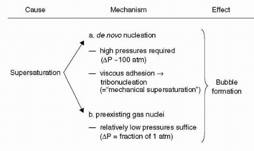

Henry’s Law

William Henry (1775-1836) was an English chemist who, in 1803, published his findings that the amount of a gas in a solution varies directly with the partial pressure of that gas over the solution. This relationship explains why dissolved nitrogen transitions to a gas phase in blood and tissues during decompressions sufficient to result in supersaturation. The resulting bubbles of nitrogen with minor amounts of oxygen, carbon dioxide, and water vapor can cause decompression sickness (DCS).


Charles and Gay-Lussac’s Law

Jacques Alexandre César Charles (1746-1823) was a French inventor, scientist, mathematician, and balloonist. In 1783, he made the first balloon using hydrogen gas; upon release, it ascended to a height of approximately 3 km (2 mi). In 1787, he discovered the relationship between the volume of gas and temperature, known variously as Gay-Lussac’s law or Charles’s law.

V1/V2 = T1/T2 or V1/T1 = V2/T2

Charles did not publish his findings and Joseph Louis Gay-Lussac first published the finding in 1802, referencing Charles’ work. The temperature is in Kelvin degrees, where °K = °C + 273. At absolute zero, −273°C, the Kelvin temperature is 0°K. The distinction between Boyle’s law and Charles’ law is what is held constant, whereas the other two parameters are varied. Boyle’s law describes changes in volume with respect to pressure when temperature is held constant. Charles’ law describes how volume changes with temperature when pressure is held constant. Although Charles’ law is very important from an engineering and chemistry standpoint, the temperature of human body is usually rather constant at body temperature, limiting its effect on physiology. Changes in all three parameters (volume, pressure, and temperature) are better described by the Ideal Gas law, which includes the three parameters in one equation with other factors to improve accuracy.

PV = nRT

where P = pressure, V = volume, T = temperature, n = number of moles, and R = universal gas constant = 8.3145 J/mol K.


Gaseous Diffusion

Experiments of Thomas Graham (1805-1869), a British chemist, showed that the diffusion of a gas is inversely proportional to the square root of its molecular weight. Therefore, gases of lower molecular weight diffuse more rapidly than gases of higher molecular weight. Diffusion of a gas is also affected by its solubility in the surrounding media and the difference in concentration of the gas between two adjacent volumes. A larger difference in concentration produces greater diffusion. A gas with greater solubility in its solvent, for example tissue or fluids, means more molecules of it will be available to diffuse as limited by the other factors. Gaseous diffusion is fundamental to the physiologic processes of lung and cellular respiration. It further applies to the process of denitrogenation, removal of nitrogen from the body, by breathing 100% oxygen.


Chronic Hypoxia


Terrestrial Environment

The historical distinction between hypoxia in the terrestrial and extraterrestrial/aerospace environment has become increasingly blurred in recent decades. The time at a certain pressure (i.e., altitude) and the time and modality to get to that pressure govern the physiology that will be discussed. The advent of ultra-long-haul flight operations in environments of decreased ambient pressure in civilian air transport operations (1) and potentially in future exploration class spaceflight missions, as well as rapid transport of civilian and military personnel to and prolonged sojourn in high-altitude environments make it very important for the aerospace medicine practitioner to be familiar with the concepts of operational significance that can play a role in those environments. The following paragraphs will review operational considerations and relevant clinical terrestrial syndromes.


Acclimatization

Altitude acclimatization is a process that occurs upon exposure to a hypobaric and hypoxic environment. Different processes occur with the common goal to protect the body tissues against the hypoxic challenge of the environment and
to allow for continued performance. Many of these processes occur at different speeds and can be summarized in different groupings (Table 3-3).


TABLE 3-3


Processes of Acclimatization and Relevant Terminology


Acute Acclimatization (Accommodation)


Minutes


Rise in Heart Rate, Increased Ventilation


Chronic acclimatization


Days


Increase in hemoglobin (initial decrease in plasma volume followed by increased red cell mass), increased capillary density


Adaptation


Years


Alterations in hypoxic ventilatory response


Owing to the processes discussed in the preceding text, it is difficult to answer the simple question as to how much time is needed to acclimatize to a given altitude, but the key aspects in acclimatization rest in the cardiorespiratory system and the blood that adapt in days to few weeks.

Unfortunately, there is no one single parameter that allows us to physiologically quantify and assess the level and degree of acclimatization. In addition to the lack of a single reliable parameter to assess acclimatization, we are faced with significant interindividual variation in speed and degree of acclimatization. A good clinical rule is to always inquire about past performance at altitude and presence or absence of signs and symptoms of acute mountain sickness (AMS); past performance is a guide to future performance in similar environments and exposures. Special attention needs to be given to any preexisting cardiac or pulmonary disease or conditions that may be exacerbated by exposure to a hypoxic environment.

There are only scarce data addressing acclimatization and the effects of age and gender. There does not appear to be a significant difference in acclimatization between men and women, and older age appears to confer some protection from AMS.

Preacclimatization is a technique used to achieve some degree of acclimatization preceding the exposure in the high-altitude environment. This can be accomplished by using an analog environment such as an altitude chamber or sojourns at altitude; good data to show consistent acclimatization benefit of intermittent hypoxic exposures with nitrogen admixture to the breathing gas (e.g., sleeping in a hypoxic environment) are lacking at this point. The benefits of acclimatization appear to dissipate over a period of 2 to 3 weeks, and it should be noted that pulmonary edema has been described in native highlanders with reexposure to altitude after as little as 12 days at low altitude (2).


Operational Considerations


Reduced Exercise Capability

Aerobic performance is significantly impaired as altitude and maximal oxygen consumption in acclimatized subjects falls from 4 to 5 L/min to approximately 1 L/min at the altitude of Mount Everest. The demands of the hypoxic environment lead to a significant reduction in exercise capacity and many an account of expeditions at extreme altitudes, especially without supplemental oxygen, is filled with vivid descriptions as to the extreme difficulty of exercise (3).

Any attempt to exercise or be physically active at high altitude is accompanied by markedly elevated levels of ventilation. It is noteworthy that ventilation is usually expressed with reference to ambient pressure, body temperature, and with the gas saturated with water vapor [referred to as body temperature and pressure saturated (BTPS)]. This measurement reference takes into account more accurately the volume of gas moved by the chest and lungs. Another measurement condition is STPD, which stands for the measurement of ventilation in conditions of Standard Temperature, Pressure, and Dry gas. The latter shows much smaller volume changes at altitude and has no overt relationship to the actual mechanics of breathing (lung/chest wall movements). Oxygen consumption and carbon dioxide production are traditionally reported in STPD reference units, such that the values are altitude independent.

Ventilation measurements at high altitudes can reach near maximum voluntary ventilation levels driven by the powerful hypoxic drive through the peripheral chemoreceptors; during the 1981 Everest expedition at 8,300 m (Pb 271 mmHg), Pizzo recorded maximum ventilation with a respiratory rate of 86 breaths/min and a tidal volume of 1.26 L/min resulting in a mean ventilation of 107 L/min (4,5).


Reduced Cognitive Ability

The exposure to any hypoxic environment has operational ramifications in that it can sharply reduce the effectiveness of an operator, especially in the first few days following insertion into a high-altitude terrestrial environment. Especially in the first week at altitude, consideration should be given to adequate rest periods (taking into account the temporary degradation of sleep quality at altitude), decreased task intensity, and if possible decreased operational tempo. In addition to the known decrements in cognitive performance associated with varying degrees of hypoxia, the development of severe headaches and neurologic symptoms and signs, as well as pulmonary symptomatology may be harbingers of a clinically relevant high altitude-related illness such as high-altitude cerebral edema (HACE) or high-altitude pulmonary edema (HAPE).


Relevant Clinical Terrestrial Syndromes Related to High Altitude

The emphasis in the discussion of high altitude-related clinical syndromes must be prevention. The hostile environment of extreme high altitudes coupled with the intense desire to accomplish a set goal (e.g., climbing a mountain, executing a
mission) in the context of highly motivated and driven team members may at times be a dangerous combination.

The education of all team members about disease entities and their symptoms that can arise at high altitudes (6,7) is of importance such that everybody may be able to observe their team members and peers. The emergence of any concerning signs or early behavioral alterations such as falling behind, change in attitude, lethargy, and so on should prompt heightened vigilance and early evaluation. Monitoring the dynamics of any signs and symptoms will allow the team to avoid bad outcomes, and to enable any team member with worsening symptoms to descend while they are still able to walk.


Acute Mountain Sickness

AMS is a syndrome encompassing headache, anorexia, lassitude, nausea, and a feeling of malaise. It can be encountered in 15% to 30% of Colorado resort skiers (8) and in up to 67% of climbers on Mount Rainier (9). Many people become symptomatic even at intermediate altitudes of 6,000 to 6,500 ft. Rapid ascent to altitude (flying, driving) may markedly exacerbate the risk. Symptoms usually manifest within hours to first few days at altitude.

The scoring of AMS can be accomplished by using Lake Louise consensus scale or a subset of questions of the environmental symptom questionnaire (ESQ). The ESQ consists of 67 questions in its ESQ III version. Clinically, it is most relevant to insist that headache be present for the diagnosis of AMS. Most practitioners prefer the Lake Louise scoring system due to its simplicity, consisting of a self-assessment (most important), clinical assessment, and afunctional score. Symptomatic therapy with nonsteroidal anti-inflammatory over-the-counter medications relieves the symptoms of headaches. Use of acetazolamide (a carbonic anhydrase inhibitor) is useful in the treatment of symptoms, but more importantly in the prophylaxis of the condition. The latter is advisable if historically a subject has had past episodes of severe mountain sickness or rapid exposure to a significant altitude is expected. Carbonic anhydrase inhibitors will facilitate acclimatization to altitude. Other agents can be used for symptom control, such as dexamethasone or other steroids. The disadvantage of using steroids for the treatment of mountain sickness is their lack of effect on acclimatization and their side effect profile. A rebound effect, that is, reoccurrence of mountain sickness after cessation of steroids at altitude is possible.

If available oxygen will alleviate symptoms of mountain sickness, severe cases may benefit from use of a portable hyperbaric chamber (10), especially in the setting of high-altitude expeditions.

The occurrence of the ataxia in a subject with severe mountain sickness should be taken very seriously as it may be a harbinger of early high-altitude cerebral edema, which if present may preclude safe self-evacuation by going to lower altitude.


High-Altitude Cerebral Edema

HACE usually occurs several days after altitude exposure in the context of mountain sickness. The differentiation between severe mountain sickness and HACE rests in the development of ataxia, impaired cognition, and higher cortical functions (hallucinations, inability to make decisions, severe mental slowing, irrational behavior, errors) as well as neurologic deficits in addition to the symptoms of severe mountain sickness as described earlier (11).

The occurrence of HACE in a hostile high-altitude environment will incapacitate the patient and lead to the need of an evacuation, thereby putting other participants potentially at risk. Avoidance of passive transport to extreme altitude and avoidance of ascending with symptoms of mountain sickness are important factors to avoid unnecessary bad outcomes. HACE may occur together with HAPE. Treatment of HACE consists of descent, the administration of steroids (e.g., dexamethasone), oxygen, and if available, use of a portable hyperbaric chamber with the goal of rendering the patient ambulatory, thereby allowing for further descent from altitude.


High-Altitude Pulmonary Edema

HAPE is a noncardiogenic pulmonary edema, which can occur in up to 1% to 2% of subjects at 12,000 ft (3,650 m), and there appears to be a genetic predisposition in some patients. A careful history will allow identification of this subpopulation. Significant exaggerated elevations of pulmonary arterial pressures in susceptible subjects in response to hypoxia at altitude appear to be causal factors in the pathogenesis of this condition.

Incidence depends on rate of ascent and peak altitude reached; reports from Pheriche (4,243 m) showed an incidence of 2.5% (12); Indian troops flown to an altitude of 3,500 m had an incidence of 0.57% (13).

Symptoms of HAPE are breathlessness, chest pain, headache, fatigue, and dizziness. Signs include mild elevation of temperature, dry cough (especially on exertion), hemoptysis, tachycardia, tachypnea, and cyanosis.

X-rays frequently reveal a pattern of irregular, patchy, later confluent infiltrates in both lower- and mid-lung fields, whereas the apices can be spared at times.

Lowering the pulmonary arterial pressures to provide relief can be accomplished with oxygen and vasodilators such as nifedipine and other agents such as phosphodiesterase inhibitors, which are currently under study for clinical use in this condition.

For individuals that have genetic disposition to this condition, use of prophylactic nifedipine may be a viable option to prevent HAPE.

Alternatively, the ambient pressure can be increased in a portable hyperbaric chamber to achieve improvement and thereby allow for transportability to lower altitude.


Chronic Mountain Sickness (Monge’s Disease)

Chronic mountain sickness is a disease entity that can be found in populations remaining at altitude for many years. The key findings include erythrocytosis and related symptoms such as headaches, dizziness, physical fatigue and mental slowing, anorexia, and dyspnea on exertion, cyanosis, and a ruddy complexion. Pulmonary hypertension and right
heart failure may also be present. Obvious contributing causes would be chronic obstructive lung disease, obstructive sleep apnea or sleep-disordered breathing conditions causing hypoxia, and other pulmonary pathology, making the patient more hypoxic and thereby enhancing the erythrocytosis even further. Relocation to low altitude in the absence of pulmonary pathology or other contributing causes is usually curative.

Laboratory investigations reveal an increased red cell count, hemoglobin concentration, and packed red blood cell volume. PaO2 is decreased and PCO2 is elevated. The increase in alveolar-arterial oxygen tension gradient is likely attributable to increased blood flow to poorly ventilated areas. The electrocardiogram shows right ventricular hypertrophy and increased pulmonary arterial pressures as well as blood viscosity (14, 15, 16, 17).


High-Altitude Retinal Hemorrhages

High-altitude retinal hemorrhages (HARH) may be seen in many climbers at very high altitude. The hemorrhages are typically without symptoms and tend to disappear spontaneously over a couple of weeks upon return from altitude. There appears to be a correlation between retinal hemorrhages and HACE (16,17). The subject may develop symptoms if these hemorrhages are close to the macula. The distribution of these hemorrhages and cotton wool spots is of a periarteriolar and perivenous distribution. Typically, no treatment is required and recovery is spontaneous.


High-Altitude Deterioration

Extended stays at altitudes greater than 5,000 m typically result in significant weight loss. Field studies and observations from expeditions certainly introduce a variety of confounding factors, such as cold, limited food supplies or lack of palatable food, or the increased need to burn calories for activities of climbing or walking. It is remarkable that similar observations were made in altitude chamber studies, such as the Operation Everest studies that were 40 days in length, and revealed, despite an unlimited diet and comfortable environmental conditions, that the subjects still lost weight. An increase in basal metabolic rate has been invoked as a causal factor. Furthermore, changes in intestinal absorption of carbohydrates, protein, and fat in the context of hypoxia may also play a role above 5,000 m (15).


Extraterrestrial Environment

Activity outside of the habitat will be required on a regular basis from any Moon- or Mars-based facility to accomplish the objectives of exploration. The pressure suit used during exploration must keep the explorer functional in the absence of an atmosphere on the Moon and near vacuum (4.5 mm Hg) on the surface of Mars. The suit should have as little negative impact on the mission as possible, which means freedom of movement and minimal fatigue. Current National Aeronautics and Space Administration (NASA) EVA suits employ 100% oxygen at 4.3 psia (226 mmHg) (18,19), which provide more oxygen than available in sea level air. These current suits are too restrictive and heavy for use on Mars or the Moon, and unless dramatic advances in suit technology are achieved, a 4.3 psia EVA suit pressure may not be feasible. Therefore, a much lower suit pressure may need to be considered. Avoiding DCS during the transition from habitat pressure to a suit pressure below approximately 3.7 psia (192 mm Hg) would also require a lower habitat pressure. A hypoxic environment in the habitat and during exploration could be experienced on a daily basis. Some physiologic changes will occur, which are analogous to terrestrial altitude-induced changes.


Adaptive Changes to a Hypobaric Hypoxic Environment

Adaptation to a low level of hypoxia in an artificial habitat environment (acclimation) could be tolerated the same way acclimatization allows thousands of humans to visit or live at high-terrestrial altitudes (3,100 m; 519 mm Hg, 10,200 ft) without supplemental oxygen. Although considerable improvement in function occurs after a few days of exposure to 3,100 m, ventilatory acclimatization would take about a week (20,21). Low gravitational forces on the Moon and Mars may reduce the workload and effect of any hypoxia during routine activity. Although the lower gravity on Mars (38% of Earth) may reduce the impact of pressure suit weight, mass is still a potential problem in terms of momentum and balance during exploration. Many factors will determine the potential atmospheres of Moon and Mars habitats, although some degree of acclimation to hypoxia is likely to be necessary.


Lower Total Pressure

Any reduction in total pressure reduces the effectiveness of electronic cooling fans and complicates atmospheric control and circulation. The engineering challenge must meet the need for close tolerances on levels of humidity, carbon dioxide, and oxygen levels to maintain comfort and physiologic function. Detection and removal of pollutants should be an extrapolation and refinement of the progress made during the International Space Station (ISS) habitation.


Water Balance

Maintenance of a comfortable level of humidity in a low-pressure habitat, for example 40% relative humidity and a temperature at 20°C (68°F), could help to reduce respiratory losses of water. This level of humidity with relatively full coverage clothing would also aid in reducing insensitive water loss.


Operational Considerations


Reduced Cognitive Ability

Acute exposure to 3,048 m (523 mmHg, 10,000 ft) in Earth’s atmosphere produces documented decrements in some cognitive tests (22,23), particularly those involving learning new tasks. In another study, 12-hour exposures to 10,000 ft (3,048 m) with rest or mild exercise produced no significant negative impact on cognitive function, but minor negative effects were observed on night vision
goggle performance under operational lighting (starlight) conditions. Increased reports of headache during the resting exposures at altitude may indicate imminent mild AMS (24). The USAF does not require its aircraft pilots to use supplemental oxygen at or below 10,000 ft during their routine acute exposures.


Reduced Exercise Capability

Even after acclimatization, maximal oxygen uptake is lower at 10,000 ft than at sea level for any individual (25). However, the effect on the submaximal effort during extraterrestrial exploration is unknown.


Communication

A reduced total pressure for a Moon or Mars habitat and pressure suits will affect vocal cord efficiency in sound development, although above a total pressure of 226 mmHg (4.4 psi; 30,000 ft), verbal communication has not been a problem. Because communication between pressuresuited explorers will require electronic transfer, appropriate amplification and filtration could compensate for lower vocal cord efficiency at suit pressures in the 141 mmHg (2.7 psia; 40,000 ft) range.


Fire Safety

The National Fire Protection Association (NFPA) has developed an equation that allows calculation of the maximum percentage of oxygen that avoids designation as an atmosphere of increased burning rate. The NFPA 99B: Standard for Hypobaric Facilities (2005;3.3.3.3) defines atmosphere of increased burning rate on the basis of a 12 mm/s burning rate (at 23.5% oxygen at 1 atmospheres absolute (ATA). The equation defining such an atmosphere (NFPA 99B Chapter 3 Definitions; 3.3.3.3) is:

23.45/(Total Pressure in Atmospheres)(0.5)

The factor 23.45 is the highest percentage of oxygen at sea level, which does not create an atmosphere of increased burning rate.

Even if a pressurized transportation system were used on the surface, continuous wear of a pressure suit would likely be required to provide adequate safety in the event of pressurization failure. The pressure suit must be designed to provide a sufficient level of oxygen and total pressure (minimum of approximately 141 mmHg O2; 2.7 psia) to allow normal physiologic function of an acclimated individual and for extensive mobility and maneuverability. If a pressure suit employing as much as 4.3 psi differential cannot be made to meet these requirements, a lower suit pressure may need to be considered.

Decompression illness (DCI) is a term used to encompass DCS and arterial gas embolism (AGE). DCS is a clinical syndrome following a reduction in ambient pressures sufficient to cause formation of bubbles from gases dissolved in body tissues. DCS follows dose-response characteristics at each involved tissue-site, the pathophysiological sequence that may or may not follow, and clinical symptoms that may occur subject to multiple moderating factors (environmental and operational tissue factors as well as marked individual susceptibility).


Historical Aspects

Sir Robert Boyle did pioneering work in the field of high-altitude medicine and was the first to observe bubble formation in vivo in one of his experimental animals during decompression in a hypobaric chamber

“I shall add on this occasion… what may seem somewhat strange, what I once observed in a Viper… in our Exhausted Receiver, namely that it had manifestly a conspicuous Bubble moving to and fro in the waterish humour of one of its Eyes.”

Subsequent clinical evidence of DCS in humans came from air-pressurized mineshaft operations. M. Triger, a French mining engineer, reported in 1841 pain and muscle cramps in coal miners (26). In 1854, two French physicians, B. Pol and T. J. J. Watelle, gave an account of the circumstances in which the disease develops upon exiting the compressed air environment: “One pays only on leaving” and recognized as well that recompression ameliorated the symptoms. They were the first to use the term caisson disease named for the compressed air environment the workers were exposed to—analogous to the diving bells (caissons) (27).

In 1869, the French physician L. R. de Mericourt published the first comprehensive medical report on DCS in divers (27). The French physiologist Paul Bert described in his classic treatise La pression barométrique (1878) the relationship between bubbles and symptoms of DCS during rapid decompression (28).

The advent of balloons and aircraft with sufficient performance to attain significant altitudes brought the clinical syndrome into the realm of aerospace medicine. In 1906, H. Von Schrötter described in his book Der Sauerstoff in der Prophylaxe und Therapie der Luftdruckerkrankungen the symptoms he experienced in a steel chamber after ascending in 15 minutes to 8,994 m (29,500 ft) closely resembling caisson disease (29). Von Schroetter discounted that hypothesis, but Boycott, Damant, and Haldane reviewed his account and wrote in an article in 1908 (30):

“Although he concludes that these symptoms could not have been due to caisson disease, we think in view of the data given by Damant and ourselves, that he was probably mistaken, and that the risk of caisson disease at very low pressure ought to be taken into account.”

This is the first clear reference to altitude DCS in the literature. In 1917, Professor Yandell Henderson provided a detailed theory in which he postulated that it would be possible to get DCS from altitude exposure (31).

J. Jongbloed described in his thesis in 1929 (32,33) the effects of simulated altitude on human subjects and called attention to the similarities of compressed air illness and DCS of altitude. In 1931, Barcroft et al. (34) described pain in the knees experienced in the hypobaric chamber while exercising at altitudes of 9,160 m (30,000 ft), which in hindsight were
most likely manifestations of DCS. In the United States, Dr. H. Armstrong researched the effects of decreased PB on the aviator and described in 1939 bubble formation that he experienced himself while at altitude in the hypobaric chamber (35):

“… Then I noticed a series of small bubbles in the tendons of my fingers … I was certain in my mind they represented aeroembolism..”

In 1938, Boothby and Lovelace reported a case of transient paraplegia in a fellow physiologist (Dr. J. W. Heim) during an ascent to 10,670 m (35,000 ft) while on oxygen; the paraplegia disappeared upon repressurization to ground level. This case illustrated the potential for serious neurologic DCS at altitude and spurred more research (36).

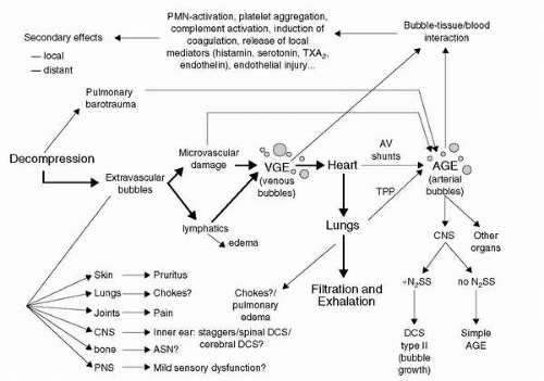
The recent decades of research have introduced new monitoring capabilities, which have allowed investigation of the bubble manifestation of the disease under controlled laboratory conditions. Ultrasonic echo-imaging Doppler measurements as an index for gas evolution have enhanced our capability to investigate the in vivo venous gas phase. The degree of venous bubbles present is graded on a numerical scale referred to as a venous gas emboli (VGE) score. The first such scoring system was devised by Spencer in 1976 (37); on this 0 to 4 scale a score of 0 refers to no bubbles and a score of 4 refers to an observation with numerous bubbles obscuring the heart sounds.

The experimental work carried out in the 1970s and 1980s has shown that bubbles can be detected in the circulation of healthy individuals after decompressions without any clinical signs of DCS (38). This confirms the early hypothesis by Behnke (1947), who postulated the existence of “silent bubbles” (39). The paradigm of “bubbles = DCS” appears not to be true in many, if not most cases, and recent research focuses on the pathophysiological cascade that can be started by the in vivo gas phase (bubbles) in the different tissue compartments and the dose-response relationships leading to clinically evident DCS manifestations. This explains why the demonstration of VGE in the cardiac chambers correlates poorly with the development of DCS. A significant proportion of subjects do have detectable VGE, but do not develop DCS and some develop DCS without evident VGE (38). As more research data becomes available, there is a trend to recognize certain degree of DCS as a normal physiological response to a defined time-pressure profile environment with the caveat of possible individual predisposition and other factors.


Terminology

The clinical syndrome of DCI was first recognized in the diving/compressed air environment and later found recognition in the area of aerospace medicine; this explains the wide variety of terms used to describe the disorder and certain specific clinical manifestations. The term decompression sickness is a direct translation from the German term Druckfallkrankheit, which was introduced by Benzinger and Hornberger in 1941 (40). Currently, altitude decompression sickness or simply decompression sickness is the term most widely used and accepted in the aerospace medicine literature.

Older terms include aeroembolism, aeropathy, dysbarism, high-altitude diver’s disease, high-altitude caisson disease, mechanicobaropathy, aerobullosis, and aeroarthrosis. Decompression illness is a term that was introduced to encompass DCS and arterial gas embolism. There is also the distinct possibility for VGE to become arterialized either by crossing the pulmonary filter or crossing through a shunting mechanism from right to left side of the heart (41). The term DCI should not be used synonymously with DCS to avoid confusion in an area with already broad terminology.

The typical clinical manifestations of DCS have received idiomatic descriptions over time, the classical limb and joint pains are referred to as bends, a term that was used by fellow workers to describe the particular gait—“doing the Grecian bend”—of workers emerging from caisson work during the construction of the piers of the Brooklyn Bridge in the 1870s (26). Respiratory disturbances are commonly referred to as the chokes, skin irritation as creeps or divers itch, and disturbances of the central nervous system (CNS) with vestibular involvement have been labeled with the term staggers.

There are distinct and very important differences between hyperbaric (diving) DCS and hypobaric (altitude) DCS, despite many shared commonalities in history, pathophysiology, and nomenclature (Table 3-4). This is a very important point as there is a tendency to indiscriminately transfer information and inferences from one field of research to another, which at times may be a valid thing to do, more often than not though may be unwise and not justified, thereby leading to potentially erroneous conclusions.

The operational significance of DCS is different in hyperbaric versus hypobaric operations in that a diver will get DCS after mission completion (ascent to the surface from depth), whereas an aviator will experience DCS during his mission at altitude. Furthermore, the aviator will have the potential to endanger others if he or she loses control of his aircraft due to DCS, whereas the diver will likely be putting only himself as an individual at risk.


BUBBLE FORMATION: THEORETIC CONSIDERATIONS

The physical principle responsible for bubble formation with decreases in ambient pressure is the concept of supersaturation, which is based on Henry’s gas law that states that the amount of gas dissolved in any liquid or tissue is proportional to the partial pressure of that gas with which it is in contact. A good example that illustrates the physical characteristics of Henry’s law as it applies to DCS is the opening of a bottle of carbonated beverage. Before opening, few, if any bubbles are visible in the liquid as the gas pressure above the liquid is in equilibrium with the liquid,
on lowering the pressure above the liquid by opening the bottle, the liquid-gas system re-equilibrates to the lowered ambient pressure by offgassing bubbles.


TABLE 3-4


Differences between Hypobaric (altitude) and Hyperbaric (Diving) Decompressive Stress (DCS)


Relevant Differences between Altitude and Diving DCS


Altitude DCS


Diving DCS


1.


Decompression starts from ground level tissue nitrogen saturated state


1.


Upward excursions from saturation diving are rare


2.


Breathing gas is usually high in O2 to prevent hypoxia and promote denitrogenation


2.


Breathing gas mixtures are usually high in inert gas due to oxygen toxicity concerns


3.


The time of decompressed exposure to altitude is limited


3.


The time at surface pressure following decompression is not limited


4.


Premission denitrogenation (preoxygenation) reduces DCS risk


4.


The concept of preoxygenation is generally not applicable


5.


DCS usually occurs during the mission


5.


DCS risk is usually greatest after mission completion


6.


Symptoms are usually mild and limited to joint pain


6.


Neurologic symptoms are common


7.


Recompression to ground level is therapeutic and universal


7.


Therapeutic chamber recompression is time limited and sometimes hazardous


8.


Tissue PN2 decreases with altitude exposure to very low levels


8.


Tissue PN2 increases with hyperbaric exposure to very high levels


9.


Metabolic gases become progressively more important as altitude


9.


Inert gases dominate


10.


There are very few documented chronic sequelae


10.


Chronic bone necrosis and neurologic damage have been documented


Pilmanis AA, Petropoulos L, Kannan N, et al. Decompression sickness risk model: development and validation by 150 prospective hypobaric exposures. Aviat Space Environ Med 2004;75:749-759.


The mechanisms that are involved in the hyperbaric diving environment as well as the hypobaric altitude environment are thought to be the same in regard to DCS, although the bubble dynamics appear to be different. Decompression to altitude results in a slower release of bubbles compared to the same absolute ratio of pressure change in a hyperbaric environment (42).

The current theories of bubble formation involve two main mechanisms. De novo formation of bubbles also referred to as de novo nucleation, requires very high degrees of supersaturation and formation of bubbles from preexisting gas nuclei (bubble nuclei), which requires pressure differentials of only fractions of an atmosphere.

The current working hypothesis for the formation of in vivo bubbles favors the gas nuclei mechanism. Viscous adhesion, which is the mechanism by which negative pressures are generated in a liquid between moving surfaces (e.g., joints), is of sufficient magnitude to cause de novo bubble formation. This mechanism has been invoked to explain vacuum phenomena in joints, the cracking of joints, and the formation of autochthonous (in situ) bubbles in the white matter of the spinal cord (43). The following discussion will highlight these mechanisms and factors influencing bubble growth.

The mechanisms and moderating factors in the human body tissues that lead to bubble formation/propagation, clinical symptoms of DCS, and moderating variables are still incompletely understood.


Factors Influencing Bubble Formation


Bubble Nuclei

The conceptual idea of the presence of “bubble nuclei” or “bubble formation centers” in the tissues derives from the physics limiting bubble growth. Very small bubbles should have a propensity to dissolve and disappear due to their very high surface tension. Surface tension is inversely proportional to the bubble radius (law of LaPlace), which would raise the internal pressure of the microbubbles above the external absolute pressure, thereby leading to their dissolution. This would suggest that larger bubbles should not be able to exist if there are no smaller bubbles that precede them, which is an obviously wrong conclusion. If we assume that we would need to create bubbles de novo, then experimental evidence shows that forces of approximately 100 to 1,400 ATA are needed. We know that bubble formation occurs at much lower pressure differentials in animals and humans (fractions of 1 ATA). Bubbles can form in fluids at low levels of supersaturation if forces act to pull objects apart which are in close proximity, a process called tribonucleation. Furthermore there is experimental evidence showing that compression of animals (shrimp, crabs, and rats) before hypobaric exposure markedly decreases bubble formation and DCS (44). These observations are consistent with the existence of gas nuclei that allow bubble formation at much lower pressure gradients (fractions of 1 ATA). The current understanding of the dynamics of bubble nuclei is that they are generated by motion (and possibly other factors, see section Mechanical Supersaturation in subsequent text) and that there is a dynamic equilibrium between generation and destruction of gas nuclei in the tissues (45).


Supersaturation

During decompression from any atmospheric pressure, some quantity of inert gas in the tissues must diffuse into the blood, travel to the lungs, and leave the body in the expired air because the quantity of inert gas that can remain dissolved in tissue is directly proportional to the absolute ambient pressure.

Supersaturation is defined by the following equation:

Supersaturation =ΣPg+ΣPv−ΣPa


Pg: sum of the tensions of all dissolved gases; Pv: sum of any vapor pressure (e.g., water); Pa: local absolute pressure.


FIGURE 3-3 Synopsis of the different mechanisms involved in bubble formation.

In summary, we can say that supersaturation can occur when either local absolute pressure is low or when the sum of the dissolved gases and vapor pressures is high.

During ascent, the reduction in PB creates a condition whereby the tissue inert gas tension (PN2 = nitrogen gas partial pressure) is greater than the total PB. This situation is called supersaturation. Therefore, if the decompression exceeds some critical rate for a given tissue, that tissue will not unload the inert gas rapidly enough and will become supersaturated. The probability of bubble formation increases with increasing supersaturation.

Supersaturation due to negative pressure occurs in many mechanical processes resulting in a local reduction of absolute pressure in a liquid system. The flow of a liquid through a local narrowing in a tube, for example, results in a local drop in pressure (Bernoulli principle), which in turn can lead to transient bubble formation (Reynold’s cavitation). Sound waves are well known to cause acoustic cavitation in liquid systems. A further mechanism of biological interest is viscous adhesion (Figure 3-3). Viscous adhesion describes the forces generated when two surfaces in a liquid are pulled apart; the negative pressures that can be generated can reach thousands of negative atmospheres. The amount of supersaturation that can be mechanically generated by viscous adhesion is directly proportional to the liquid viscosity and indirectly proportional to the cube of the distances between the two surfaces. Bubbles that are generated by this mechanism are said to be generated by tribonucleation. This mechanism is invoked in the generation of the cracking of joints by pulling apart their articular surfaces, resulting in the generation of a vapor-filled bubble, which collapses upon release of the traction, as well as in the appearance of vacuum phenomena (gaseous cavitation) in joints under traction and in the spinal column within disks, facet joints, and vertebrae (43,45).


Critical Supersaturation

Apparently, a level of supersaturation is reached which the body can tolerate without causing the inert gas to come out of solution to form bubbles. Once the critical supersaturation ratio is reached, however, bubbles develop which can lead to DCS. The English physiologist J. S. Haldane first described the concept of critical supersaturation in 1908 (30). Haldane was commissioned by the British Admiralty to investigate and devise safe decompression procedures for Royal Navy divers, and his work demonstrated that humans could be exposed to hyperbaric pressures and subsequently decompressed without having DCS as long as the total pressure reduction was no greater than 50%. Haldane devised the concept of tissue half-time to define the ability of a particular tissue to saturate/desaturate with nitrogen by 50% (i.e., a tissue would be saturated 50% after the passing of one tissue half-time). He postulated that the body tissues with different perfusion rates can be adequately represented by half-times of 5, 10, 20, 40, and 75 minutes (five-tissue model, constant allowable ratio). Haldane argued that the human body could hypothetically tolerate a 2:1 decrease in ambient pressure without getting DCS symptoms (“2-to-1” rule). Further operational research showed that the Haldane diving tables consisting of Table 1 for shorter dives up to 30-minute decompression time and, up to a depth of 204 ft sea water (fsw) and Table 2 for longer dives (with bottom times > 1 hour and decompression times of >30 minutes) were overly conservative for Table 1 and not safe enough for Table 2. Current theory is based on variable allowed ratio for different tissues; this is influenced by tissue nitrogen half-time, time, and ΔP. This is the reason why no current decompression schedules use Haldane’s 2-to-1 rule, but it is discussed here to show a mathematical concept. If Haldane’s 2-to-1 relationship of allowable total pressure change is converted to a PN2 to PB relationship, the critical supersaturation ratio (R) would be:


For example,


In fact, there are apparently a number of critical supersaturation ratios for the various mathematical compartments, representing different tissues.

A person living at sea level and breathing atmospheric air will have a dissolved PN2 of 573 mm Hg in all body tissues and fluids, assuming that PB equals 760 mm Hg; PAO2 equals 100 mmHg; PACO2 equals 40 mmHg; and PAH2O equals 47 mmHg.


If that person is rapidly decompressed to altitude, a state of supersaturation will be produced when an altitude is reached where the total PB is less than 573 mm Hg, a condition that occurs at an altitude of 2,287 m (7,500 ft). Therefore, the altitude threshold above which an individual living at sea level would encounter supersaturation upon rapid decompression is 2,287 m (7,500 ft).

The lowest altitude where a sea-level acclimatized person may encounter symptoms of DCS may be lower than 3,962 m (13,000 ft) (46). However, recent data revealed a 5% threshold at 5,944 m (19,500 ft) (47) using a probit analysis of more than 120 zero-prebreathe, 4-hour exposures with mild exercise to generate an onset curve showing less than 0.001% DCS at 13,000 or below. The degree of supersaturation at 5,489 m (18,000 ft) can be expressed as a ratio, as follows:


If the tissue PN2 equals 573 mm Hg and PB equals 372 mm Hg, then R equals 573/372, or 1.54. This value approaches the critical supersaturation ratio expressed by Haldane. The incidence of altitude DCS reaches 50% by 7,010 m (23,000 ft) with zero prebreathe and mild exercise at altitude (47).

Symptoms can occur at much lower altitudes when “flying after diving” or “diving at altitude and driving to higher altitude” (diving in mountain lakes). Many cases of DCS have been documented in divers who fly too soon after surfacing. Altitudes as low as 1,524 to 2,287 m (5,000 to 7,500 ft) may be all that is necessary to induce bubble formation in a diver who has made a safe decompression to the surface. The problem involves the higher tissue PN2 that exists after diving. The Undersea and Hyperbaric Medical Society’s recommended surface interval between diving and flying ranges from 12 to 24 hours depending on the type and frequency of diving (48).


Factors Influencing Bubble Growth

Upon decompression to altitude, the factors causing a bubble to grow are as follows:


  • Boyle’s law (P1V1 = P2V2) expansion due to reduced pressure


  • Entrance of nitrogen from tissues in the state of supersaturation


  • Entrance of O2 and CO2 (negligible effect during decompression from hyperbaric exposures, significant in hypobaric exposures)


Boyle’s Law Effects

Once a bubble is formed, its size will increase if the total pressure is decreased (Boyle’s law: P1V1 = P2V2). During hyperbaric therapy, bubble size is reduced during compression. The surface tension of a bubble is inversely related to bubble size and opposes bubble growth. Therefore, as total pressure within the bubble is increased, the surface tension opposing bubble growth also is increased. Once a critically small bubble size is achieved, the surface tension is so high that the bubble can no longer exist. The bubble collapses, and its gases are dissolved (44,49).


Gaseous Composition

Nitrogen, or another inert gas, is generally considered to be the primary gas involved in symptomatic bubbles. If nitrogen were the only gas initially present in the newly formed bubble, an immediate gradient would be established for the diffusion of other gases into the bubble. Hence, a bubble will quickly have a gaseous composition identical to the gaseous composition present in the surrounding tissues or fluids. When bubbles are produced upon decompression from hyperbaric conditions, gases other than nitrogen represent only a small percentage of the total gas composition of the bubble. Exposure to a hypobaric environment decreases the partial pressures of all gases including nitrogen. The partial pressures of O2 and CO2 at the tissue level are close to independent from hypobaric or hyperbaric conditions because appropriate levels are a prerequisite for life. If we assume that a bubble were to be present at sea level (1 ATA) with O2 and CO2 representing 6%, respectively, of its total volume and pressure and we decompress to an altitude of 5,487 m (18,000 ft, 1/2 ATA), then O2 and CO2 would each account for 12% (together 24%) of the total volume and pressure of the bubble. If the decompression were instantaneous then O2 and CO2 would each account for 6% of the bubble volume, but the partial pressure would be half of the sea level partial pressures, which would cause an immediate influx of O2 and CO2 into the bubble accounting for 12% growth in bubble volume in this example. This example (45) illustrates the importance of the metabolic gases O2 and CO2 in hypobaric exposures; at this point it is also valuable to remember the important contribution of water vapor to bubble formation and gas behavior in general in a hypobaric environment (Figure 3-9B), especially as we approach water vapor pressure (47 mmHg) and, therefore Armstrong’s line (zone) at 63,000 ft.


Hydrostatic Pressure

The tendency for gases to leave solution and enlarge a seed bubble can be expressed by the following equation introduced by Harvey in 1944 (50):


where ΔP is the differential pressure or tendency for the gas to leave the liquid phase, t is the total tension of the gas in the medium, and Pab is the absolute pressure (i.e., the total PB on the body plus the hydrostatic pressure).

Within an artery at sea level, t equals 760 mm Hg. The absolute pressure, Pab, is 760 mmHg plus the mean arterial blood pressure (100 mmHg), or 860 mmHg. Therefore,


When the value of ΔP is negative, there is no tendency toward bubble formation or growth. If the value for ΔP becomes zero or positive, bubble formation or growth is likely to occur.

Within a great vein at sea level, PO2 equals 40 mmHg, PCO2 equals 46 mmHg, and PH2O equals 47 mmHg; therefore, t equals 706 mmHg and PN2 is 573 mm Hg. Absolute pressure, Pab, is 760 mmHg plus the mean venous pressure (which in the great veins in the chest may be 0 mmHg). Therefore,


By suddenly exposing a person to an altitude of 5,490 m (18,000 ft) without time for equilibration at the new pressure, venous ΔP would have a large positive value:


The value for t in the earlier equation can also be increased in local areas by high levels of CO2 production. Hence, in muscular exercise, a high local PCO2 associated with a reduction in PB causes higher positive values of ΔP than with a reduction in PB alone. It is important to appreciate that this is a highly localized process, unless the exercise is at anaerobic levels; situations of this nature are not likely to occur for more than a few minutes in the operational aerospace environment due to the ensuing fatigue.

Hydrostatic pressure is, therefore, considered to be a force opposing bubble formation or bubble growth and includes not only blood pressure and cerebrospinal fluid pressure but also local tissue pressure (turgor), which varies directly with blood flow.


Influence of Tissue Perfusion and Diffusion

The rate of inert gas washout from tissues is dependent on perfusion; therefore, factors that alter tissue perfusion influence inert gas washout. Studies in the hypobaric environment have shown that exercise before exposure reduces the risk of DCS while prebreathing oxygen (DCS incidence decreased from 90% to 20%) (51). The putative mechanism for these effects is the increase in cardiac output with increased peripheral circulation as well as vascular volume shifts to the chest during immersion. Negative pressure breathing has similar effects to immersion with increases in cardiac output and increased inert gas washout (52). Changes in body position do have similar influence on inert gas washout; supine position has similar effects to immersion compared to the erect body position. Effects of temperature—in the context of tissue perfusion—are mediated by changes in vascular tone, as warm temperature will result in vasodilatation and enhanced inert gas washout, whereas lowering of the temperature results in vasoconstriction and decreased inert gas washout.


Pathophysiology of Bubbles

The presence of bubbles in tissues has direct and indirect effects. The location of bubbles is important in this context; extravascular bubbles can cause local tissue distortion, dysfunction, and possibly local ischemic changes. The painful sensations of joint pain are thought to be related to compressive effects on periarticular, peripheral nerve fibers. Intravascular bubbles are of lesser importance in the context of hypobaric DCS unlike their role in diving DCS, they may—depending on their location within the vasculature and the tissue—cause symptoms due to local relative hypoperfusion. The indirect effects of bubbles are more complex in nature; the interaction of cells (blood, tissue, endothelium) with the bubble surface leads to the release of mediators, which in turn may influence chemotaxis for leucocytes (polymorphonuclear neutrophils) with subsequent generation of oxygen radicals, complement activation, activation of the intrinsic coagulation pathway, generation of arachidonic acid metabolites, release of endothelium-derived mediators to name just a few. The modulation of these tissue reactions depends furthermore on the target tissues, the bubble load (dose-response), local factors such as degree of ischemia, collateral circulation, reperfusion injury, and environmental factors, for example, hypoxia, exercise, temperature, and rapidity of ambient pressure change (44,49,53).

The putative fate of bubbles formed during decompression is summarized in Figure 3-4. It is important to emphasize that our knowledge is far from complete and that the extravascular bubble dynamics are of more relevance to altitude DCS compared to the intravascular bubbles of diving DCS.


Target Organs of Bubbles Created during Decompressive Stress


Lungs

VGE results in a dose-dependent increase of pulmonary artery pressure and subsequent increase in pulmonary vascular resistance (54). These changes can be attributed to mechanical obstruction of the pulmonary vascular bed and vasoconstriction; hypobaric exposures of greater than 24,000 ft (7,315 m) did not result in appreciable increases in pulmonary arterial pressures (55). In cases with large gas loads that overwhelm the capacity of the pulmonary circulation filter, the embolization of the pulmonary vascular bed results in ventilation-perfusion mismatching leading to decreased peripheral arterial O2 saturation and decreased end-tidal CO2 levels (56).


FIGURE 3-4 Synopsis of the putative pathophysiological pathways of bubbles in decompression sickness (DCS), bold arrows indicate the major physiological pathway for elimination of inert gas, question marks indicate lack of scientific literature proving the postulated relationship. PMN, neutrophilic leucocytes TXA2, thromboxane A2; VGE, venous gas emboli; AV, arteriovenous; AGE, arterial gas emboli; TPP, transpulmonary passage; CNS, central nervous system; N2SS, nitrogen supersaturation; DCS type II, serious neurologic (cerebral) decompression sickness; ASN, aseptic bone necrosis; PNS, peripheral nervous system.

Only gold members can continue reading. Log In or Register to continue

Stay updated, free articles. Join our Telegram channel

Aug 29, 2016 | Posted by in ENDOCRINOLOGY | Comments Off on Physiology of Decompressive Stress

Full access? Get Clinical Tree

Get Clinical Tree app for offline access