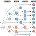
Numerous cells are able to ingest foreign materials, but the ability to increase this activity in response to opsonization by antibody and/or complement, so as to acquire antigen specificity, is restricted to cells of the myeloid series, principally polymorphs, monocytes and macrophages; these are sometimes termed ‘professional’ phagocytes.
Apart from some variations in their content of lysosomal enzymes, all these cells use essentially similar mechanisms to phagocytose foreign objects, consisting of a sequence of attachment (top), endocytosis or ingestion (centre) and digestion (bottom). In the figure this process is shown for a typical bacterium (small black rods). In general, bacteria with capsules (shown as a white outline) are not phagocytosed unless opsonized, whereas many non-capsulated ones do not require this. There are certain differences between phagocytic cells; e.g. polymorphs are very short-lived (hours or days) and often die in the process of phagocytosis, while macrophages, which lack some of the more destructive enzymes, usually survive to phagocytose again. Also, macrophages can actively secrete some of their enzymes, e.g. lysozyme. There are surprisingly large species differences in the proportions of the various lysosomal enzymes.
Several of the steps in phagocytosis shown in the figure may be specifically defective for genetic reasons (see Fig. 33), as well as being actively inhibited by particular microorganisms (see Figs 27–32). In either case the result is a failure to eliminate microorganisms or foreign material properly, leading to chronic infection and/or chronic inflammation.
Chemotaxis
The process by which cells are attracted towards bacteria, etc., often by following a gradient of molecules released by the microbe (see Fig. 7).
Pinocytosis
‘Cell drinking’; the ingestion of soluble materials, including water, conventionally applied also to particles under 1 µm in diameter.
Hydrophobicity
Hydrophobic groups tend to attach to the hydrophobic surface of cells; this may explain the ‘recognition’ of damaged cells, denatured proteins, etc. (see Fig. 29).
Pattern-Recognition Receptors
Phagocytic cells have surface and phagosomal receptors that recognize complementary molecular structures on the surface of common pathogens (for details see Fig. 5). Binding between pathogens and these receptors activates intracellular killing and digestion, as well as the release of many inflammatory chemokines and cytokines (see Figs 23 and 24).
C3 Receptor
Phagocytic cells (and some lymphocytes) can bind C3b, produced from C3 by activation by bacteria, etc., either directly or via antibody (for details of the receptors see Fig. 6).
Fc Receptor
Related posts:


Stay updated, free articles. Join our Telegram channel

Full access? Get Clinical Tree


