Fig. 5.1
Percutaneous ethanol injection: procedure of treatment. (a) Fluid drainage. (b) Needle tip within the fluid area of nodule. (c) Near total fluid removed. (d) Ethanol infusion
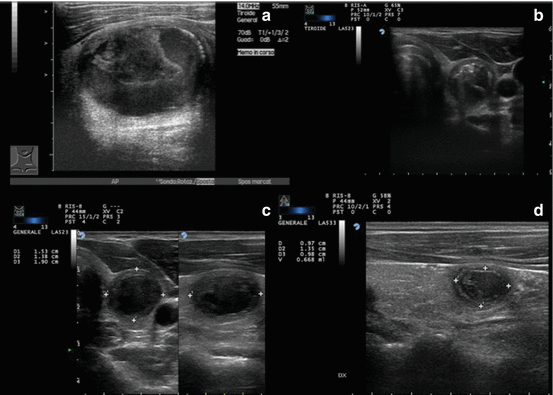
Fig. 5.2
Long-term results of percutaneous ethanol injection treatment of a large cystic lesion of the thyroid gland. (a) Baseline volume: 19 cc. (b) Ethanol injection after fluid drainage. (c) 6 months after treatment nodule volume: 2.1 cc. (d) 24 months after treatment nodule volume: 0.6 cc
Needle insertion may be performed according to two different approaches: using a needle pointing device (US-guided) or with a free-hand (US-assisted) technique [30]. No head-to-head studies have been carried out to compare the outcomes of PEIT performed with either of these two approaches. US-assisted technique usually requires well-trained operators because the needle path cannot be assessed before the procedure and the progression of the needle tip toward the target lesion needs a continuous monitoring. The use of a needle pointing device allows a more rapid and safe insertion but obliges the operator to a fixed entry site and does not allows changes in needle positioning. This approach may represent a limit during the procedure, especially in case of complex cystic lesions of the thyroid, which often require a repositioning of the needle during the manoeuvre. Alternatively, after the use of the needle pointing device for guiding needle insertion, the tool may be removed from the US probe to widen the range of needle movements within the target lesion.
5.2.1.1 Cystic Nodules
The first step is the near complete drainage of the fluid component from the lesion. Fluid aspiration is facilitated by the use of a syringe holder (e.g. the Cameco pistol) that makes easier to reach an effective negative pressure. After fluid extraction, the aspirating syringe is substituted with one containing 95 % sterile ethanol. Alcohol is carefully and gently injected into the remaining, near virtual, cavity of the cyst. In order to limit the risk of needle displacement during syringe changes, a 20 cm plastic tube connecting the syringe to the needle, possibly furnished with a T clamp system, can be used. During US monitoring, the infusion of ethanol is easily visualized as a hyperechoic cloud slowly refilling the cyst cavity. The ethanol infused into the cavity of thyroid cysts can either be removed within a short period (e.g. 3–5 min) of time or may be left inside the cyst, to ensure a more protracted activity. The second option is widely adopted because re-aspiration of the injected ethanol seems not necessary for decreasing the risk of ethanol leakage and peri-thyroidal fibrosis [21]. No major differences in efficacy between the two treatment modalities are reported, while ethanol removal was reported to be associated to a higher rate of discomfort for the patient [22].
The volume of injected ethanol generally corresponds to 35–50 % of the volume of the fluid drained from the cystic lesion. Cho et al. reported a significantly better outcome when the amount of ethanol injected into the thyroid cysts was > 10 ml [22]. However, the exact ethanol amount should be tailored according to the size and structure of the lesion, the operator opinion during the procedure and the patient compliance.
5.2.1.2 Viscous Cystic Nodules
About 30 % of the large and about 10 % of the small-size cystic nodules are characterized by a highly viscous content [13, 14]. When the fluid collection is dense, the use of a 18-gauge needle should be considered for its drainage. If colloid material cannot be aspirated even with a 18-gauge needle, cyst decompression is very difficult to be attained and surgery remains the main therapeutic option [23]. Alternatively, in very viscous cysts, a two-step treatment has been suggested: first, a small amount of ethanol (e.g. 0.5–1.0 ml) is injected in the attempt to make the dense content more fluid. Then, 2 weeks later, a regular treatment of cystic nodule may be attempted after the occurrence of an at least partial colloid fluidification [24, 25]. This two-step PEIT procedure for viscous nodules is anedoctically reported to be effective, with results apparently comparable to those obtained in pure fluid nodules [23, 24], but no controlled trials are available. A second approach is the drainage of the dense colloid collection, after a local anaesthesia with xylocaine, by means of a large (16-gauge) needle connected to a suction pump. The injection of ethanol is postponed to 10 min, using an amount of alcohol corresponding to 50 % of the aspirated fluid. This approach was reported to attain a significant decrease of the volume of thyroid cysts with viscous component (78 % and 95 % at 1- and 6-months examinations, respectively [26].
5.2.1.3 Complex Nodules
In complex thyroid lesions, PEIT procedure is similar to that employed for “pure” cystic nodules, but multiple treatments are often required because the cavity is usually multiloculated. The volume reduction appears in these cases less striking to the persistence of the solid component of the lesion.
5.2.1.4 Solid Nodules
In solid lesions, a pre-existent cavity is lacking and the diffusion of the injected ethanol is erratic [30]. Hence, multiple areas of the nodule should be treated with minimal amounts (0.1–0.3 mL) of ethanol during each session. This approach is necessary to prevent the seepage of ethanol into the thyroid capsule and the surrounding cervical tissues and the consequent occurrence of sharp neck pain and of possible fibrosis. For solid cold nodules, the injection of a total ethanol dose of 20–30 % of the pretreatment nodular volume is suggested. Ethanol injection should be immediately stopped in case of uneven intranodular diffusion of alcohol, ethanol seepage outside the nodule, or complaint of severe pain during the procedure [27].
On the basis of the reported series, the volume reduction of cold nodules after PEIT is not strictly related to the amount of ethanol injected. A similar reduction (about 50 %), in fact, may be achieved in cold nodules with the injection of a volume of ethanol corresponding either to 20 %, 25 % or 50 % of the baseline nodule volume [28]. To achieve a more relevant volume reduction (up to about 75 %), Caraccio et al. used a high cumulative ethanol dose (up to 130 % of the pretreatment nodule volume) [29]. This report confirms the absence of a precise correlation between the volume of injected ethanol and the obtained nodule shrinkage. The use of high cumulative doses of ethanol to obtain an additive reduction of 25 % versus the baseline nodule volume seems questionable in clinical practice. Even if the efficacy of multiple sessions of PEIT for very large thyroid nodules has been claimed [30], in clinical practice the need of repeated PEIT sessions makes the treatment of solid thyroid lesions as scarcely cost-effective when compared to surgery.
Once the injection has been completed, the final infusion (0.5–1 ml) of saline solution or lydocaine may prevent, by flushing the needle, the transient but sometimes sharp pain that may follow the needle extraction, due to ethanol leakage into the thyroid capsule or the surrounding cervical tissues.
In skilled hands, PEIT is devoid of major side effects, provided some points of caution are accomplished: (a) the needle tip should be clearly seen along the entire phase of ethanol infusion; (b) the procedure should be immediately stopped if the needle position is not clearly defined; (c) the occurrence of progressive resistance to the injection of ethanol should stop the manoeuvre and the needle position should be verified before a further ethanol injection; (d) the occurrence of cough or pain should make immediately stop the infusion [31]. A complete patient information and his/her reassurance before the procedure are of pivotal importance for the tolerability and safety of PEIT [13]. The experience of the operators, the patients’ compliance and the quality of US imaging influence the final outcome of the procedure and the risk of complications. These factors may explain the reduced rate of side effects and complications reported in more recent studies [32, 33].
5.2.2 Assessment of the Response to PEIT
Nodule volume reduction is the outcome more frequently reported in studies focused on PEIT treatment of thyroid lesion. The size of thyroid nodules is usually assessed by ultrasound and is calculated by means of the ellipsoid formula (D1 × D2 × D3 × 0.52) [34] before and 6–12 months after the procedure. For pure cystic lesions, devoid of a solid component, a volume reduction from 50 to 89 % may be defined as a partial response, while a volume reduction greater than 90 % may be considered as a complete response [30, 35].
Pressure and/or cosmetic symptoms should be assessed before and 6–12 months after PEIT. Patients are asked to rate local symptoms on a ten-step visual analogue scale, while physical findings are registered by the physician on a 1–4 cosmetic score based on palpation and inspection [28, 36, 37]. Even if volume decrease is usually correlated to the amelioration of the symptoms/cosmetic scores of the patients [27], these last parameters should always be separately considered to define the actual treatment efficacy and the improvement of the quality of life.
In AFTN or “toxic” nodules, the response to PEIT may be defined as a positive if, together with the reduction of the volume and of the pressure and/or cosmetic symptoms, serum TSH and thyroid hormones (FT3, FT4) return within normal levels 6–12 months after treatment [35]. PEIT treatment may be considered “complete” when all the nodules appear perfused from the injected ethanol with no colour-Doppler signal detectable after the procedure. Anyway, in some occasions, hyperfunctioning tissue is still present at scintiscan even when colour-Doppler signals are undetectable [38]. An abundant vascularization of the nodule is a negative predictor of successful response, as it may cause a rapid drainage of ethanol (“syphon effect”), resulting in a partial neutralization of the chemical damage and a consequent lower success rate of the procedure [39]. In case of AFTN, total disappearance of the “hot” nodule and a complete recovery of extranodular uptake at scintiscan may be viewed as a complete response to PEI [30]. The failure of treatment is indicated by still undetectable TSH levels during the post-treatment follow-up [35].
5.2.3 Side Effects
The tolerability of PEIT is fairly good and its side effects are few and transient. Discomfort and pain are reported as similar to those experienced by patients undergoing FNAUS. Only a minority of patients treated with PEIT complain of a slight to moderate pain, lasting for several minutes after the procedure and of a subsequent local tenderness lasting 2–3 days [27].
Pain is rare when predominantly cystic lesions are treated [40]. Instead, treatment of solid nodules, especially when deeply located or close to the thyroid capsule, may be very painful because of extranodular ethanol leakage. This event may be followed by a perilesional fibrosis that could negatively influence a successive surgical treatment, if required [41]. During needle withdrawing, a small amount (0.5–1.0 ml) of xylocaine or saline solution may be injected to avoid the back-diffusion of alcohol along the needle tract and to prevent the occurrence of local pain [42]. Some authors suggested to add 1 g of oral paracetamol or salicylate to the local anaesthesia with lidocaine before ethanol injection [27], but this measure seems not to be necessary in ordinary practice.
Dysphonia is a rare adverse effect in PEIT treatment of thyroid cysts. This complication, on the other hand, is not infrequent (1–4 %) when solid nodules are treated. Dysphonia is due to the chemical injury induced to the laryngeal nerve by the extracapsular diffusion of the injected ethanol. This complication, fortunately, in most cases recover in few weeks after a short course of steroid therapy [35]. To early diagnose a potential nerve injury, the patient is asked to pronounce some words immediately after the conclusion of ethanol injection [42]. To minimize the risk of leakage, the operator should inject ethanol slowly and at a low pressure, especially in case of already treated, fibrotic lesions, and carefully monitor the location of the needle tip within the nodule. The sudden onset of intense pain or cough, in the absence of US signs of haemorrhagic swelling inside or around the nodule, may signal the extrathyroidal leakage of ethanol [31].
Two cases of injury of cervical sympathetic chain, followed by transient Horner’s syndrome, have been reported after PEIT treatment of thyroid nodules [43].
In euthyroid patients, PEIT is not followed by significant changes of serum TSH and thyroid hormone levels [31]. The onset of hyper- or hypothyroidism, associated with development of serum auto-antibodies, was occasionally reported [27, 41]. A case of Graves’ ophthalmopathy in a patient previously treated with PEIT was anedectotally reported. No evidence, however, demonstrates a possible relationship between ethanol injection and induction of an anti-thyroidal autoimmune response [44].
When treating toxic nodules with PEIT, a prophylactic treatment with β-blockers may be appropriate to prevent the possible exacerbation of thyrotoxic symptoms, usually caused by a transient increase in serum levels of thyroid hormones [35]. Blood pressure should be monitored after the administration of β-blockers, because hypotension, due to the combined effect of the drug and of the peripheral vasodilatation induced by metabolites of ethanol (acetaldehyde acetate), has been reported in few cases [45]. Other major side effects such as cervical haematoma, ipsilateral facial dysesthesia, jugular vein thrombosis and septic complications have been occasionally described. The most relevant complication up to now reported is a single case of chemical necrosis of the larynx combined with necrosis of cervical tissues in a patient treated with PEIT for an AFTN [46]. Severe side effects and major complications, however, are always the consequence of inappropriate modalities of treatment and surveillance of the procedure.
5.3 PEIT of Thyroid Cysts
5.3.1 Rationale
Thyroid nodules may be defined as cystic if the fluid component is >60 % of their volume. Pure cysts and, more frequently, pseudocystic lesions represent a common finding (likely 25–30 % of all thyroid nodules detected by US) in clinical practice [31]. The majority of large cystic thyroid lesions is caused by intralesional bleeding and/or degeneration of a pre-existing nodule and their fluid content is made of colloid, blood and necrotic debris. Haemorrhagic cystic lesions present suddenly with neck tenderness, pain and pressure symptoms. Their spontaneous regression is possible but most of these lesions persist, frequently grow over time, and usually recur after aspiration [47–51]. Since drainage results in a stable regression only in a minority of cases, further modalities of treatment should be considered [36]. LT4 suppression therapy was demonstrated as scarcely effective in prevent cyst recurrence in several randomized trials. Prolonged TSH suppression, moreover, may cause minor adverse effects, osteoporosis or atrial arrhythmia [52, 53]. Surgery has therefore long been viewed as the standard therapeutic approach for cystic nodules, especially if large and symptomatic [18]. In the last two decades, due to the excellent clinical results provided by a number of different studies [21, 23, 30, 35–37, 40, 50, 51, 54–57], PEIT has become the first-line treatment for thyroid cystic lesions [13, 58, 59]. Various agents have been proposed for the sclerotherapy of thyroid cysts (sodium tetradecyl sulphate, hydroxypolyaethoxydodecan, tetracycline, OK-432) [51, 60] but ethanol injection should be considered as the most cost-effective of them.
5.3.2 Results
PEIT treatment may be defined as successful if it results in a ≥50 % reduction of baseline cyst volume. This outcome is achieved in the vast majority (about 70–90 %) of patients, with a mean volume decrease ranging from 65 to 95 % [35]. Interestingly, a correlation between baseline and final volume is not generally observed in cystic lesions [35]. In addition to volume measurement, many studies report other parameters, like pressure or cosmetic symptoms, rated accordingly to specific scale values. Amelioration of local symptoms and cosmetic complaints usually mirrors the entity of volume decrease and is commonly observed in more than 2/3 of the treated patients [27, 36]. Stable symptoms improvement occurs more frequently after PEIT than after simple cyst drainage [35, 40, 56]. This evidence was provided by a prospective randomized study which compared the results of ethanol injection versus isotonic saline flushing, both followed by fluid aspiration. US control indicated a clear superiority (82 % vs. 48 % cure rate) of PEIT [40]. PEIT outcomes are persistent in the long term, with an exceedingly low recurrence rate (3.4 % and 6.5 % over 5 and 10 years, respectively) [23, 35].
It has long been debated if PEIT has a similar efficacy in case of large (>40–50 ml) cysts as well [30, 40]. A large initial volume, anyway, is not clearly correlated with an unsuccessful outcome [21, 23, 35, 54, 57]. According to some studies, the reduction of the lesion volume is directly correlated to the amount of drained fluid and to that of instilled ethanol [21, 51]. On the other hand, the nodule structure exerts a main impact on the final outcome. As a rule, complex cysts and “mixed” nodules with a large solid component are less susceptible of a marked and stable volume reduction as compared to (nearly) “pure” cysts. Moreover, the first ones usually require a prolonged treatment with multiple PEIT sessions [30, 35, 51, 57, 61]. Hence, when dealing with complex cysts, the best results are obtained in smaller lesions [30, 35]. Another possible obstacle to a successful PEIT procedure is the quality of the cyst content [21, 37, 51]. A high viscosity of the fluid, usually predicted by the US appearance of the colloid collection, should suggest the use of large bore needles for the drainage. The chemical and physical properties of the fluid content, however, may change and the finding of a content too viscous to be fully drained does not invariably predict a poor treatment outcome (see the technique section for further details) [24–26]
5.3.3 Indications for Clinical Practice
PEIT should be used as the first-line treatment for thyroid cystic lesions after the risk of malignancy has been ruled out [13, 59, 62]. The impact of this approach in clinical practice is relevant because pseudocystic lesions are frequent (25–30 % of all thyroid nodules detected by US) and are frequently growing over time [48]. In comparison with surgery, PEIT is extremely less expensive, presents a nearly negligible risk of permanent complications and is not followed by the need of lifelong substitution therapy.
5.4 Autonomously Functioning Thyroid Nodules (AFTNs)
5.4.1 Rationale
AFTNs are benign neoplasms, usually monoclonal, associated to a variable degree of thyroid hyperfunction. The disease may be classified in two subgroups according to thyroid hormone levels: (a) pretoxic nodules, when TSH is undetectable but thyroid hormone are within the normal limits, and (b) toxic nodules, when undetectable TSH is accompanied by T3 and/or T4 levels above the normal range. Classical therapeutic options are surgery or radioiodine treatment. In most cases radioiodine, although complicated by a non-negligible rate of late hypothyroidism, is the first choice of treatment but its use in young patients is controversial. Surgery is commonly indicated for toxic nodules larger than 4–5 cm (that are frequently associated to pressure symptoms) because these lesions require high activities of radioiodine and necessitate protective measures in several European countries [63, 64]. Due to the potential complications and costs of surgery and the limited effects of radioiodine treatment on nodule volume, PEIT has been proposed since 1990 as a novel treatment for AFTNs [9–11].
5.4.2 Results
Data published during the first 10 years of use of PEIT reported a high percentage (about 90 %) of patients completely or partially cured by PEIT [9, 11, 12, 65, 66]. A major limit of these trials was the short (usually ≤1 year) follow-up period and the heterogeneous criteria of enrolment and treatment. Studies based on long-term surveillance reported up to 35 % prevalence of recurrences of thyrotoxicosis [30, 33, 35]. Predictive factors of PEIT efficacy were assessed in the large multicentric Italian study. Pretoxic nodules showed better results than toxic nodules, with a cure rate of 83.4 % vs 66.5 % [65]. A more favourable outcome for pretoxic vs toxic nodules has been confirmed by subsequent trials [35]. Moreover, a ≥30 % fluid component and a ≤15 ml pretreatment volume were associated with a better outcome, independently from the baseline functional status. The best responses were observed in small nodules (≤5 mL volume) with only partial inhibition of the extranodular uptake [35]. In this, as in most studies, cure rate was inversely correlated to baseline volume. Yet, a satisfactory, even if rarer, outcome has been reported also for large (≥40 ml) toxic nodules [32, 38, 66, 67].
A complete ablation of hyperfunctioning tissue is not always obtained after PEIT. In a study on 117 patients with a median follow-up of 2.5 years, a favourable hormonal outcome was reported in 87 % of cases, but radioisotope scan revealed the effacement of the hot areas in only 10 % of patients [38]. Differences in the volume and US structure of the treated lesions and variability of PEIT procedures (e.g. number of treatment sessions, volume of ethanol injected and schedule of follow-up) may account for the inconsistency of results reported by the various authors.
PEIT was proposed in combination with radioiodine as a multi-modality therapy for large AFTNs. Zingrillo et al. treated two groups of patients with AFTNs larger than 4 cm alternatively either with radioiodine alone or with radioiodine after 2–4 preliminary sessions of PEIT. After 12 months of follow-up, nodules treated with combined therapy had the same hormonal normalization rate but a significantly higher reduction of volume and local symptoms compared with nodules treated with radioiodine alone [68]. This approach, which combines the advantages of a limited number of PEIT sessions and of a reduced 131I dose, could avoid surgery in patients with large symptomatic AFTN and might induce a lower rate of late hypothyroidism [58].
5.4.3 Indications for Clinical Practice
In the majority of patients with small-size “hot” thyroid nodules, PEIT may result in normal thyroid function and significant volume reduction. Unfortunately, these results are usually obtained with multiple sessions of therapy, each one associated with non-negligible discomfort and risk of side effects. Moreover, the risk of recurrence of hyperthyroidism in the long term has not been completely defined. Thus, PEIT of hyperfunctioning thyroid nodules should be considered in selected cases only: Symptomatic patients who are not candidate to traditional treatments (as pregnant women), small pretoxic AFTNs in young patients who exhibit a low progression to overt hyperthyroidism and large AFTNs as an adjunctive tool in multi-modality treatment.
5.5 Cold Nodules
5.5.1 Rationale
The assessment of the a priori risk of malignancy is the first step in the management of “cold” thyroid nodules [13] Once malignancy and abnormal thyroid function have been excluded, patients may be followed up with clinical and US surveillance unless local compression or cosmetic symptoms are present [13].
When treatment is required, long-term LT4 suppressive therapy demonstrates a limited efficacy and is associated with potential side effects [13, 69]. Hence, surgery is the traditional treatment of symptomatic or steadily growing thyroid nodules. However, due to the potential complications and costs of surgery, PEIT was the first minimally invasive US-guided ablation treatment proposed for the shrinkage of large or growing benign thyroid nodules [27].
5.5.2 Results
Several studies demonstrated that PEIT may induce a volume decrease from 40 to 70 % vs baseline in cold thyroid nodules [27, 28, 30, 51, 70]. Large volume seems not to be an absolute obstacle for PEIT efficacy. In a series of cold thyroid nodule larger than 10 ml, a >50 % reduction was reported in 90 % of the treated cases [71]. Similar results were reported in a second study, with a 75 % mean volume reduction that was maintained up to 36 months. Volume decrease was clinically significant (≥50 % of baseline volume) in 88.9 % of patients [30]. In a well-designed randomized, prospective study on solitary “cold” thyroid nodules treated either with PEIT or LT4 suppressive therapy, Bennendbaek et al. reported the achievement of a significantly greater nodule volume reduction (47 % vs. 9 %) in the PEIT versus the LT4 group. PEIT, moreover, resulted in a significantly higher (56 % vs. 32 %) rate of relief of pressure and cosmetic complains. The decrease of the volume of perinodular thyroid tissue (about 20 % versus baseline) was reported only in the LT4-treated group [27]. The association of PEIT with LT4 is not followed by additional volume decrease of the lesions [70]. The structure of the nodule, as assessed by the preliminary US evaluation, is a good predictor of the outcome of PEIT. Long-term decrease is remarkably lower in solid nodules as compared to complex or cystic lesions. These data indicate that a solid, densely parenchymatous structure is less successfully affected by ethanol injection than a spongiform or multiloculated tissue [51].
5.5.3 Indications for Clinical Practice
In “cold” benign solid thyroid nodules, PEIT is definitely less effective than in cystic lesions. Moreover, the risk of side effects or complications is higher and the schedule of treatment is more complex and time consuming. A further limit is the concern for the possible presence of an occult malignancy. False negative cytological results are infrequent but possible (from 1 to 3 % in the various series), and histological evidence of papillary thyroid cancer has been occasionally reported in patients previously treated with PEIT who finally underwent thyroid surgery [2].
Related posts:


Stay updated, free articles. Join our Telegram channel

Full access? Get Clinical Tree


