Pathophysiology of Chemotherapy-Induced Peripheral Neuropathy


Pathophysiology of Chemotherapy-Induced Peripheral Neuropathy


Xiao-Min Wang

Jane M. Fall-Dickson

Tanya J. Lehky


INTRODUCTION

Chemotherapy-induced peripheral neuropathy (CIPN) is a common dose-limiting side effect of taxanes, platinum compounds, vinca alkaloids, epothilones, bortezomib, thalidomide, and lenalidomide. CIPN commonly occurs in 30% to 40% of patients undergoing chemotherapy treatment, ranging from 10% to 90% of patients (1). CIPN is one of the main reasons that patients decide to stop chemotherapy treatment before completion. Early termination of chemotherapy negatively affects patient outcomes because current oncology practice uses more aggressive single agent or combination regimens to decrease the risk of recurrence and improve patient’s survival rates (2,3,4). CIPN remains a challenging treatment sequela for both patients and clinicians. For patients, CIPN can be a life-long burden, particularly for younger cancer survivors. CIPN limits the ability of oncologists to administer the optimal and aggressive regimens that improve cancer survival. Although a variety of neuroprotective approaches have been investigated in both experimental studies and clinical trials, there is no available preventive strategy or effective treatment for chemotherapyinduced neurotoxicity because its etiology has not been fully elucidated. Therefore, defining the mechanisms of CIPN is critical to develop preventive and treatment strategies and to enhance health-related quality of life.

Most chemotherapeutic drugs penetrate the blood-brain barrier poorly, but readily penetrate the blood-nerve barrier (BNB) and bind to the dorsal root ganglia (DRG) and peripheral nerves (5). Experimental studies reveal that chemotherapeutic drugs preferentially accumulate and bind in the DRG cells and peripheral nerves (5). This mechanism of action may be related in part to the relative deficiency and higher permeability of the BNB at the areas of the DRG and nerve terminals (6). Additionally, endoneural compartments have no lymphatic system to remove toxins (7). These factors increase peripheral nerve vulnerabilities to toxicity when compared with the central nervous system. Thus, chemotherapy-induced neurotoxicity mainly targets the peripheral nervous system (PNS) and manifests as distal peripheral neuropathy. Although there are cases of central nervous system toxicity manifested as encephalopathy, these cases are very uncommon and mostly unpredictable (8). For this reason, this chapter mainly focuses on chemotherapy-induced neuropathophysiology in the PNS, focusing on the localized lesions, followed by description of the known mechanisms underlying CIPN.


WHAT IS CIPN?

CIPN is primarily a polyneuropathy, with simultaneous malfunction of many peripheral nerves. The symptoms most commonly associated with CIPN are related to sensory neuropathy. Numbness, burning, tingling, pain, and weakness in the limbs are the most common complaints reported by oncologists. The onset of symptoms can be subacute, such as paclitaxel acute pain syndrome (P-APS) (9), or may gradually progress over time. Initially, patients frequently feel abnormal sensations like tingling, burning pain, or numbness. These symptoms often start symmetrically in the toes and fingers and spread proximally in a “stocking and glove” distribution. Many patients complain of difficulty in walking, dropping things, or feeling as if they are wearing gloves or stockings when they are not. If internal organs are affected, patients may experience diarrhea or constipation, low blood pressure, irregular heartbeat, or difficulty breathing. The incidence and severity of CIPN are influenced by multiple factors, including chemotherapy dose intensity, cumulative dose, duration of infusion, and combination regimen, as well as age, and preexisting conditions, such as diabetes, vitamin B12 deficiency, alcohol abuse, and prior chemotherapy exposure. These factors may have a negative effect on the progression of neuropathic symptoms.

CIPN presents with unique clinical characteristics that are different from those seen with peripheral nerve injury in diabetes, metabolic neuropathy, stoke, or trauma. Table 6.1 summarizes the characteristics of peripheral neuropathy induced by the most common chemotherapeutic agents. These features include the following:


  • Unlike neuropathic pain associated with diabetes, which starts in the feet and spreads to the hands over months or years, neuropathic pain caused by chemotherapy often begins in the feet and the hands acutely.


  • Presentation is predominantly dose-dependent sensory symptoms (especially pain) in both frequency and severity, rather than motor symptoms.


  • There exists a length-dependent “dying back” distribution, with the earliest symptoms occurring at fingertips and toes, followed by a progression of symptoms proximally along the limbs as the neuropathy progresses. This pattern of CIPN has been attributed to the fact that the longest fibers have the greatest surface area exposed to CIPN drugs and hence are at risk for greater toxicity.


  • Known onset and extent of neuronal or nerve injury induced by chemotherapeutic drugs provides an opportunity for translational work through both basic and clinical research to test preemptive trials for CIPN.


  • Histological findings have indicated that, unlike painful peripheral neuropathies due to trauma and diabetes, CIPN-related pain occurs in the absence of axonal degeneration in peripheral nerves.


TABLE 6.1 Characteristics of peripheral neuropathy induced by common chemotherapeutic agents


Anticancer Drugs


Taxanes (Paclitaxel, Docetaxel)


Platinum (Cisplatin, Carboplatin, Oxaliplatin)


Vinca Alkaloids (Vincristine, Vinblastine, Vinorelbine, Vindesine)


Bortezomib


Thalidomide


Treatment


Breast, ovarian, non-small cell lung cancer


Testicular, ovarian, lung, bladder, colorectal cancers


Hematological cancers Pediatric sarcomas


Multiple myeloma


Multiple myeloma


Incidence


30-60% (overall)


30-100% (overall) Early (90%)a, late (60%)b


30-50% (overall)


30-55% (overall) 10-20% (severe)


25-80% (overall) 28% (severe)


Symptoms


Symmetrical painful paresthesias or numbness in a stocking-glove distribution, sensory loss Motor symptoms at high dose


Symmetrical painful paresthesias or numbness in a stocking-glove distribution Early: cold allodynia and hyperalgesia Later: loss of motor function Long-term chronic sensory neuropathy


Symmetrical sensorimotor painful neuropathy: tingling paresthesias, proprioceptive loss, areflexia, and ataxia Constipation Muscle weakness Gait dysfunction


Hallmark: sensory painful neuropathy, resistant to treatment


Symmetrical distal paresthesias, dysesthesias Sensory painful neuropathy Muscle cramps


Main toxic targets


Axons and Schwann cells


Dorsal root ganglia


Axons


Axons


Axon Dorsal root ganglia


Possible mechanisms


Microtubule disruption Mitochondrial dysfunction Neurofilament accumulation DRG damage Damage blood supply to PNS


Bind to DNA adducts →apoptosis Anterograde axonal neuropathy Myelin sheath damage Channelopathy (Na+, Ca2+, K+) Damage blood supply to PNS


Dysfunctions of mitochondria and endoplastic reticulum Microtubule disruption Autoimmune Inflammation


Binds to the proteasome complex, leading to cell cycle interruption and apoptosis Mitochondrial disturbance Microtubule disruption


Antiangiogenesis Direct toxic effects on the DRG Neurotrophin dysregulation


Moleculargenetic profiles


Matrix metalloproteinase-3 and CD163


IL-1, TNF, and CD11b


Voltage-dependent calcium channel α2δ-1(dorsal spinal cord) ITGBL1


TRPM8


Voltage-dependent calcium channel α2δ-1 (DRG)


TRPM8


ITGBL1, AURKA, MK167


RHOBTB2, CPCT1C ITGBL1, SOX8,


Singlenucleotide polymorphisms


ABCB1


ERCC1, GSTP1, C118T, GSTP1


PARP1, LTA, GLI1 ABCC1, DPYD, ADRB2, CAMKK1, CYP2C9, NFATC2, ID3, SLC10A2, CYP2C8


ALOX12, IGF1R, SOD2, MYO5A, MBL2, PPARD, ERCC4, ERCC3


ABCA1, ICAM, PPARD), SLC12A6, SERPINB2, SLC12A6, LIG4,


DRG, dorsal root ganglia; PNS, peripheral nervous system; IL-1, interleukin 1, TNF, tumor necrosis factor.


a Early onset—after first cycle of induction of treatment;b Late onset—after two to three cycles of induction treatment.


PATHOPHYSIOLOGY OF CIPN

Various CIPN mechanisms have been proposed based on the findings of in vitro and in vivo animal models. However, the pathophysiology of CIPN has not yet been fully established and can vary with different classifications of chemotherapeutic agents. An elucidation of the underlying mechanisms of peripheral neuropathy is imperative to identify potential targets for the prevention and treatment of CIPN. At the histological level, it has been generally accepted that chemotherapeutic drugs commonly induce (1) axonopathy or distal axonal neuropathy, causing a “dying back” axonal degeneration; (2) ganglionopathy, affecting cell bodies in the DRG; and (3) myelinopathy with primary segmental demyelination (10). At the cellular level, chemotherapeutic agents damage microtubules and interfere with microtubulebased axonal transport; interrupt mitochondrial function; or directly target DNA (11). Peripheral nerve degeneration or small fiber neuropathy occurs that leads to sensitization and spontaneous activity of these fibers through an increase in voltage-gated sodium and calcium channels, which then facilitates the release of substance P and glutamate and leads to hyperexcitability of these fibers. Figure 6.1 illustrates the proposed targets of chemotherapy-induced neurotoxicity in the PNS.

Taxane compounds, including paclitaxel and docetaxel, bind to β-tubulin subunits, stabilize polymerization, and interfere with microtubule dynamics. Tubulin is a primary component of microtubules and the basis of cellular cytoskeletal structure. Microtubules are fundamental to axonal transport processes, providing trophic factors and energy for the long axons of peripheral nerves. In vitro, paclitaxel exposure induces marked microtubule aggregation in large myelinated axons (12) and DRG (13), and correspondingly paclitaxel interferes with anterograde axonal transport (14). It has been postulated that taxanes elicit neurotoxic action through interaction with microtubules in the long axons of peripheral nerves, perhaps because microtubules are the key components in axonal transport. Thus, microtubule damage and subsequent dysfunction of axonal transport have long been associated with taxane-induced axonopathy (15).

Vinca alkaloid compounds include vincristine, vinblastine, vinorelbine, vindesine, and vinflunine. Axonal sensorimotor neuropathy induced by vincristine often occurs early during treatment and manifests itself by paresthesias followed by motor weakness. Histological studies show that vincristine causes main lesions of axonal degeneration in both small and large fibers by disorientation of microtubules and disruption
of the myelin sheath, leading to a decrease in nerve conduction velocity (16). Vincristine-induced neuropathic pain is characterized by abnormal spontaneous discharges in the A-fiber and C-fiber primary afferent neurons (17), but pain is not a prominent feature of vincristine-induced peripheral neuropathy (18).


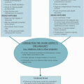
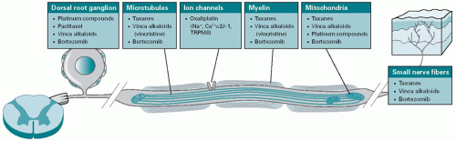
Figure 6.1. A proposed diagram illustrating the targets of most commonly used chemotherapy-induced neurotoxicity in the peripheral nervous system.

Similar to taxanes, vinca alkaloids bind to tubulin and inhibit microtubule dynamics (19). The affinity for tubulin differs among vinca alkaloid compounds (vincristine, vinblastine, vinorelbine, and vinflunine in descending order). For example, vincristine produces significant alterations in axonal cytoskeletal structure, including microtubule disorientation and neurofilament accumulation (20,21), which contribute to interruption of axonal transport (22) and result in Wallerian-like axonal degeneration and axonopathy (20,21,23). In addition, these compounds have also been reported in vitro to produce direct axonal toxicity (24). Both taxanes and vinca alkaloids affect the stability of microtubules and disrupt the axonal transport of growth factors and molecules essential to normal nerve function. However, this does not explain why platinum-based compounds, which induce DNA adducts in the nucleus, also cause the painful sensory neuropathy that taxanes and vinca alkaloid compounds do.

Platinum compounds, including cisplatin, carboplatin, and oxaliplatin, predominantly target and accumulate in the DRG. This preferential accumulation might be partially due to the relative deficiency of the BNB in peripheral nerves, especially in the DRG area, or their high affinity for the DRG cells (25). These compounds form DNA intrastrand adducts and interstrand crosslinks that lead to DNA derangement, morphologic changes, and subsequent apoptosis (26,27). Platinum compounds exert direct damage on neurons and non-neuronal cells in the DRG, and the levels of platinum in DRG of treated patients correlate with the severity of CIPN (11,28,29,30). Histological study reveals axonal loss associated with a secondary DRG atrophy. Therefore, platinum compound-associated symptoms, such as cisplatin-induced peripheral neuropathy, are described as a primary neuronopathy rather than an axonopathy (31). Additionally, the oxidative stress and mitochondria dysfunction are involved in triggering neuronal apoptosis (32). Cisplatin can also disrupt axonal microtubule growth that may also contribute to the cisplatin-induced peripheral neuropathy (33).

Only gold members can continue reading. Log In or Register to continue

Stay updated, free articles. Join our Telegram channel

Aug 25, 2016 | Posted by in ONCOLOGY | Comments Off on Pathophysiology of Chemotherapy-Induced Peripheral Neuropathy

Full access? Get Clinical Tree

Get Clinical Tree app for offline access