Fig. 7.1
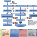
Schematic representation of the cAMP signaling pathway involved in the control of cortisol secretion in primary adrenal Cushing’s syndrome. The binding of ACTH to MC2R leads to dissociation of Gs-α subunit and activation of adenylate cyclase generating cAMP from ATP. The binding of cAMP to specific domains of the regulatory subunits of protein kinase A (R1 α) dissociates the tetramer, thereby releasing the catalytic subunit (C α), which phosphorylates different intracellular targets, including the transcription factor CREB; the latter activates the transcription of cAMP-responsive element-containing genes in the nucleus including cholesterol transporters and steroidogenic enzymes. Specific PDEs are responsible of the degradation of the intracellular cAMP in order for the two R1 α and C α subunits of PKA to be reassembled to return to their inactive state. Genetic defect in this pathway leading to its constitutive activation can underlie tumor development and excess hormone production. BMAH or CPA cells can express several functional aberrant GPCR. Activation of these receptors by their natural ligands induces the activation of intracellular cascade similar to the one activated normally by the binding of ACTH to MC2R, thereby stimulating the release of both cortisol and locally produced ACTH (in BMAH tissues) which also triggers cortisol production through autocrine and paracrine mechanisms involv ing the MC2R. ARMC5 is a new indirect or direct regulator of steroidogenesis and apoptosis. ARMC5 inactivating mutations induce a decreased steroidogenic capacity and a protection against cell death. ARMC5 Armadillo repeat-containing 5, BMAH bilateral macronodular adrenal hyperplasia, CPA cortisol-producing adenoma, GPCR G-protein-coupled hormone receptors, MC2R melanocortin type 2 receptor, PDE phosphodiesterases, PPNAD primary pigmented nodular adrenocortical disease
Primary adrenal causes account for 20–30% of overt endogenous hypercortisolism and include unilateral adrenal adenomas (10–20%), carcinomas (5–7%), or rarely bilateral adrenal hyperplasias (BAH) (<2%) [14]. BAH can be macronodular (nodules >1 cm) or micronodular (nodules <1 cm) [2]. Micronodular subtype includes the pigmented form of primary pigmented nodular adrenocortical disease (PPNAD) and the nonpigmented form of micronodular adrenocortical disease (MAD) [13, 14]. PPNAD presents either as isolated disease or as part of Carney complex (CNC).
Cortisol-Producing Adenomas (CPA)
Somatic mutations in the gene encoding the catalytic subunit of PKA (PRKACA) represent the most frequent mechanism of cortisol-secreting adenomas [15] (Table 7.1). They occur in patients diagnosed with CS at a younger age (45.3 ± 13.5 vs. 52.5 ± 11.9 years) [16] with a female predominance [17]. The first two mutations identified in a cohort of ten cortisol-producing adrenal adenomas were shown to inhibit the binding of the R subunit making the Cα subunit constitutively active [15]. A combination of biochemical and optical assays, including fluorescence resonance energy transfer in living cells, showed that neither mutants can form a stable PKA complex, due to the location of the mutations at the interface between the catalytic and the regulatory subunits [18]. The most common mutation p.Leu206Arg was present in 37% of these adrenal tumors [15]. It consists of substitution of a small hydrophobic leucine with a large positively charge hydrophilic arginine at position 206. It is located in the active cleft of the C subunit, and it inactivates the site where the regulatory subunit RIIβ binds, leading to cAMP-independent PKA activation. The second mutation (Leu199_Cys200insTrp) involves the insertion of a tryptophan residue between the amino acid 199 and 200 and was present in one case only. Later, two novel mutations were identified in a study of 22 adrenal adenomas with CS with p.Cys200_Gly201insVal and p.Ser213Arg + p.Leu212_Lys214insIle-Ile-Leu-Arg being found in three and one adenomas, respectively. They indirectly interfere with the formation of a stable PKA holoenzyme by impairing the association between the catalytic and the regulatory subunits [19]. Other groups confirmed the presence of these mutations in unilateral adrenal adenomas with overt hypercortisolism at a rate of 23–65% [16, 17, 19–21]. However, they are seldom present in adenomas with mild cortisol secretion, which might justify why subclinical CS rarely becomes overt CS [15, 19, 20]. These observations suggest that subclinical CS may have a different genetic etiology than overt CS rather than being a part of the same pathophysiological spectrum [22]. Isolated somatic GNAS mutations can also occur in 5–17% of cortisol-secreting adenomas [16, 21, 23]. Finally, Bimpaki et al. demonstrated that cortisol-secreting adrenal adenomas could have functional abnormalities of cAMP signaling, independently of their GNAS, PRKAR1A, PDE11A, and PDE8B mutation status most probably due to epigenetic events or other gene defects [24] (Table 7.1). Although β-catenin (CTNNB1) mutations are mainly observed in larger and nonsecreting adrenocortical adenomas, suggesting that the Wnt/β-catenin pathway activation is associated with the development of less differentiated tumors, Bonnet et al. described CTNNB1 mutations in 6 and 8 out of 19 and 46 subclinical and overt cortisol-producing tumors, respectively [25]. Recently Goh et al. identified CTNNB1 mutations as responsible for 16% of the cortisol-secreting adenomas [16]; they were also noted by other groups in some cases of adrenal adenomas with CS or SCS [17, 26, 27] (Table 7.1). Exome sequencing of 74 cortisol-secreting adenomas without initial identification of PRKACA mutations identified several mutations in cAMP/PKA pathway (including three novel PRKACA mutations), in the WNT/β-catenin pathway, and in the Ca2+-signaling pathway [28].
Table 7.1
Molecular mechanisms implicated in adrenocortical tumors
CPA | PPNAD | BMAH | PA | ACC | |
|---|---|---|---|---|---|
A. Genetic alterations | |||||
1. cAMP/PKA signaling pathway | – | – | MC2R a (missense) | ||
GNAS a | – | GNAS a | |||
PRKAR1A b (allelic losses) | PRKAR1A b (LOH) | – | PRKAR1A b | ||
PRKACA a (missense or insertion) | PRKACA c | PRKACA c | |||
PDE8B b | PDE8B b | PDE8B b | |||
PDE11A b | PDE11A b | ||||
2. Armadillo proteins | – | – | ARMC5 b (LOH, nonsense or missense) | ARMC5 b | |
CTNNB1 a AXIN2 a | CTNNB1 a | – | CTNNB1 a | ||
3. Other | – | – | MEN1 b , FAP b , FH b , EDNRA, DOTL1, HDAC9, PRUNE2 | KCNJ5, CACNA1D, CACNA1H, ATP1A1, ATP2B3, chimeric 11β-hydroxylase/aldosterone synthase gene | IGF2, TP53, ZNRF3, TERT, RPL22, TERF2, CCNE1, NF1 |
B. Abnormal protein expression | GPCR | – | GPCR | GPCR | |
PRKAR1A | – | PRKAR1A | |||
– | PRKACA | – | |||
– | Glucocorticoid receptor | – | |||
– | Estrogen receptor | – |
Primary Bilateral Macronodular Adrenal Hyperplasia (BMAH)
In contrast to somatic mutations causing cortisol-secreting adenomas, very rare germline complex genomic rearrangements in the chromosome 19p13.2p13.12 locus, resulting in copy number gains that includes PRKACA gene, rarely caused either micronodular or macronodular hyperplasia depending on the extent of gene amplification [15, 29, 30]. MC2R mutations are extremely rare causes of adrenal hyperplasia or tumor formation [31, 32]. In only two patients with BMAH , constitutive activation of the MC2R with consequent enhanced basal receptor activity resulted either from impaired receptor desensitization secondary to a C-terminal MC2R mutation (F278C) [33] or from synergistic interaction between two naturally occurring missense mutations in the same allele of the MC2R: substitution of Cys 21 by Arg (C21R) and of Ser 247 by Gly (S247G) [34] (Table 7.1).
Somatic activating mutations of the Gs-α subunit of heterotrimeric G protein also termed gsp mutations (GNAS) were the first identified in a particular bilateral nodular form of primary adrenal CS [35, 36]. It occurred in a mosaic pattern in some fetal progenitor cells during early embryogenesis resulting in the constitutive activation of the cAMP pathway in cells of various tissues which developed from the affected progenitor cells. This mutation was identified initially in the McCune-Albright syndrome (MAS) where a minority of patients develops nodular adrenal hyperplasia and CS among other more common manifestations such as café au lait spots and bone fibrous dysplasia or other endocrine tumors causing ovarian precocious puberty, acromegaly, or hyperthyroidism [35, 37]. In MAS patients with CS, GNAS mutations are found in the cortisol-secreting nodules, whereas the internodular adrenal cortex which is not affected by the mutation becomes atrophic as ACTH becomes suppressed. Isolated somatic GNAS mutations can also occur in rare cases of BMAH [38, 39] without any other manifestations of MAS. This suggests that the somatic mutation in MAS occurs at an early stage of embryogenesis in cells which are precursors of several tissues. In isolated BMAH, the somatic mutation probably occurs in mosaic pattern in more differentiated adrenocortical progenitor cells only which will migrate to generate bilateral macronodular adrenal glands; a somatic GNAS mutation giving rise to a unilateral adenoma occurs later in life in a single committed zona fasciculata cell [16, 21, 23].
Inactivating germline mutations in ARMC5 gene were first described in apparently sporadic cases of primary BMAH [40] (Table 7.1). Armadillo proteins form a large family of proteins that are characterized by the presence of tandem repeats of a 42-amino acid motif with each single ARM-repeat unit consisting of three α-helices [41]. The most well-known protein of this family is β-catenin, which is crucial in the regulation of development and adult tissue homeostasis through its two independent functions, acting in cellular adhesion in addition to being a transcriptional co-activator. Deregulation in the Wnt/β-catenin signaling pathway is involved in the pathogenesis of adrenocortical adenomas and carcinomas. Armadillo repeat-containing 5 (ARMC5) is a novel Armadillo (ARM) repeat-containing gene and encodes a protein of 935 amino acids; its peptide sequence reveals two distinctive domains: ARM domain in the N-terminal and a BTB/POZ in the C-terminal (Bric-a-brac, Tramtrack, Broad complex/Pox virus, and Zinc finger) [42].
The bilateral nature of macronodular hyperplasia as well as its long and insidious onset motivated the search for a genetic predisposition that could result in earlier diagnosis and better management to avoid bilateral adrenalectomy. Single-nucleotide polymorphism arrays, microsatellite markers, and whole-genome and Sanger sequencing were applied to genotype leucocyte and tumor DNA obtained from patients with BMAH. The search for the responsible genes was conducted in apparently sporadic and familial cases [40, 43–47]. The initial germline mutation in the ARMC5 gene, located at 16p11.2, was detected in 18 out of 33 apparently sporadic tumors, 55% of cases of BMAH with Cushing’s syndrome [40]. Further studies in sporadic cases found that the prevalence of germline ARMC5 mutations was closer to 25% [43, 45, 46]. Inactivation of ARMC5 is biallelic, one mutated allele being germline and the second allele being a somatic secondary event that occurs in a macronodule; these findings are consistent with its role as a potential tumor suppressor gene according to Knudson’s two-hit model [40, 43]. Correa et al. demonstrated that ARMC5 has an extensive genetic variance by Sanger sequencing 20 different adrenal nodules in the same patient with BMAH [48]. They found the same germline mutation in the 20 nodules (p.Trp476* sequence change) but uncovered 16 other mutation variants in the 16 nodules. This suggests that the germline mutation is responsible for the diffuse hyperplasia, but second somatic hits are required to enhance adrenal macronodular formation [40, 48]. In the first large BMAH family studied, a heterozygous germline variant in the ARMC5 gene (p.Leu365Pro) was identified in all 16 affected Brazilian family members as well as other mutations in two of three other families [43]. Interestingly, only two mutation carriers had overt CS, and the majority had subclinical disease, and one carrier had no manifestations despite being 72 years old. In addition, in one third of the affected individuals, only unilateral adrenal lesion was present as progression of the full-blown disease, needing many years and requiring the occurrence of additional somatic mutations in several macronodules. This raised the question of the prevalence of ARMC5 mutation in apparently unilateral incidentalomas in the general population. Recent screening of sporadic cases of patients with bilateral incidentalomas revealed a low frequency (1 out of 39 patients) of ARMC5 mutation [49].
Other families with BMAH have also been identified with ARMC5 mutations or alterations [44, 47, 50–52]. In all cases the pattern of inheritance is autosomal dominant. A germline deletion rather than mutation of ARMC5 was reported in a family presenting with vasopressin-responsive SCS and BMAH [52]. By applying droplet digital polymerase chain reaction, the mother and her son had germline deletion in exon 1–5 of ARMC5 gene locus. Furthermore, Sanger sequencing of DNA from the right and left adrenal nodules as well as peripheral blood of the son revealed the presence of another germline, missense mutation in ARMC5 exon 3 (p.P347S) [52].
The presence of ARMC5 mutation in patients with BMAH and aberrant GPCR has been reported, but the relationship has not been well established yet. The most frequent aberrant responses were to upright posture, isoproterenol, vasopressin, and metoclopramide tests [40, 45, 51]. In contrast, none of the patients with food-dependent CS carried ARMC5 mutations [40, 45]. ARMC5 inactivation decreases steroidogenesis, and its overexpression alters cell survival, which could argue why relatively inefficient cortisol overproduction is seen despite massive adrenal enlargement [2, 53, 54]. Despite this, the index cases operated for Cushing’s syndrome and ARMC5 mutation carriers presented more severe CS than cases operated for Cushing’s syndrome without ARMC5 mutation; carrier patients had a more severe clinical phenotype and biochemical profile as well as larger adrenals on imaging with a higher number of nodules [45, 46]. ARMC5 mutations appear to be the most frequent genetic alteration in BMAH with 61 different mutations, 27 germinal and 30 somatic, found all along the protein in different domains. Thus, genetic counseling and screening for these mutations are highly encouraged in family members of patients with BMAH even without the evidence of a clinical disease [42, 43, 54]. As ARMC5 appears to be a tumor suppressor gene and is widely expressed in many tissues other than the adrenal, it is of interest to examine whether mutation carriers could develop other tumors. In a few families with BMAH, the occurrence of intracranial meningiomas was described, and a somatic ARMC5 mutation was found in a meningioma of a patient with familial BMAH with a germline ARMC5 mutation suggesting the possibility of a new multiple neoplasia syndrome [44]. Finally, ARMC5 mutations have been identified in primary aldosteronism where 6 patients of 56 (10.7%, all Afro-Americans) had germline mutations in the ARMC5 gene; among these 6 patients, 2 suffered from BMAH [55].
Several other gene mutations have been reported in patients with CS mainly presenting with BMAH including the multiple endocrine neoplasia type 1 (MEN1), familial adenomatous polyposis (APC), and type A endothelin receptor (EDNRA) [39, 53, 56, 57]. Furthermore, somatic mutations other than ARMC5 have also been found in patients with BMAH such as the DOT1L (DOT1-like histone H3K79 methyltransferase) and HDAC9 (histone deacetylase 9) genes; these two nuclear proteins are involved in the transcriptional regulation; however, their mutations were found at a much lower frequency than ARMC5 [17] (Table 7.1).
In addition to genetic alterations, the abnormal expression of several proteins was found to regulate steroidogenesis in cortisol-secreting tumors and hyperplasia [58]. In particular, the aberrant adrenocortical expression of various G-protein-coupled receptors (GPCR) can be increased such as the ectopic ones for glucose-dependent insulinotropic peptide (GIPR), β-adrenergic receptors (B-AR), vasopressin AVP (V2-V3 R), serotonin (5-HT7R), glucagon (GCGR), and angiotensin II (AT1R). Other eutopic receptors can be overexpressed such as those for vasopressin (V1R), luteinizing hormone/human chorionic gonadotropin (LHCGR), or serotonin (5-HT4R) [59]. Five systematic studies have demonstrated abnormal expression of more than one type of GPCR in 80% of patients with BMAH. Multiple responses within individual patients occurred with up to four stimuli in 50% of the patients; AVP and 5-HTR4 agonists were the most prevalent hormonal stimuli triggering aberrant responses in vivo [39, 60–63]. In addition, more recently, local paracrine production of ACTH was identified in clusters of BMAH cells, and ACTH production was stimulated by aberrant GPCR [64]. Thus, cortisol production is controlled by aberrant GPCR as well as by ACTH produced within the BMAH adrenocortical tissue, amplifying the effect of the aberrant receptors’ ligands. The percentage of aberrant responses in patients with unilateral adenoma and mild CS or SCS was similar to those in BMAH patients [63]. However it was less frequent in patients with unilateral adenomas and overt CS [62] most probably due to higher prevalence of PRKACA mutations in these patients [15].
Micronodular Adrenal Hyperplasia
PRKAR1A is an ad renocortical tumor suppressor gene according to in vitro and transgenic mouse studies. Its inactivation leads to ACTH-independent cortisol secretion by the resulting bilateral micronodules [9, 65]. PKA activation due to PRKAR1A mutations results either from reduced expression of the RIα subunits or from impaired binding to C subunits [66] (Table 7.1). Loss of RIα is sufficient to induce ACTH-independent adrenal hyperactivity and bilateral hyperplasia and was demonstrated for the first time in vivo in an adrenal cortex-specific PRKAR1A KO mouse model referred to as AdKO. Pituitary-independent CS with increased PKA activity developed in AdKO mice with evidence of deregulated adrenocortical cell differentiation, increased proliferation, and resistance to apoptosis. Moreover, RIα loss led to regression of adult cortex and emergence of a new cell population with fetal characteristics [65]. In vitro and in vivo models of PPNAD (AdKO mice) showed that PKA signaling increased mTOR complex 1, leading to increased cell survival and possibly tumor formation [67]. Tumor-specific loss of heterozygosity (LOH) involving the 17q22-24 chromosomal region harboring PRKAR1A and inactivating mutations of PRKAR1A are responsible for CS in isolated or familial PPNAD and CNC [66, 68–70]. They are found in more than 60% of patients with CNC and in up to 80% of CNC patients who develop CS from PPNAD [70, 71]. Furthermore, somatic allelic losses of the 17q22-24 region and inactivating mutations in PRKAR1A were identified in 23% and 20% of adrenocortical tumors, respectively [72]. Although PRKAR1A mutations are not found in BMAH, somatic losses of the 17q22-24 region and PKA subunit and enzymatic activity changes show that PKA signaling is altered in BMAH similarly to what is found in adrenal tumors with 17q losses or PRKAR1A mutations [73]. CS presenting in persons younger than 30 years of age with bilateral, small (usually 2–4 mm in diameter), black-pigmented adrenal nodules are all characteristics of PPNAD. A distinctive feature of PPNAD compared to BMAH is the presence of atrophy in the internodular adrenal tissue. CNC is a familial autosomal variant that includes PPNAD among other tumors such as atrial myxomas, peripheral nerve tumors, breast/testicular tumors, and GH-secreting pituitary tumors along with skin manifestations [74]. Patients with CS due to PRKAR1A mutations tend to have a lower BMI with evidence of increased PKA signaling in periadrenal adipose tissue, which is in concordance with the role of PKA enzyme in the regulation of adiposity and fat distribution [75].
Phosphodiesterases (PDE) play a role in the hydrolysis of cAMP. There are two types of PDE8 enzymes coded by two distinct genes, PDE8A and PDE8B, which are highly expressed in steroidogenic tissues such as the adrenal, the ovaries, and the testis as well as in the pituitary, thyroid, and pancreas [76, 77]. Genetic ablation of PDE8B in mouse models or long-term pharmacological inhibition of PDE8s in adrenocortical cell lines were shown to increase the expression of steroidogenic enzymes such as StAR and p450scc (CYP11A); furthermore, they potentiated ACTH stimulation of steroidogenesis by increasing cAMP-dependent PKA activity [78]. A PDE8B missense mutation (p.H305P) was described in a young girl with isolated micronodular adrenocortical disease (iMAD), which is a nonpigmented micronodular hyperplasia without PRKAR1A [79]. HEK293 cells transfected with the PDE8B mutant gene exhibited higher cAMP levels than with wild-type PDE8B, indicating an impaired ability of the mutant protein to degrade cAMP [79]. Other inactivating mutations in phosphodiesterase 11A isoform 4 gene (PDE11A) and 8B (PDE8B) have been also described in adrenal adenomas, carcinomas, and BMAH [14, 23, 24, 78, 80–82].
In a Carney complex patient without Cushing’s syndrome but with skin pigmentation, acromegaly, and myxomas, gene triplication of chromosome 1p31.1, including PRKACB, which codes for the catalytic subunit beta (Cβ) resulted in increased PKA activity (Fig. 7.1). It is likely that whereas the loss of RIα leads to the full Carney complex phenotype, the gain of function in Cα leads to adrenal adenomas and Cushing’s syndrome, while in this case, amplification of Cβ resulted in certain nonadrenal manifestations of CNC [83]. Finally, somatic CTNNB1 mutations were also found in 2 out of 18 patients with PPNAD (11%). In both cases, the mutations occurred in relatively larger adenomas that had formed in the background of PPNAD [27, 84].
A distinctive feature of PPNAD is the paradoxical increase in urinary-free cortisol during the 6-day dexamethasone suppression test (Liddle test), which was found in 69–75% of two small series of patients with PPNAD [85, 86]. The glucocorticoid receptor (GR) is largely overexpressed in PPNAD nodules [87], which mediates stimulation of PKA catalytic subunits [85]. In a patient with PPNAD, who increased cortisol secretion during pregnancy and oral contraceptive use, β-estradiol (E2) stimulated cortisol secretion in a dose-response manner [88] via activation of overexpressed estrogen receptors ERα and G-protein-coupled receptor 30 (GRP30) [89].
Recently, it was shown that PPNAD tissues overexpress the 5-HT synthesizing enzyme tryptophan hydroxylase type 2 (Tph2) and the serotonin receptors, types 4, 6, and 7, leading to formation of an illicit stimulatory serotonergic loop whose pharmacological inhibition in vitro decreases cortisol production. PRKAR1A mutations led to PKA activation and induction of serotonin and fuctional aberrant serotonin receptors partly regulating cortisol excess [90].
Primary Aldosteronism (PA)
Potassium homeostasis and maintenance of intravascular volume are controlled by the binding of free aldosterone to the mineralocorticoid receptor in epithelial cells, resulting in increased intestinal and renal Na+ and Cl− absorption and reabsorption, respectively [91]. Aldosterone excess in PA results in sodium retention and hypertension and can also result in hypokalemia [92]. Under resting physiological conditions, the strongly negative resting membrane potential of ZG cells is maintained by the concentration gradient of K+ between the intracellular and extracellular space which is generated by the activity of the Na+, K+-ATPase. Angiotensin II and increased K+ lead to cell membrane depolarization which opens voltage-dependent Ca2+ channels. Furthermore, angiotensin II acts through the angiotensin II type 1 receptor (AT1R) inducing Ca2+ release from the endoplasmic reticulum. Consequently, the increase in intracellular Ca2+ concentration activates the calcium signaling pathway, which triggers activation of CYP11B2 transcription [92].
The mechanisms implicated in the pathophysiology of PA are not entirely clarified. Gene rearrangement or mutations in ion channel genes regulating intracellular ionic homeostasis and cell membrane potential were described in sporadic and familial cases of PA [93–96]. On the other hand, several hormones seem to activate variable levels of eutopic or ectopic aberrant receptors [97], and autocrine and paracrine regulatory mechanisms [98] can increase aldosterone secretion in PA independently from the suppressed RAS.
Genetic Alterations in PA
Bilateral adrenal hyperplasia (BAH) and aldosterone-producing adenoma (APA) are the two most common subtypes of PA (BAH in 50–70% of the cases and APA in 30–50%); less frequent causes include primary (unilateral) adrenal hyperplasia (5%), aldosterone-producing adrenocortical carcinoma (<1%), familial hyperaldosteronism (1%), and ectopic aldosterone-producing adenoma or carcinoma (<0.1%) [99]. Familial hyperaldosteronism (FH) type 1, previously known as glucocorticoid-remediable aldosteronism (GRA), is suspected in young patients whose relatives suffer from cerebrovascular accidents and who present with PA that is relieved by dexamethasone [100]. It is an autosomal dominant disease whereby the promoter of the chimeric 11β-hydroxylase/aldosterone synthase gene belongs to the 5′ end of CYP11 B1 (11β hydroxylase) and drives the expression of the 3′ end of CYP11 B2 (aldosterone synthase) ectopically in ZF cells under the main regulation by ACTH [101]; the diagnosis in these is used to be based on the drop in aldosterone secretion following dexamethasone administration by more than 80% or to <4 ng/dL [102]; however, nowadays genetic analysis is the main diagnostic tool. FH type 2 is more frequent (1.2–6%) than FH type 1 (≤1%); it is diagnosed in a PA patient who has a first-degree relative (parent/sibling/offspring) with established PA but without FH type 1 gene rearrangement. To date no culprit gene has been identified but linkage analysis has mapped FH type 2 to chromosome 7p22 [103]. FH type 3 and FH type 4 are caused by germline mutations in KCNJ5 [104] and CACNA1D/CACNA1H genes, respectively, encoding a potassium channel GIRK4 and voltage-gated calcium channels [105, 106], respectively (Table 7.1).
Somatic mutations in KCNJ5 were identified in almost 30–40% of APA with higher prevalence in the Japanese population [107]. KCNJ5 can alter channel selectivity allowing enhanced Na+ conductance; Na+ influx results in cell depolarization, the activation of voltage-gated Ca2+ channels, aldosterone production, and cell proliferation [94, 108]. Mutations in CACNA1D gene result in channel activation and less depolarized potentials causing increased Ca2+ influx, aldosterone production, and cell proliferation in affected ZG cells [96, 105]. Mutations in CACNA1H gene were discovered in children with PA; they result in impaired channel inactivation and activation at more hyperpolarized potentials, producing increased intracellular Ca2+ and aldosterone excess [106]. Mutations in ATP1A1 gene (encoding the Na+/K+ ATPase α subunit) and ATP2B3 gene (encoding the plasma membrane Ca2+ ATPase) were identified in 5.2% and 1.6%, respectively, of patients in a series of APA [93] (Table 7.1). The pathophysiology of progression from normal adrenal to APA and the causes of diffuse bilateral hyperplasias, either as BAH or in mild form adjacent to the dominant aldosteronoma, are still unknown. In fact, these somatic mutations appear to be as independent events since different mutations in the genes described above are found in different aldosterone-producing nodules from the same adrenal [109]. Nishimoto et al. found that aldosterone-producing cell clusters (APCCs), which are nests of cells below the adrenal capsule with an increased expression of CYP11B2, are common in normal adrenals. They protrude into cortisol-producing cells that are usually negative for CYP11B2 expression. He also speculated that APCCs could be a precursor for APA because they harbor a different mutational spectrum compared to APA [110]. Moreover, nodule formation and excess aldosterone production constitute two dissociated events, complying with the two-hit hypothesis for APA formation [111, 112] because no mutations of any of the above ion channel genes were found in BAH or in ZG hyperplasia adjacent to the dominant aldosteronomas [93, 108, 113]; the first hit may result from a somatic mutation in one of the genes described above, at least in approximately 60% of cases, causing a unilateral aldosteronoma or a dominant nodule adjacent to ZG hyperplasia. Possible causes of the second hit that results in dysregulation in cellular proliferation/apoptosis accelerating adenoma formation could be due either to dysregulation of PKA pathway or to gene mutations such as ARMC5 [55] or to activation of the Wnt/β-catenin pathway [114, 115]. The activation of the latter pathway could be mostly related to downregulation of a negative regulator of this pathway, named SFRP2 caused by a high methylation of SFRP2 promoter [116].
Aberrant Expression or Function of G-Protein-Coupled Receptors or Their Ligands
Adrenal production of aldosterone in APA and BAH was found to be under the influence of aberrant G-protein-coupled receptors (GPCR) and their ligands, as demonstrated by in vivo and in vitro studies [117, 118]. Whether these aberrant regulatory secretory mechanisms and the overexpression of GPCR in PA are secondary to unknown proliferative mechanisms or are primary and at least partially responsible for the abnormal proliferation, the initiation of diffuse BAH is currently unclear. Yet, they obviously play a role in aldosterone production in PA which is relatively autonomous from the suppressed RAS but regulated by the ligands of various aberrant hormone receptors.
In zona glomerulosa cells, binding of ACTH or other hormones to their specific GPCR activates the cAMP/PKA pathway [8], the cAMP response element-binding protein (CREB), and the StAR expression. This induces a slow but sustained calcium influx through the L-type calcium channels. The subsequent increase in intracellular calcium activates calcium/calmodulin-dependent protein kinase and steroidogenesis [119, 120].
Aldosterone secretion is regulated mainly by angiotensin II and potassium and to a lesser extent by ACTH and serotonin. However, the latter two play an increased role in PA most probably due to the overexpression and function of melanocortin-2 receptor (MC2R) and serotonin receptor (5-HT4 R), respectively [99]. An acute ACTH administration resulted in an exaggerated increase in plasma aldosterone concentration in PA patients with APA or BAH compared to normal individuals. Endogenous ACTH can be an important aldosterone secretagogue in PA as demonstrated by the diurnal increase in aldosterone in early morning and its suppression by dexamethasone. Screening using a combination of dexamethasone and fludrocortisone test reveals a higher prevalence of PA in hypertensive populations compared to the aldosterone to renin ratio [99]. RT-PCR or transcriptome studies demonstrated eutopic overexpression of MC2R in resected aldosteronomas as compared to cortisol-secreting adenomas, nonfunctional adenomas, and adrenocortical carcinomas [121–123]. Levels of MC2R mRNA were increased by a mean of 3.9-fold in 15 adrenal tumors tissues (14 APA and 1 BAH) compared to normal adrenal [97]. However, great variability was noticed in the level of expression in each tumor as four had lower levels than normal (0.3–0.7-fold), while those with increased expression varied between 1.4- and 20.6-fold compared to normal. As most patients with BAH are not operated, there is almost no data on MC2R expression in BAH, but in the only case with BAH studied by this group, MC2R was 20-fold increased. The variable level of MC2R overexpression in each APA or in the adjacent zona glomerulosa hyperplasia may explain the discordant results of adrenal vein sampling between basal levels and post ACTH administration in the determination of source of aldosterone excess [124].
Stay updated, free articles. Join our Telegram channel

Full access? Get Clinical Tree


