Pathogenesis and Progression of Squamous Cell Carcinoma of the Head and Neck


Pathogenesis and Progression of Squamous Cell Carcinoma of the Head and Neck


Ryan M. Aronberg

Natalia Issaeva

Wendell G. Yarbrough


Over the past few decades, advances in cellular and molecular biology have led to an accelerated understanding of the pathogenesis and hallmarks of neoplastic disease. More recently, the development of array technologies and highthroughput genetic sequencing have helped to identify many of the underlying molecular defects involved in carcinogenesis and resistance to therapeutics. These capabilities have led to an overwhelming volume of data, requiring advances in bioinformatics to keep pace. The wealth of information being produced has laid the foundation to develop new treatments targeting these defects. Future goals will be the identification of biomarkers to guide diagnosis and therapy, the development of new targeted and combined therapies, and the personalization of therapy based on the molecular characteristics of individual tumors. It will become ever more important for clinicians to understand the molecular characteristics of the disease in caring for their patients. Overall, recent advances in the understanding of tumor biology and related fields (e.g., immunology) make this an exciting time of discovery that should translate into increased survival and improved quality of life for our patients.

This chapter will provide a framework to be used as a basis for exploring the pathogenesis of neoplasia, with an emphasis on the latest findings in head and neck squamous cell carcinoma (HNSCC). We will first introduce general concepts of carcinogenesis and then review the characteristics of the neoplastic phenotype and genotype seen in HNSCC. Along the way, recent biologic insight and therapeutic applications for HNSCC will be explored.


THEORIES OF CARCINOGENESIS


Clonal Evolution and Molecular Progression Models

It is widely accepted that an accumulation of alterations in several genes ultimately leads to a transition from normal to dysplastic to a neoplastic phenotype. Clonal evolution theory, proposed by Peter Nowell in 1976, likens cancer to an evolutionary process involving clonal proliferation, genetic diversification, and subclonal selection.1 Random mutational events, in conjunction with selective pressures within the tumor environment (tissue barriers, the immune system, induction of programmed cell death, anticancer therapeutics), allow genetic diversification and drift. The cumulative loss of tumor suppressor genes or activation of oncogenes leads to changes in cellular behavior, which confers a survival or proliferative advantage over other cells, ultimately resulting in territorial expansion.2 Eventually, the further accumulation of defects can confer new traits such as immortality, angiogenesis, or the ability to invade.

Colon cancer represents the first and most comprehensive molecular progression model.3 In the model, events including oncogene activation and tumor suppressor inactivation lead to progression from normal mucosa, to benign adenomatous growth, to carcinoma in situ, to invasive carcinoma. As in colon cancer, it is the accumulation of these events, rather than an ordered occurrence, that leads to HNSCC,4 and a similar histologic progression occurs from normal mucosa, to dysplastic mucosa, to carcinoma in situ, to frank invasive carcinoma.


Field Cancerization

For decades, it has been observed that the “normal” mucosa adjacent to head and neck cancers has histologic and genetic alterations not unlike the cancer itself. Additionally, it is not uncommon for satellite lesions or second primaries to occur in HNSCC. These observations led to the “field cancerization” hypothesis that an entire field of mucosa, which is exposed to the same environmental factors, is at risk for carcinogenesis (Fig. 2.1).5 This theory proposes that although a cancer develops from a small localized segment of the mucosal field, the surrounding cells within a larger field of mucosa exist on a dysplastic spectrum and share some genetic alterations with the cancer. Early on, lesions can appear clinically and histologically normal, but molecular signatures can help identify altered cells at risk for progression to cancer.


Cancer Stem Cells

When tumor cells are grown in vitro or in a xenograft model, only a small fraction of the cells have the ability to form a new tumor.6,7 In HNSCC, isolation of a population of cells expressing surface marker CD44 and aldehyde dehydrogenase (ALDH) was shown to have significant tumorigenic potential, whereas CD44(-) cells did not.8 The cells that possess the necessary characteristics of self-renewal and differentiation have been termed “cancer stem cells.” They constitute a minority of the cells within the tumor itself but are responsible for much or all of its tumorigenicity. The ability of these cells to selfrenew can provide a near-endless supply of new tumor cells, and the ability to differentiate in phenotypically diverse ways allows them to produce a heterogeneous population of cells. Their capacity to differentiate and produce cells with new properties has linked them to cancer initiation, treatment resistance, local tumor recurrence, and metastasis.9 Meanwhile, due to their slow growth and ability to adapt, these cells are not easily targeted by radiotherapy or traditional chemotherapies. For example, after irradiation of breast or glioblastoma
xenografts, cancer stem cells were found to be enriched in the surviving tumor tissue.10,11 These surviving cancer stem cells were found to possess fewer reactive oxygen species (ROS) (mediators of radiation-induced damage) and activated DNA damage response/repair pathways in response to the radiotherapy. Knowledge of the biologic nature and response of stem cells has led to the hope of targeting these resistance mechanisms therapeutically.


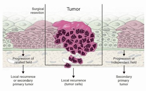
Figure 2.1. Field cancerization. Field cancerization is defined as the presence of one or more mucosal areas consisting of epithelial cells that have cancer-associated genetic or epigenetic alterations. A preneoplastic field (shown in light pink) is monoclonal in origin and does not show invasive growth or metastatic behavior, which are the hallmarks of an invasive carcinoma (dark pink). Field cancerization has been supported by molecular data and provides a theoretical explanation for multiple primaries. (Adapted from Leemans CR, Braakhuis BJ, Brakenhoff RH. The molecular biology of head and neck cancer. Nat Rev Cancer. 2011;11(1):9-22.)


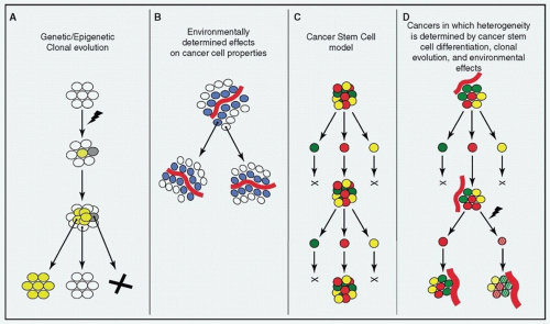
Figure 2.2. Tumor heterogeneity. Heterogeneity can arise within tumors through: (A) the stochastic process of clonal evolution, (B) extrinsic environmental differences within tumors, and (C) the presence of cancer stem cells that variably differentiate. These processes are not mutually exclusive, but rather synergistic in producing a heterogeneous population of cells (D). (From Magee JA, Piskounova E, Morrison SJ. Cancer stem cells: impact, heterogeneity, and uncertainty. Cancer Cell. 2012;21(3):283-296, with permission.)


Tumor Heterogeneity

Like most cancers, HNSCCs are not simply an aggregate of a genetically identical cell population, but are composed of cells with marked genetic and cellular heterogeneity (Fig. 2.2).12 This unexpectedly high degree of heterogeneity is thought to result from a combination of genomic instability, clonal evolution, and the effects of diverse, highly selective, microenvironments within a cancer. Recent evidence indicates that cancer stem cells
may be principally responsible13,14 for creating a heterogeneous population of cells, but that clonal evolution and the effects of the tumor’s microenvironment act in a synergistic manner (i.e., the cancer stem cells themselves may undergo clonal evolution and be affected by local influences in the tumor environment). Higher levels of intracancer heterogeneity have also been correlated to tumor progression, poorer survival, and adverse outcomes in patients with HNSCC. Recently, levels of tumor heterogeneity, when factored with HPV status, were found to be useful in predicting clinical outcome15 in HNSCC.

Intratumor heterogeneity makes it unlikely that a single biopsy will fully capture the histologic or genomic landscape of a patient’s cancer. New methods have attempted to measure heterogeneity in an attempt to incorporate it into diagnostic workup and treatment, with the applications such as predicting metastatic potential, identifying treatment resistance, and predicting responses to targeted therapies. While performing multiple samples of a cancer (spatially or temporally spaced) may prove challenging and potentially dangerous, future efforts may focus on collecting the DNA of circulating cancer cells or even using molecular imaging to survey multiple areas of the cancer.16


RISK FACTORS AND ETIOLOGIC AGENTS

HNSCC has traditionally been, and continues to be, believed to be a cancer caused by environmental elements. Until the 1990s, almost all cancers of the head and neck were thought to be caused by tobacco-related carcinogens. Over the last 20 years, a remarkable shift has taken place, with the human papillomavirus (HPV) becoming responsible for a growing proportion of cancers of the head and neck, specifically of the oropharynx. HPV(-) and HPV(+) HNSCCs are now widely recognized as having distinct etiologies, risk factors, patient populations, clinical attributes, responses to therapy, and prognosis (Table 2.1). Given these observed clinical differences, it may come as no surprise that recent molecular analyses of hundreds of their cancers show a clear and marked distinction between HPV(+) and HPV(-) HNSCCs with regard to mutational profile, gene expression, methylation patterns, and signaling pathway activation.21,22,23,24 The molecular and clinical differences between HPV(+) and HPV(-) tumors are clear indicators that we must no longer consider HNSCC as a single disease. Going forward, subtypes of HNSCC defined by the molecular characteristics of the tumor, as well as the genetic background of the patient, will guide therapy, with the goal of personalized cancer treatment. Accordingly, we explore HPV(-) and HPV(+) HNSCC separately in this chapter.


Table 2.1 Distinct Clinical Features of HPV(+) and HPV(-) HNSCC


Characteristic


HPV(-) HNSCC


HPV(+) HNSCC


Etiology/risk factors


Smoking, alcohol


HPV (high-risk sexual practices)


Incidence


Decreasing


Increasing


Age


Older


Younger


Gender


Male


Male


Race/ethnicity


AA > Caucasian > others


Caucasian > AAa


Socioeconomic status (SES)


Lower SES


No predilection


Site


All head and neck


Oropharynx


AJCC staging (TNM)


Higher T, lower N (79)


Lower T, higher N


Histopathology


Most commonly moderately to well-differentiated SCC, producing keratin (70)


Most commonly poorly differentiated or basaloid SCC, with koilocytes


p53


Inactivating mutations


Inhibited by E6


Rb


17p LOH, p16 mutation, deletion, or promoter hypermethylation


Inhibited by E7


p16 expression


Decreased


Increased


Chemoradiotherapy response


Good, but with high rate of recurrence


Good, with low rate of recurrence


Survival/recurrence


Poor for advanced stages


Good, but worse if smokers


a Data show a difference in racial/ethnic population affected by HPV(+) compared to HPV(-) HNSCC, though precise incidence rates have not been reported because data are from small cohort studies. (Gillison ML, et al. Distinct risk factor profiles for human papillomavirus type 16-positive and human papillomavirus type 16-negative head and neck cancers. J Natl Cancer Inst. 2008;100(6):407-420; Settle K, et al. Racial survival disparity in head and neck cancer results from low prevalence of human papillomavirus infection in black oropharyngeal cancer patients. Cancer Prev Res (Phila). 2009;2(9):776-781. References 17, 18.)
AA, African American; AJCC, American Joint Committee on Cancer; LOH, loss of heterozygosity; N, lymph node stage; Others, Asian/Pacific Islander + American Indian/Alaska native + Hispanic; SCC, squamous cell carcinoma; SES, socioeconomic status; T, tumor size stage. (Data sources: www.cdc.gov; Westra WH. The morphologic profile of HPV-related head and neck squamous carcinoma: implications for diagnosis, prognosis, and clinical management. Head Neck Pathol. 2012;6(suppl 1):S48-S54; Ang KK, Sturgis EM. Human papillomavirus as a marker of the natural history and response to therapy of head and neck squamous cell carcinoma. Semin Radiat Oncol. 2012;22(2):128-142. References 19, 20.)


Environmental Toxins

The vast majority of HPV(-) HNSCC are caused by exposure to environmental carcinogens. Fifty-five thousand HNSCC cases were estimated to occur in the United States in 2014.25 The exact incidence of HPV(-) HNSCC is difficult to determine because HPV testing is not universal and reporting is not required. Long considered the traditional risk factors, tobacco and alcohol are known to dramatically increase the risk of head and neck cancer.26,27 While the risk from consumption of alcohol alone is modest, it synergistically increases risk when combined with
tobacco.28,29 In addition to the direct trauma to mucosal surfaces induced by these agents, tobacco products are composed of dozens of known carcinogenic compounds, including polycyclic aromatic hydrocarbons (PAHs), oxidizing substances, and free radicals. Following metabolic activation by endogenous enzymes (often cytochrome p450s), these carcinogens form covalent DNA adducts and/or induce epigenetic changes. These DNA adducts must be repaired by designated DNA repair machinery or else risk causing errors in replication (resulting in mutations).

Because many more people use tobacco than develop cancer, there are probably individual factors that moderate the risk of cancer development following exposure to the more than 60 known carcinogens in tobacco smoke.30 The role of individual factors as modulators of the risk of cancer development has been examined with a focus on enzymes that metabolize the carcinogens. For example, the increased incidence of HNSCC in first-degree relatives of patients who have HNSCC supports a role for genetic predisposition that could be related to carcinogen metabolism.31 Studies of gene-environment interactions are difficult and frequently underpowered, and in the case of tobacco carcinogen detoxification, the genes implicated exist in large families, which functionally overlap. Despite these constraints, polymorphisms in glutathione-S transferase (GST) and uridine 5′-diphosphate-glucuronosyltransferase (UGT) have been identified as possible risk factors.32,33 That being said, the overall increased risk attributable to the presence or absence of any detoxifying enzyme polymorphism is modest, and mechanisms for translating knowledge of polymorphisms into decreased risk are not clear. As has been proven by recent decreases in cancer incidence,25 a more fruitful area for impact is advocacy and education to decrease the use of tobacco.


Human Papillomavirus

HPV was first linked to cervical carcinogenesis in the 1970s by Professor Harald zur Hausen. The idea of a virus causing cancer went against the prevailing views of that time, and he was awarded the Nobel Prize in Medicine for this important discovery in 2008.34 Soon after, an association between HPV and head and neck malignancies was demonstrated when HPV antigens were detected in preserved histologic specimens.35 However, it remained unclear if the HPV in these cancers was truly a causative agent or simply a passenger or contaminant. More recently, multiple lines of evidence have shown that HPV can be causative of HNSCC, particularly those arising in lymphatic-associated epithelium of the palatine and lingual tonsils. Epidemiologic data show that since the 1980s, there has been a decrease in the incidence of cancers of the head and neck in many developed countries, directly mirroring the decline in tobacco consumption. However, the incidence of cancer of the head and neck in nonsmokers has increased dramatically, along with the incidence of HPV-related cancers.36 High-risk HPV is now causatively linked to the majority of oropharyngeal squamous cell carcinomas (OPSCCs).37 Known aspects of HPV biology and mechanism of malignant transformation, as well as differences between HPV(+) and HPV(-) HNSCCs, will be discussed in depth later in this chapter.


Familial Disorders

As opposed to the modestly increased risk associated with polymorphisms in carcinogen-metabolizing enzymes, the risk of developing cancer in patients with familial cancer syndromes is dramatically increased. Fanconi anemia (FA) is an autosomal recessive disorder caused by mutations in any of a number of DNA repair genes (including the FANC and BRCA genes) that are primarily responsible for double-strand break repair. Disruption of these genes leads to chromosomal instability, an abnormally large number of mutations, and susceptibility to DNA-damaging agents. About 3% of patients with FA develop HNSCC, which represents a 700-fold increase over the general population.38 Fanconi patients are also at a 50-fold increased risk for all cancers combined and are particularly susceptible to cancers caused by HPV,39 leading to the hypothesis that the DNA damage response may be required for repairing DNA defects caused by HPV. Alternatively, defective DNA repair could accelerate HPV-driven tumorigenesis, HPV replication, or tolerance of HPV DNA. While DNA damage is considered an important component in the development of all types of solid tumors, it is unclear why HNSCCs represent such a high proportion of cancers in these individuals. Other familial disorders that predispose to HNSCC are Bloom syndrome, Lynch II syndrome, xeroderma pigmentosum, ataxia telangiectasia, and Li-Fraumeni syndrome—all of which are associated with DNA damage repair deficiencies (Table 2.2). This underscores the critical role that DNA damage plays in HNSCC carcinogenesis.

The CDKN2A gene encodes a protein (p16INK4a) important in cell cycle regulation, as detailed later. Loss of functional p16INK4a by deletion, mutation, or promoter methylation is found in more than half of all cancers of the head and neck. Interestingly, families with germline p16INK4a mutations also have a very high incidence of malignancies, including melanoma, pancreatic cancer, and HNSCC.41,42,43,44,45 Overall, due to the rarity of these predisposing syndromes and germline mutations, patients with HNSCC with these syndromes constitute a very small percentage of all HNSCC.


TYPES OF GENETIC ALTERATIONS IN HNSCC

As is true for all cancers, genetic defects are at the root of carcinogenesis in the head and neck. Genetic defects leading to cancer can be inherited or acquired through defective
DNA replication or repair, exposure to mutagens/carcinogens, or infection by microorganisms and viruses. The initiation and progression of cancer involves a stepwise accumulation of these genetic insults (or “hits”). These “hits” are usually alterations in tumor suppressor genes or oncogenes. They can accumulate in many forms, including mutations, copy number variations (CNVs), epigenetic changes, and others. We will discuss the alterations that contribute to development or progression of HNSCC (Fig. 2.3). While genes such as p53 and p16 are altered in the vast majority of HNSCCs, most affected genes in
HNSCC occur in fewer than 30% of the cancers. Despite the enormous number of potential alterations, they tend to cluster in a limited number of biologic pathways, which helps to organize and understand the pathogenesis, and will be discussed afterward.


Table 2.2 Familial Syndromes and HNSCC


Syndrome


Gene Affected (Function)


Risk of HNSCC


Other Associated Cancers


Other Characteristics


Fanconi anemia


FANC (DNA repair)


500-fold increased


Hematologic


Growth retardation, café au lait spots, skeletal malformations, bone marrow failure, renal anomalies


FAMMM


CDKN2A (cell cycle control)


Described rarely in families


Melanoma, pancreas


Familial melanoma/atypical nevi, early-onset pancreatic cancer


Bloom syndrome


BLM (DNA helicase)


Moderately increased


Leukemia, lymphoma, other carcinomas


Dwarfism, sun-induced skin rash, café au lait spots, facial abnormalities (normal IQ), immunodeficiency


Xeroderma pigmentosum


XP-A to XP-G (DNA repair)


Moderately increased


UV-induced skin cancer


Severe photosensitivity, ocular problems, neurologic problems (retardation, neuropathies)


Ataxia telangiectasia


ATM (DNA damage detection/repair)


Moderately increased


Leukemia, lymphoma


Progressive ataxia, telangiectasias, immunodeficiency


Li-Fraumeni


p53 (cell cycle, DNA damage response, others)


Moderately increased


Sarcoma, breast, glioblastoma, leukemia, lymphoma


No other associated abnormalities


FAMMM, familial atypical multiple mole melanoma syndrome; UV, ultraviolet radiation. (From van Monsjou HS, et al. Head and neck squamous cell carcinoma in young patients. Oral Oncol. 2013;49(12):1097-1102. Reference 40.)


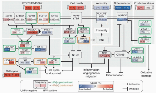
Figure 2.3. Pathways affected in HNSCC. Signaling pathways frequently altered in HNSCC, based on recent TCGA analysis. The frequency (%) of genetic alterations for HPV(-) and HPV(+) tumors is shown separately within subpanels and highlighted. (From The Cancer Genome Atlas N. Comprehensive genomic characterization of squamous cell carcinoma of the head and neck. Nature. 2015;517(7536):576-582, with permission.)


Mutations

Mutations describe alterations in the sequence of DNA itself and can occur in the form of nucleotide substitutions, deletions, or insertions. Their effect on the function of the protein is variable, as they may be categorized as silent (causing no change in the encoded protein), missense (leading to an altered amino acid sequence), or nonsense (truncation of the protein). The mutational landscape of HNSCC is being increasingly revealed by high-throughput, “next-generation sequencing.” HNSCC is associated with one of the highest mutation rates of any cancer, possibly due to the association of these tumors with environmental carcinogens known to induce DNA damage. Though there are a few characteristic mutations in HNSCC (Table 2.3), there is a large amount of genetic variability between tumors. Tumor suppressors including p53 (71%), CDKN2A (22%), FAT1 (23%), and NOTCH1 (20%) are the most frequently mutated genes in HNSCC, with only one oncogene (PIK3CA, 21%) having a mutation rate >20%.21 These frequently mutated genes map to a diverse set of biologic pathways including DNA repair (p53), cell cycle regulation (p53, CDKN2A), apoptosis (p53, PIK3CA), and cell differentiation (NOTCH1). Importantly, the mutational landscapes of HPV(-) and HPV(+) HNSCCs are quite different (Table 2.3),46 as will be discussed later.


Table 2.3 Common Gene Defects in HNSCC


Oncogenes


% HPV(+) tumors


% HPV(-) tumors


PIK3CA


20 + 28 + 8 = 56


16 + 14 + 4 = 34


CCND1


3


31


EGFR


6


11 + 3 + 1 = 15


MYC


3


14


FGFR1


10


ERBB2


3


3 + 2 = 5


HRAS


5


Tumor suppressor genes


% HPV(+) tumors


% HPV(-) tumors


TP53


3


83 + 1 = 84


CDKN2A


33 + 25 = 58a


PTEN


3 + 3 = 6


2 + 10 = 12


NOTCH1


19


7


TRAF3


14 + 8 = 22


NSD1


10 + 2 = 12


Amplification; Activating mutations; Amplification and activating mutation; Inactivating mutation; Deletion; Protein down-regulation; Total. The Cancer Genome Atlas N. Comprehensive genomic characterization of head and neck squamous cell carcinomas. Nature. 2015;517(7536):576-582. Reference 21.


a A significant proportion of HPV- tumors also display CDKN2A promoter hypermethylation (not shown).


Overall, it is clear that mutational changes drive cancer initiation and progression, but we are just beginning to understand the functional significance of many of them. Additionally, each tumor may have hundreds of mutations, and it can be difficult to determine the effect of each on the encoded protein, as well as whether a given mutation is a “driver” (directly causing carcinogenesis) or a “passenger” (having little impact on tumor cell fitness but co-occurring with driver mutations).


Copy Number Variations

A CNV consists of a structural change in a chromosome effecting a gain or loss of a portion of that chromosome, which can involve a single or multiple contiguous genes. They can come in the form of deletions, duplications, inversions, or translocations. Though prevalent in the normal human genome, CNVs that involve a loss of tumor suppressors or gain of oncogenes can predispose to neoplastic transformation. They can also result in a gene moving to come under the control of abnormal promoters or regulatory elements (e.g., BCR-Abl—the Philadelphia chromosome). CNVs in cancer cells have historically been detected using cytogenetic techniques such as chromosomal banding or fluorescent in situ hybridization (FISH), but nextgeneration sequencing has accelerated detection of CNVs and fostered an understanding of their crucial role in cancer.

CNVs are important in the pathogenesis of HNSCC. Each HNSCC harbors on average over 100 altered copy number segments, indicating a high degree of genomic instability. In HNSCC, the most common alterations are gains in chromosomal regions 3q, 5p, and 8q and loss of 3p and 8p.21 These particular segments are gained or lost with such frequency because of the growth/survival advantage they confer (due to loss of tumor suppressors or gain of oncogenes). In HNSCC, deleted chromosomal segments include known tumor suppressors such as FAT1, NOTCH1, SMAD4, and CDKN2A. Recurrently amplified regions often include receptor tyrosine kinases (RTKs) involved in growth signaling (growth hormone receptors EGFR, FGFR1, and ERBB2). The 3q region that is frequently amplified contains oncogenes important for survival, squamous differentiation, and stemness (PIK3CA, p63, hTERT, and SOX2). Many CNVs observed in HNSCC are common to epithelial cancers occurring elsewhere in the body, suggesting a common underlying pathophysiology. For many CNVs, the driver behind the change is unknown. Often, several contiguous genes are affected, making it difficult to decipher which genes are drivers versus passengers.

HPV(+) tumors display a distinct pattern of CNVs compared to HPV(-) HNSCC. The differences in CNVs reflect unique selective pressures that occur in HPV(+) versus HPV(-) HNSCC. For example, HPV(+) tumors express the oncoproteins E6 and E7, which through their inactivation of p53 and Rb diminish the pressure to delete either p53 or p16 in HPV(+) cancers. On the other hand, some CNVs (such as 3q amplifications and 3p deletions) are shared between HPV(+) and HPV(-) HNSCCs and are thus likely required for maintenance of squamous cells regardless of the etiologic agent.


Epigenetics

Epigenetics is a broad term that refers to self-perpetuating changes in gene expression that do not affect the actual sequence of DNA. The most well-characterized epigenetic alterations are DNA methylation and histone modifications.
By altering the nonsequence structure of DNA and the histones that package it, epigenetic modifications can make genes more or less accessible to activators of transcription and therefore modulate their expression. Methylation is the most frequently studied epigenetic change, partly because there are well-established methods to examine it. Promoter methylation, as well as global hypermethylation, has been shown to facilitate tumorigenesis by the silencing of tumor suppressors. In HNSCC, promoter methylation of genes such as CDKN2A (encoding p16INK4a and p14ARF), DAPK, RASSF1A, RARB2, APC, and MGMT is often an early event during neoplastic progression.47,48,49 It has been postulated that epigenetic changes are complementary to the genetic changes, for example, silencing a wild-type tumor suppressor allele when the other is inactivated by mutation.

Additionally, whole-exome sequencing studies have revealed that several of the genes responsible for histone modifications are recurrently mutated in HNSCC (e.g., EZH2, MLL2, MLL3, NSD1).21,50,51 Mutations of these genes result in aberrant chromatin structure and gene regulation. This finding also underscores the role that epigenetics plays in HNSCC tumorigenesis.

Studies have shown that HPV(+) HNSCCs have increased global methylation compared to HPV(-), and clustering based on methylation can predict HPV status.52 The clinical implications of epigenetics in HNSCC have yet to be firmly established, but it is likely that certain methylation patterns will be prognostic of tumor aggressiveness and/or predictive of therapeutic response.52 Increasing knowledge about epigenetics has also helped to produce a new group of rational therapeutics, targeting histone deacetylases (HDACs) and DNA methyl-transferases (DNMTs), aimed at reversing aberrant epigenetic changes.

In addition to epigenetic alterations, gene expression can be modified at the transcriptional and translation level by microRNAs (miRNAs). These are small ˜20 nucleotide RNA oligonucleotides, which complementarily bind mRNA, altering its fate through one of several mechanisms.53 Although miRNAs are a normal, evolutionarily conserved process in plant and animal cells, tumor cells up- or down-regulate certain miRNA, which can enhance malignant properties. In HNSCC, expression of specific miRNA are consistently altered to deregulate expression of genes involved in cell cycle regulation (e.g., PTEN, p21)54 and other cancer-related processes (e.g., KRAS). Presence or absence of certain miRNA has been correlated to prognosis, metastatic likelihood, and resistance to treatment; however, most miRNAs have several targets,55,56 and their analysis is complex. In the future, miRNA signatures may be applied to identify tumor-specific subtypes or to guide treatment. Techniques for efficient delivery of miRNAs are being developed in hopes that they can be used therapeutically.


LINKING GENETICS TO PATHWAYS

The recent developments of whole-exome sequencing and other high-throughput techniques have added a wealth of data to the large preexisting body of work in the molecular biology of HNSCC. Although these data have allowed a more complete picture of defects in HNSCC, they have also highlighted the complexity of its pathogenesis. Understanding the role of even a single gene requires integrating the various types of genetic and epigenetic changes that affect it in the cancers of different patients, the molecules it interacts with, and how the gene is affected over time and in spatially distinct areas of each patient’s cancer. For example, though p53 itself is found to be mutated in around 70% of HNSCCs, it can also be deactivated in the remaining tumors by overexpression with or without amplification of MDM2 (which facilitates p53 degradation) or by expression of the HPV E6 oncoprotein. As the realm of tumorrelated data has expanded to include mutations, amplifications/deletions, mRNA and protein expression profiles, microRNA expression, immune profiling, and epigenetic events, multiplatform data must be simultaneously considered to determine the drivers of carcinogenesis and direct therapy. Recent analyses suggest that data from different platforms carry overlapping information and omission of one type of data from multiplatform analyses does not necessarily alter classification.57

Categorizing defects into cancer-related biologic pathways can be a useful approach to organize and assign meaning to the wealth of data. For example, PIK3CA, a cell survival and growth gene, is mutated in 21% of a recent HNSCC cohort21; but when copy number amplifications were considered, that number rose to 36%, and when “hits” to other genes in the PIK3CA pathway were included, its pathway was affected in the majority of tumors.21,58 For this reason, studies have shown that mutational data may be more useful when placed into pathways.59 Though most of the early therapeutic successes in targeted cancer therapy have been based on individual mutations (e.g., BRAF in melanoma or EGFR in lung adenocarcinoma), understanding the mechanisms and pathways involved may help illuminate the most promising molecular targets for future treatments.

In the following section, we describe biologic pathways that are most commonly affected in HNSCC. Increasing knowledge of the normal function and interactions of these genes has laid a framework to fit the observed alterations into a narrative of cancer initiation and progression (Fig. 2.3). However, there is significant overlap and interaction between these pathways, and some pathways may be more affected in one tumor versus another. Genetically speaking, there are many different routes to cancer, which is why treating the disease in the future will likely require an equally sophisticated approach.


HALLMARKS OF HEAD AND NECK CANCER

In HNSCC, the transition of epithelial cells from normal to neoplastic involves a multistep process of accumulated genetic changes, which produces characteristic changes in biologic pathways that can be observed at the phenotypic level. The characteristic phenotypic changes, or “hallmarks,” seen in cancer cells have been frequently described and updated in recent years (Fig. 2.4).60 We use these events as a framework to review the recent developments in the study of HNSCC. While we review the hallmarks with a focus on HNSCC, a comprehensive review can be found elsewhere.60 The hallmarks discussed here include genomic instability, cellular proliferation, invasion and metastasis, angiogenesis, resisting cell death, replicative immortality, and reprogrammed metabolism. Additionally, interplay with the immune system (evasion of immune detection and tumor-promoting inflammation) will be discussed.


Genomic Instability

Normal cells possess a very high-fidelity system for DNA replication, such that errors occur once per 100,000 copied bases. Even those “rare errors” are corrected 99% of the time

by DNA repair machinery, bringing the final error rate to one per ten million bases. A wide variety of systems known as “caretakers” detect and repair errors in the genome or, if unsuccessful, freeze the cell cycle and activate cell death.61,62 Some mechanisms directly inactivate or intercept mutagens even before they damage DNA. The “clonal evolution theory” suggests that many alterations are needed for cells to gradually transform into cancer. Although the chances of this happening in a normal cell are infinitesimally small, damage to caretakers through genetic or epigenetic changes can lead to instability of the genome, increasing the error rate and allowing propagation of defects to subsequent generations of cells. Thus, by allowing the series of alterations required for neoplastic change, genomic instability is considered an enabling characteristic common to nearly all cancers.63 The quintessential tumor suppressor p53 plays a central role in guarding the genome by activating DNA repair, cell cycle arrest, or apoptosis in response to genetic damage or cellular stresses.64


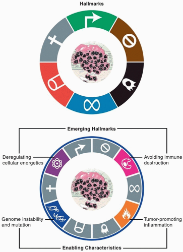
Figure 2.4. Hallmarks of cancer. Hanahan and Weinberg initially described, then expanded, attributes needed for cancer development. (Adapted from Hanahan D, Weinberg RA. Hallmarks of cancer: the next generation. Cell. 2011;144(5):646-674.)

Endogenous and environmentally induced defects accumulate much faster without these proofreading capabilities, and although many of those genomic defects may be phenotypically silent, some (e.g., affecting tumor suppressors or oncogenes) will be involved in the carcinogenic processes described in the following sections. Across all cancers, HNSCC has among the highest levels of mutations, chromosomal rearrangements, and copy number alterations, highlighting the importance of instability within the genome to the pathogenesis of this disease.65


Dysregulation of Proliferative Signaling

Cells within normal tissues have a tightly regulated balance of proliferative and antiproliferative signaling that govern cellular growth and replication, thus ensuring homeostasis between cell population and host resources. Progrowth signals most often come in the form of growth factors in the cells’ environment that bind receptors on the cell surface and initiate signaling cascades within the cell. There also exist mechanisms to inhibit growth, ensuring that mitogenic signals are only transient and even inhibited in the presence of certain stimuli (such as DNA damage or absence of sufficient resources). Even in the presence of growth signals, cellular proliferation is tightly regulated, and cells must progress through many checkpoints and phases in the cell cycle in order to duplicate their DNA and divide into two daughter cells. In cancer cells, cell cycle checkpoints are universally circumvented to allow for aberrant cellular proliferation. One of the most fundamental characteristics of neoplastic cells is their ability to sustain proliferation, by either acquiring autonomous proliferative signaling or evading inhibitory mechanisms.


Progrowth Proliferative Signaling

Normal cells require a basal level of growth signals in order to survive and proliferate. Typically, growth factor molecules are released by distant or neighboring cells and bind to tyrosine kinase receptors on the cell surface, which relay that signal to a branching network of downstream effectors. By producing a growth factor ligand autonomously, increasing the quantity/efficiency of growth factor receptors, or constitutively activating downstream effectors, cancer cells routinely acquire autonomous growth signals.66 In HNSCC, the epidermal growth factor receptor (EGFR) and its pathway are altered in a significant proportion of HNSCCs. Whereas mutations to the EGFR gene are uncommon, amplification and overexpression of EGFR are very common.67 Other RTKs that are amplified or overexpressed and less frequently affected include hepatocyte growth factor receptor (MET), fibroblast growth factor receptor (FGFR), and insulin-like growth factor (IGFR).21 Chief downstream effectors of growth factor receptors are also directly implicated in the pathogenesis of HNSCC, such as the PI3K/Akt/PTEN/mTOR,58 JAK/STAT,68 and RAS/RAF/MEK/MAPK pathways, each of which initiates a complex cascade of proliferative, survival, metabolic, or related functions.

Loss of growth inhibition is also common. Transforming growth factor beta (TGF-β) acts as an antiproliferative signal for normal epithelium, and its downstream effector, SMAD4, is down-regulated in up to 20% of HNSCC.21 As part of their greater functions as tumor suppressors, both Rb and p53 help to suppress proliferative activity as well. Although there is not a single growth factor or receptor that is universally altered in HNSCC, summation of various insults to the compilation of growth signaling pathways supports the concept that aberrant growth factor signaling is required for HNSCC development.


Cell Cycle

The normal cell cycle, the process by which cells replicate their DNA and divide, is guarded at various checkpoints to ensure that cells divide only when it is appropriate to do so (Fig. 2.5). The cell cycle consists of four phases: G1 (gap phase 1), S (DNA synthesis), G2 (gap phase 2), and M (mitosis). Progression through the cell cycle is mediated by activity of cyclin-dependent kinases (CDKs), interacting with cyclins. Additional regulation is introduced by expression of CDK inhibitors (CDKi) that can result in stalling of the cell cycle or permanent arrest. The quantity and phosphorylation status of cyclin-CDK complexes, as well as expression of CDKi, largely determine if a cell will initiate DNA replication and begin a round of the cell cycle. Proteins that regulate cell cycle progression are known as “gatekeepers” that prevent the cell from replicating unless conditions are appropriate. The tumor suppressors Rb and p53 are the canonical gatekeepers that halt cell proliferation in response to extracellular growth arrest signals or intracellular signals of DNA damage and resource limitations. They operate within complex circuits, which can also activate DNA repair or cell death if necessary. Gatekeepers must be circumvented for the aberrant cell cycle progression that occurs in cancer cells.

As will be discussed later, p53 itself is mutated or deleted in roughly 70% of HNSCC. In most of the remaining 30% of cases, p53 is inactivated by the HPV E6 oncoprotein. Similarly, Rb activity is diminished through a variety of mechanisms in HNSCC. Although the Rb gene is mutated or deleted in only 5% of HNSCC, it is often directly inhibited by the HPV oncoprotein E7 or by alterations to its regulators (cyclin D1, p16INK4a). The nearly ubiquitous, though heterogeneous, insults to the gatekeeper circuits indicate that this is one of the key requirements for development or progression of HNSCC.69


Abnormal Differentiation: Invasion and Metastasis

As part of their normal function, squamous epithelial cells express specific adhesion molecules in order to create a tightly packed functional sheet, remain external to the basement membrane, and eventually terminally differentiate, senesce, and
slough away. The normal maturation of epithelial cells involves orderly changes as they progress from the basal epithelial layer to become mature keratinocytes (Fig. 2.6). These changes are mediated by transcriptional changes driving increased or decreased expression of specific genes involved in epithelial differentiation. Disruption of this normal differentiation is thought to occur in HNSCC and other epithelial cancers, in which cell populations can attain properties and appearance of mesenchymal (connective tissue) cells. The resulting cells often lose their epithelial architecture, become less dependent on cell-cell contact, and possess increased motility, invasion, angiogenesis, and other mesenchymal-like properties. This phenomenon is referred to as epithelial-mesenchymal transition (EMT). The process of EMT is mediated by four major transcription factors: Snail, Slug, Twist, and Zeb 1/2. EMT is a normal process that is used during embryogenesis and wound healing but is hijacked during carcinogenesis.


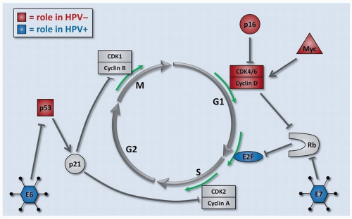
Figure 2.5. Cell cycle control. Progression through the cell cycle is tightly controlled in noncancer cells. In HNSCC, the regulators of cell cycle progression are ubiquitously altered by many different mechanisms. The most common defects in cell cycle regulators are shown for HPV- (red) and HPV+ (blue) HNSCCs.


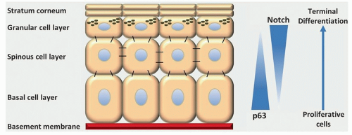
Figure 2.6. Maturation of keratinocytes. Epithelial cell proliferation is limited to the basal layer, and cells progressively differentiate as they migrate superficially before being sloughed from the surface. NOTCH and p63 play key roles in this differentiation process and are frequently disrupted during development of HNSCC.

Adherens junctions, a protein complex consisting of E-cadherin and α- and β-catenins, constitute a part of the normal cell-cell contacts between neighboring epithelial cells. They render the cells relatively immobile and prevent cellular proliferation through a process known as contact inhibition. Compared to normal epithelium, cadherins are down-regulated in HNSCC.70 Meanwhile, integrins, which help cells attach to the underlying extracellular matrix, are up-regulated in some HNSCC.71 Beyond changes of adhesion properties, cancer cells have increased motility and invasion and possess matrixdegrading enzymes (matrix metalloproteinases—MMPs)
(Fig. 2.7).72,73 These changes are needed for the destructive invasive properties of HNSCC as well as for processes associated with lymphatic and hematogenous metastases.

Only gold members can continue reading. Log In or Register to continue

Stay updated, free articles. Join our Telegram channel

Dec 18, 2016 | Posted by in ONCOLOGY | Comments Off on Pathogenesis and Progression of Squamous Cell Carcinoma of the Head and Neck

Full access? Get Clinical Tree

Get Clinical Tree app for offline access