Parenteral Nutrition


Parenteral Nutrition


Mary E. Hagle

Sharon M. Weinstein


HISTORY OF PARENTERAL NUTRITION

The term parenteral hyperalimentation was coined by Dr. Jonathan E. Rhoads and Dr. Stanley J. Dudrick over four decades ago. In a classic citation from the literature, they defined it as “the intravenous administration of nitrogen, calories, and other nutrients sufficient to achieve tissue synthesis and anabolism in patients with normal or excessive nutritional needs” (Wilmore & Dudrick, 1968). These accomplishments led to further research and application as other clinicians and researchers realized the potential of this approach to feeding. Hyperalimentation soon became a recognized specialty resulting in nutritional support teams being formed. Nutritional support provides specially formulated or delivered parenteral or enteral nutrients to maintain or restore optimal nutrition status.

In 1976, a multidisciplinary group of professionals (physicians/licensed independent practitioners [LIPs], nurses, dietitians, and pharmacists) was organized, and the American Society for Parenteral and Enteral Nutrition (A.S.P.E.N.) held its first clinical congress in Chicago.

Hyperalimentation evolved as a science in the 1980s and became more commonly known as total parenteral nutrition, and eventually as parenteral nutrition (PN), the term
used throughout this chapter. Disease-specific formulas were developed to address the particular needs of patients with renal, cardiac, or hepatic disease and became commercially available. Multiple organizations, such as the Oley Foundation and hospital-based support groups, grew out of need for the rehabilitation of patients surviving catastrophic illnesses by maintenance on home parenteral nutrition (HPN). PN has evolved into a sophisticated field of therapeutic intervention that has its own multidisciplinary specialists and a large body of established knowledge.

With the changing health care environment, including the emergence of care management, accountable care organizations, changing practice settings, and more, the field continues to change.


OVERVIEW OF PARENTERAL NUTRITION

Parenteral nutrition is the intravenous (IV) provision of nutrients in patients without a functional or accessible gastrointestinal (GI) tract. PN provides protein in the form of amino acids, carbohydrates as dextrose, fats, vitamins, minerals, and trace elements to sustain life. The goals of PN are to ease the metabolic response to stress, prevent oxidative cellular injury, and to modify the immune response in a positive way (McClave et al., 2009). In many cases, PN is lifesaving.

PN is administered via a short peripheral IV catheter or central venous access device (CVAD). Peripheral parenteral nutrition (PPN) is used when CVAD placement cannot be achieved. PPN has a lower concentration of nutrients in a dilute solution and may not meet the protein and caloric needs of the patient. The osmolarity of the PPN solution must be
<600 mOsm and the final concentration of dextrose 10% or lower. Solutions with an osmolarity >600 mOsm are known to cause phlebitis. Typically, PPN is given for 7 days or less. It is not intended for prolonged nutrition support. It is also not appropriate for patients who cannot tolerate large volumes of fluid. Risk versus benefit must be weighed before the decision to treat with PPN is made. Strategies to reduce the risk of phlebitis should be implemented if PPN is selected as a course of treatment:


  • Addition of a steroid such as hydrocortisone to the solution


  • Addition of heparin


  • Concomitant administration of fat emulsion


  • Infusing the solution using a cyclic schedule such as infusing over 12 hours per day (INS, 2011)

Parenteral nutrition is a formula that contains complete nutrition and can only be given via a CVAD with catheter tip location in the cavoatrial junction due to its high osmolarity.

Nutrients in greater concentrations with smaller fluid volumes are administered. PN can be given for a short period of time such as days to weeks. Some patients with chronic conditions may require therapy for years to a lifetime.

Home parenteral nutrition (HPN) is the term used for PN administered in the home care or alternative care setting such as a long-term care setting. Indications for HPN are chronic diseases such as Crohn’s disease where therapy may continue from months to years or a lifetime.

Enteral nutrition (tube feeding) is the administration of liquid nutrients through a tube, catheter, or stoma directly into a functional GI tract (A.S.P.E.N., 2009a, 2009b). It is the preferred method of nutrition when feasible and safe in a patient because it has the advantages over PN of decreased risk of metabolic complications, infection, reduced cost, and shorter hospital stays.


NUTRITIONAL ASSESSMENT

Nutritional assessment is necessary because inadequate nutrition increases the complications of disease, especially infection; decreases the ability to heal wounds; prolongs the length of the hospital stay; and contributes to anorexia and weakness. Malnutrition is determined through a nutritional assessment reviewing body weight, body composition, somatic and visceral protein stores, and laboratory values. Early nutritional intervention is recommended to avoid additional complications of malnutrition, including impaired respiratory muscle function, renal failure, and nonhealing wounds.

Two common types of protein-energy malnutrition include marasmus and kwashiorkor. Marasmus is characterized by catabolism of fat and muscle tissue, lethargy, generalized weakness, and weight loss. Visceral proteins are relatively preserved with serum albumin >3 g/dL. Patients are generally <80% of standard weight for height (Hammond, 2004). Kwashiorkor is characterized by edema, catabolism of muscle tissue, weakness, neurologic changes, secondary infections, stunted growth in children, and changes in hair. Visceral proteins are depressed with serum albumin <3 g/dL. Patients are generally >90% of standard weight for height (Hammond, 2004).

Another type of protein-energy malnutrition is characterized by a combination of marasmus and kwashiorkor symptoms. Patients are generally <60% of standard weight for height and have depressed visceral proteins with serum albumin <3 g/dL. This type of malnutrition usually occurs when a patient with marasmus is exposed to stress.


Nutritional Screening and Assessment Standards

Nutrition screening, assessment, and intervention in patients with malnutrition are key components of nutrition care. A.S.P.E.N. defines nutrition screening as a process to identify an individual who is malnourished or who is at risk for malnutrition to determine if a detailed nutrition
assessment is indicated. In the United States, The Joint Commission (TJC) identifies a standard to screen nutrition within 24 hours of admission to an acute care center. The goal of nutrition assessment is to identify any specific nutrition risk(s) or clear existence of malnutrition. Nutrition assessments may identify recommendations for improving nutrition status (e.g., an intervention such as change in diet, enteral or parenteral nutrition, or further medical assessment) or a recommendation for rescreening (Mueller, Compher, Druyan, & American Society for Parenteral and Enteral Nutrition (A.S.P.E.N.) Board of Directors, 2011). Nutrition assessment has been defined by A.S.P.E.N. as “a comprehensive approach to diagnosing nutrition problems that uses a combination of the following: medical, nutrition, and medication histories; physical examination; anthropometric measurements; and laboratory data” (Mueller et al., 2011). A nutrition assessment provides the basis for a nutrition intervention. A focus on mandatory screening and nutritional assessment as part of the specific nutritional care standards began with TJC in 1995. The standards emphasize an interdisciplinary approach. In many settings, the nutritional screen is completed by a health professional other than the dietitian. The goal of nutritional screening is to identify individuals who are at nutrition risk, as well as to identify those who need further assessment.

Many hospitals have established formal nutritional screening programs for all hospital admissions to identify those patients most at risk or already malnourished. An algorithm for nutritional screening is presented in Figure 16-1. Once a nutritional screen is completed, patients who are at nutritional risk are usually referred to a dietitian.

A nutritional assessment should be included in the overall assessment of patients, particularly on admission to the hospital and periodically throughout the hospital stay. It should also be a component of nursing assessment in all health care settings, with reassessment at appropriate intervals, depending on the patient’s status. Studies indicate that when admitted to acute care facilities, between 33% and 65% of all patients have some degree of malnutrition, and nutritional status deteriorates when patients are hospitalized >2 weeks (Hammond, 2004). Primary care, outpatient management, and the new standards have moved the role of nutritional assessment into the physician/LIP’s office and home care setting as well. Mechanisms for documenting nutritional screening and assessment should be established and monitored. Suggested goals of nutritional assessment include the following:


  • To identify individuals who require aggressive nutritional support


  • To restore or maintain an individual’s nutritional status


  • To identify appropriate medical nutrition therapies


  • To monitor the efficacy of these interventions (Hammond, 2004)

A nutritional assessment traditionally begins with a complete history, which includes a diet history, anthropometric measurements, physical assessment, and complete medical history, including drug-nutrient interactions and biochemical evaluation.


HEALTH, WEIGHT, AND DIETARY HISTORY

The patient’s usual and current dietary intake is helpful in identifying the adequacy of nutrients (i.e., protein, carbohydrate, lipid, vitamins, minerals, trace elements, and water) and possible nutritional deficiencies. Nutrients in the medical history should include any acute or chronic diseases that may have an impact on nutrient intake or utilization and conditions that increase metabolic needs or fluid and electrolyte losses. Surgical history,
certain medications, and social factors also have an impact on nutritional status and should be evaluated. Multiple computerized programs for dietary assessment are available, each providing different end points of data (Probst & Tapsell, 2005). Benefits of a computerized assessment include standardized questioning, immediate micronutrient information, and possible recommendations; additionally, these programs may be used for practice or research. Specific factors that may alter nutritional status are listed in Box 16-1.


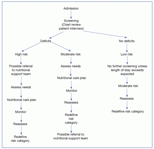
FIGURE 16-1 Nutritional screening. (Courtesy of Grant, A., & DeHoog, S. (1999). Nutritional assessment, support and management (5th ed). Seattle, WA: Authors.)


PHYSICAL ASSESSMENT

A thorough physical assessment by an experienced clinician can detect signs and symptoms suggesting nutritional deficiencies. These include but are not limited to changes in hair, eyes, skin, nails, and all organ systems (Table 16-3). Anthropometric measurements consist of simple, noninvasive, inexpensive techniques for obtaining body measurements useful

in evaluating for overnutrition or undernutrition. The patient’s actual height and weight should be obtained and used to help determine nutritional status.


Body weight can by assessed by several methods including (a) body mass index (BMI), (b) comparison with usual body weight (UBW), and (c) comparison with ideal body weight (IBW). BMI is a tool for judging body weight in relation to height and risks for weight-related health problems such as heart disease, diabetes, and certain types of cancer (Box 16-2). Electronic medical record (EMR) systems often provide BMI once height and
weight are entered. This information may be part of the clinical decision support system and provide an automatic consultation to the dietitian for specific BMI parameters indicating that a patient is at nutritional risk. The Centers for Disease Control and Prevention (CDC) also provides a Web site for calculating BMI for adults and children: http://www.cdc.gov/healthyweight/assessing/bmi.


TABLE 16-3 PHYSICAL ASSESSMENT FINDINGS IN NUTRITIONAL DEFICIENCIES


Physical Changes


Deficiency


Hair


Lackluster, thin, sparse; pigmentation changes


Protein, calorie, zinc, linoleic acid


Mouth


Angular stomatitis: cracks, redness at one or both corners of the mouth


Vitamin B


Cheilosis—vertical cracks in the lips


Riboflavin and niacin


Varicose veins under the tongue


Vitamin C


Tongue


May become purplish, red, or beefy red or may appear smooth and pale; one or more fissures, atrophy of taste buds


Vitamin B


Skin


Dryness, flakiness


Vitamin A, essential fatty acids


Petechiae or easy bruising; hemorrhagic spots on skin at pressure points; may occur in the presence of liver disease or during anticoagulation


Vitamins C and K


Musculoskeletal


Muscle wasting (especially quadriceps, deltoids, and temporalis)


Protein, calorie


Kyphosis, osteoporosis


Calcium and vitamin D


Neurologic


Confusion, listlessness


Protein malnutrition


Sensory motor, vibratory


Thiamine and vitamin B12


UBW is obtained from past medical records or subjectively from the patient or family member. Actual weight as a percentage of UBW is usually the most accurate determinant of weight loss. Minimum weight for survival is 48% to 55% of UBW (Hammond, 2004). UBW can be calculated as follows:


Percentage of IBW may also be used and can be determined with the preceding formula by substituting IBW for usual weight. This method is not as effective when assessing the nutrition risk of a critically ill patient. A normally thin person, with stable weight, may be incorrectly identified as malnourished if weight is less than IBW; the obese patient with significant recent weight loss may be overlooked if weight is still above IBW (Grant & DeHoog, 2008).

Many of the nutrition screens examine BMI, weight loss, and weight history (Mueller et al., 2011). Recording actual weight and weight history carefully will inform the team and enable appropriate interventions. Additionally, weight will be used to assess for fluid overload, along with other parameters, as well as to track progress of nutrition support. The significance for weight loss over specific time periods is outlined in Table 16-4.

Other anthropometric measurements include triceps skinfold thickness, which estimates subcutaneous fat stores, and mid-arm muscle circumference, which estimates somatic protein stores (skeletal muscle mass). These measurements can be helpful in assessing individuals over time but not in the critical and acute care settings because changes in body fluid and composition may influence the results (Hammond, 2004). Reproducibility of accurate measurements requires strict adherence to protocol and may vary widely in the same individual with different examiners.

For the patient in the ICU, the A.S.P.E.N. guideline directs an “evaluation of weight loss and previous nutrient intake prior to admission, level of disease severity, comorbid
conditions, and function of the GI tract” (McClave et al., 2009). Traditional anthropometric measures are not validated in critical care (McClave et al., 2009).


TABLE 16-4 ESTIMATING BODY WEIGHT AND PERCENTAGE LOSS IN PN


Duration of Therapy


Considerable Weight Loss


Serious Weight Loss


10 d


1%-2%


>2%


1 mo


5%


>5%


3 mo


7.5%


>7.5%


Common anthropometric measurements include weight, height, and weight/height and their comparisons to standard values. To calculate the percent weight change (PWC) in the patient receiving parental nutrition, follow this formula:


image


LABORATORY ANALYSIS (BIOCHEMICAL ASSESSMENT)

A variety of biochemical tests are used to assess nutritional status, with a broad range of sensitivity. The most commonly used, and most readily available, are visceral proteins and tests to evaluate immune function.


VISCERAL PROTEINS

Depletion of visceral proteins is characteristic of protein malnutrition, which occurs acutely in the hospitalized patient (hypoalbuminemia). An estimate of visceral protein status can be obtained from measurements of specific serum transport proteins that are synthesized in the liver. These include albumin, transferrin, prealbumin, and retinol-binding protein (Table 16-5). Serum albumin is useful for helping to determine nutritional status but has limited value in assessing acute changes in nutritional status related to its long half-life (Carney & Meguid, 2002). It is recommended that serum albumin measurements be included in the initial chemical profile when screening for malnutrition. An albumin concentration of <3.5 g/dL is associated with increased morbidity in hospitalized patients (Carney & Meguid, 2002). However, in the critically ill patient, these traditional measures (albumin and prealbumin) are not validated in the ICU (McClave et al., 2009).

Transferrin has a shorter half-life than albumin but does not respond rapidly enough to protein-energy status to be useful in acute care settings (Litchford, 2012). Retinol-binding protein also has a shorter half-life but does not accurately reflect protein-energy status in acutely stressed patients. It is also not useful for patients with vitamin A deficiency or renal failure (Litchford, 2012). Prealbumin, with a shorter half-life, seems to be a more sensitive and specific indicator for monitoring the effectiveness of nutritional support. It is more expensive and may be limited in smaller hospitals. Consideration should also be given to assessing non-nutritional factors, such as inflammation, that may affect these visceral proteins. Even though these tests are useful, new nutritional markers are needed that better identify malnourished patients and precisely monitor the effectiveness of nutritional intervention on nutritional status. Other proteins are being investigated to determine their efficacy in nutritional assessment.


TABLE 16-5 VISCERAL PROTEINS


Visceral Protein


Half-Life


Normal Range


Causes of Decrease


Causes of Increase


Albumin


Maintains plasma oncotic pressure Carrier protein (zinc, calcium, magnesium, fatty acids)


18-21 d


Normal: >3.5 g/dL


Mild depletion: 2.8-3.5 g/dL


Moderate depletion: 2.1-2.7 g/dL


Severe depletion: <2.1 g/dL


Reflects chronic, not acute change


Metabolic stress Infection, inflammation


Liver disease ↑ Losses (wounds, burns, fistulas)


Inadequate protein intake


Fluid imbalance (ascites, edema, overhydration)


Malabsorptive states


Dehydration may falsely ↑ levels Salt-poor albumin infusion


Transferrin


Carrier protein for iron


8-10 d


Normal: >200 mg/dL


Mild depletion: 150-200 mg/dL


Moderate depletion: 100-150 mg/dL


Severe depletion: <100 mg/dL


Chronic infection Inflammatory state Acute catabolic states ↑ Iron stores Liver damage Overhydration


Pregnancy


Hepatitis


Iron deficiency anemia


Dehydration


Chronic blood loss


Thyroxine-binding prealbumin (transthyretin)


Carrier protein for retinol-binding protein


Transport protein for thyroxine


2-3 d


Normal: >20 mg/dL


Mild depletion: 10-15 mg/dL


Moderate depletion: 5-10 mg/dL


Severe depletion: 5 mg/dL


Sensitive to acute changes in protein status


Limited use in situation in which there is a sudden demand for protein synthesis


Infection


Acute catabolic state


Postsurgery (5-10 mg/dL drop 1st week)


Liver disease


Altered energy and protein balance


Hyperthyroid


Chronic renal failure Corticosteroids


Retinol-binding protein


Transport retinol (alcohol of vitamin A)


10-18 h


Normal: 3-5 mEq/dL


Reflects acute change


Vitamin A deficiency


Acute catabolic states


Postsurgery


Liver disease


Hyperthyroid


Renal disease


IMMUNE FUNCTION TESTS

Alterations in the immune system may be influenced by stress, specific disease states, and malnutrition. The total lymphocyte count (TLC) is indicative of a patient’s ability to fight infection and is known to decrease with malnutrition, especially inadequate calories and protein. TLC may be calculated using the complete blood count with differential: TLC = white blood cell (WBC) count × lymphocytes. Normal levels range from 1,500 to 1,800 mm3. However, TLC is affected by many medical disease states and varies widely; thus, it is of limited value.


Cell-mediated immunity is most frequently evaluated with delayed cutaneous hypersensitivity skin tests. Intradermal injections of specific recall antigens, such as Candida, mumps, or tuberculin, are used. Patients who are immunocompetent will exhibit a positive response within 24 to 48 hours; this response consists of a small, reddened area (5 mm) around the test site. Patients who are malnourished and possibly immunosuppressed may exhibit a delayed response, a response to only one of the antigens, or no reaction. These results are classified as normal or reactive if there is a response to two or more antigens, relatively anergic if only one response occurs, and anergic if there is no response to any of the antigens. These results must be weighed with other findings in the nutritional assessment because non-nutritional factors, such as infection, sepsis, cancer, liver disease, renal failure, immunosuppressive diseases, and immunosuppressive drugs, may also affect immunocompetence.


DECISION FOR NUTRITIONAL SUPPORT

Many nutritional assessments are available for different health care providers (Brogden, 2004; Rodriguez, 2004), as well as technologies that provide information on the individual’s metabolic analyses (Stanberry et al., 2013). There is no single indicator of nutritional status. A global assessment of many indices, and the patient’s clinical status, is necessary to determine the degree of nutritional deficiency. Based on the nutrition assessment, the guideline recommendation states that nutrition support is recommended for patients identified as at risk for malnutrition or who are malnourished (Mueller et al., 2011). A small body of sound evidence identifies quality outcomes for patients who receive nutrition support when they are malnourished; outcomes include improved nutrition status, nutrient intake, physical functioning, as well as fewer hospital readmissions (Mueller et al., 2011).

Reassessment is individualized whenever the clinical status changes and periodically throughout the course of nutritional therapy. Once the nutritional assessment data have been collected and evaluated, the optimal method for nutritional support must be selected. This decision process is depicted in Figure 16-2.

A growing body of evidence supports the use of EN, rather than PN, for the critically ill patient in the ICU (Casaer et al., 2011; Jeejeebhoy, 2012; McClave et al., 2009). However, specific guidelines outline when PN is appropriate for the patient in the ICU (McClave et al., 2009).


NUTRITIONAL REQUIREMENTS

The practical outcome of the nutritional assessment is to determine the caloric and protein needs of the patient. These requirements change in acute, catabolic illness, and in some chronic conditions.


Energy Requirements

Nutrient energy requirements are usually expressed in kilocalories (kcal). Numerous methods for determining a person’s nutritional requirements are available, with varying degrees of sophistication. Indirect calorimetry is one of the most accurate ways of determining a patient’s energy needs. In the clinical setting, metabolic charts are often used to determine

the oxygen consumption and carbon dioxide production of the body over a period of time. This helps to determine actual energy expenditure. Indirect calorimetry requires expensive equipment and therefore is not available at all institutions (Hammond, 2004).


FIGURE 16-2 Decision-making process for nutritional support. (Adapted from Grant, J., & DeHoog, S. (1999). Nutritional assessment, support, and management (2nd ed.). Seattle, WA: Authors; and Livingston, A., Seamons, C., & Dalton, T. (2000). If the gut works, use it. Nursing Management, 31(5), 39-42.)

A classic equation used to estimate energy expenditure is the Harris-Benedict Equation, which was derived from healthy individuals. It estimates basal energy expenditure (BEE) in kilocalories, using weight, height, age, and sex with the following equations:


BEE(male) =(66 + 13.8W + 5H) − 6.8A

BEE(female) = (655 + 9.6W + 1.8H) − 4.7A

where W is weight in kilograms, H is height in centimeters, and A is age in years.

UBW is most commonly used, although an adjusted body weight is often recommended if the patient is obese (BMI > 30). Stress and activity factors are often added to predict energy expenditures of critically ill or injured patients, which can range from an addition of 30% to 50% above resting energy expenditure (Ireton-Jones, 2002). Actual energy requirements are then estimated by multiplying BEE × AF × IF. Correction factors (for activity [AF] and stress/injury [IF]) to estimate nonprotein energy requirements of hospitalized patients are listed in Table 16-6.

Another common method to estimate energy needs is by calculating energy per kilogram. It has been estimated that 25 to 35 kcal/kg body weight is needed for total energy
expenditure in the nonobese population, whereas only 21 kcal/kg body weight is needed in the obese critically ill population (Rychlec, Edel, Murray, Schurer, & Tomko, 2000).


TABLE 16-6 CORRECTION FACTORS FOR ESTIMATING NONPROTEIN ENERGY REQUIREMENTS OF HOSPITALIZED PATIENTS


Activity Description


Activity Factor


Chair or bed bound


1.2


Out of bed


1.3


Little movement and little or no leisure activity


1.4-1.5


Seated work with requirement to move but little strenuous leisure activity


1.6-1.7


Clinical Status


Stress Factor


Elective surgery


1-1.1


Peritonitis


1.05-1.25


Multiple/long bone fractures


1.1-1.3


Cancer


1.1-1.45


Severe infection


1.2-1.6


Closed head injury


1.


Infection with trauma


1.3-1.55


Multiple trauma


1.4


Burns


1.5-2.1


Actual energy requirements = basal energy expenditure (BEE) × activity factor (AF) × injury factor (IF).


Adapted from Rychlec, G., Edel, J., Murray, M., Schurer, D., & Tomko, M. M., (2000). Nutrition assessment of adults. In B. Hornick. (Ed.), Manual of clinical dietetics (6th ed., pp. 3-33). Chicago, IL: American Dietetic Association.


Regardless of the method chosen, it is important to avoid overfeeding. The early adage of nutritional support “the more, the better” is no longer advocated. Overfeeding, which causes hyperglycemia and has deleterious effects on respiratory and hepatic function, should be avoided, especially in the acutely ill patient. Additional calories for desired weight gain should be held until the patient recovers.


Protein Requirements

Protein requirements for healthy people are based on the amount needed to maintain nitrogen equilibrium, assuming energy needs are being met by nonprotein kilocalories. The recommended daily allowance for healthy adults is 0.8 g/kg/d. Protein requirements are increased during illness to meet stress needs for wound healing, promote immune competence, and replace losses. Critically ill patients require an increased protein intake of 1.5 to 2.0 g/kg/d (Cresci, 2002).


CALORIE-NITROGEN RATIO

A nonstressed patient can achieve nitrogen balance with adequate calorie intake and protein accounting for 8% of calories. When a patient becomes stressed, both calorie and protein requirements increase. Protein should contribute 15% to 20% of energy in a stressed patient. The optimal calorie:nitrogen ratio for critically ill patients is 150:1.


NITROGEN BALANCE

Nitrogen balance is an objective method of evaluating the efficacy of the patient’s nutritional regimen. For a patient to be in a positive nitrogen balance or anabolic state, the amount of nitrogen taken in by the patient (IV and orally) needs to be more than that excreted. A 24-hour urine collection is needed to measure the amount of urinary urea excreted. A factor of 4 g is added (2 g for fecal losses and 2 g for integumentary losses) to measure total nitrogen excretion. A nitrogen balance of 2 to 4 g of nitrogen per day indicates that the patient is in an anabolic state. It is nearly impossible to achieve positive nitrogen balance immediately after a metabolic insult. Nitrogen balance studies are not accurate for patients with impaired creatinine clearance, severe hepatic failure, massive diuresis, or abnormal nitrogen losses through diarrhea or large draining wounds (Cresci, 2002). The nitrogen balance formula and measures that reflect the severity of the catabolic state can be found in Box 16-3.


Fluid Requirements

Individual fluid requirements vary greatly and can fluctuate on a daily basis. Fluid needs should be carefully assessed when designing the PN formula. The minimum daily requirement for healthy adults is 1 mL/kcal. Factors that may increase fluid needs include fever and increased losses from diuresis, diarrhea, vomiting, and drainage from wounds and fistulas.
Environmental factors such as specialized high-air-loss beds, ultraviolet light therapy, and radiant warmers also increase fluid requirements. Humidified air reduces insensible fluid loss and results in lower fluid requirements. Preexisting excess or deficiency states and cardiac and renal function must also be evaluated.


Fluids may be required in addition to the fluids provided by PN. Because fluid requirements shift rapidly with changes in clinical status in hospitalized patients, this is best done by giving additional crystalloid IV fluids, such as dextrose 5% in water (D5W) or dextrose 5% in 0.45% sodium chloride solution by a separate lumen or route. These can be frequently adjusted as necessary without affecting nutrient delivery. They can also be used as additional vehicles for electrolyte corrections, avoiding possible wastage of the PN solution. The PN solution can be formulated using concentrated nutrients (70% dextrose, 15% amino acids, 15% lipids) to provide adequate nutrients when fluid restriction is needed.


PARENTERAL NUTRITION SOLUTIONS

The complex solutions used in PN can provide all the necessary nutrients to meet requirements for growth, weight gain, anabolism, and wound healing.


Components

Total nutrient admixtures (TNA) are formulations consisting of carbohydrates, amino acids, lipids, vitamins, minerals, trace elements, water, and other additives in a single container. The proportion of each component is individualized based on the patient’s clinical status, chronic diseases, fluid and electrolyte balance, and specific goals of PN.


CARBOHYDRATES

Glucose is the primary energy source in most PN solutions. Concentrations from 10% to 70% glucose may be used with a final solution concentration of no more than 10% to 12% for peripheral infusion and no more than 35% for central venous infusion. A carbohydrate, parenteral glucose is hydrolyzed and provides 3.4 kcal/g. Consistent with a classic study by Galica, at least 150 to 200 g of glucose is needed to meet the obligate needs for glucose by the brain, central nervous system, red blood cells, WBCs, active fibroblasts, and certain phagocytes, which normally require glucose as the sole or major energy source (Galica, 1997). No more than 5 to 7 mg/kg/min (equals a maximum of 5 to 7 g/kg/d, which is easier to calculate) should be given, which is the maximum rate of glucose oxidation. Less is given to the diabetic, hyperglycemic, or critically ill patient—usually no more than 4 g/kg/d. The incidence of hyperglycemia seems to increase if excess quantities are administered. Overfeeding carbohydrates may result in conversion of excess glucose to fat, which requires energy (increased oxygen consumption and carbon dioxide production), and leads to fatty liver changes with prolonged use. From 50% to 70% of the daily nonprotein kilocalories usually are given as glucose.


LIPIDS

Lipids (fats) provide the second source of nonprotein calories for PN solutions. IV lipid emulsions are given to prevent essential fatty acid deficiency (EFAD) and as a concentrated source of energy (each gram of fat provides 9 kcal). The only recommendation regarding specific fatty acids is that 1% to 2% of daily energy requirements should be derived from linoleic acid and about 0.5% of energy from α-linoleic acid to prevent EFAD (Kris-Etherton, Taylor, Yu-Poth, et al., 2000). EFAD has been detected as early as 3 to 7 days after initiation of fat-free PN, although clinical signs of dietary inadequacy may take 3 to 4 weeks to become evident. Clinical signs include dry or scaly skin, thinning hair, thrombocytopenia, and liver function abnormalities. Prevention of this deficiency state is desirable because EFAD is associated with decreased ability to heal wounds, adverse effects on red blood cell membranes, and a defect in prostaglandin synthesis.


Lipids provide an energy-dense source of calories and, when used, may decrease the fluid volume of PN required to achieve the caloric intake desired. Thus, use of IV lipids with glucose in a daily PN solution decreases the glucose calories and minimizes insulin requirements. Lipids should provide 30% to 50% of the nonprotein calories in PN solutions but should not exceed 60% or 2.5 g/kg or an infusion rate of 1.7 mg/kg/min (Galica, 1997). Some data support the use of a maximum of lipid of 1 mg/kg/d intravenously, especially in critically ill patients (Cresci, 2005).

Only gold members can continue reading. Log In or Register to continue

Stay updated, free articles. Join our Telegram channel

Aug 17, 2016 | Posted by in ONCOLOGY | Comments Off on Parenteral Nutrition

Full access? Get Clinical Tree

Get Clinical Tree app for offline access