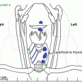
Fig. 6.1
Parathyroid lipoadenoma with proliferation of parenchymal parathyroid tissue and stromal elements with adipose tissue comprising greater than 50 % of the adenoma. The parenchymal tissue is comprised of oxyphilic cells and small numbers of chief cells organized in small groups and thin, branching cords. Hematoxylin and Eosin stain: medium power
Parathyroid glands typically weigh 20–40 mg, while those weighing more than 50–60 mg are usually abnormal [8]. Normal parathyroid glands consist of chief cells – the main source of PTH, transitional cells, oxyphil cells, and adipose tissue. Adipose tissue is the primary component of the parathyroid stroma and occupies approximately 20–30 % of an adult parathyroid gland. Also parathyroid cellularity can be highly variable within a single gland, among glands in the same patient, and between patients. It also varies by age, sex, and body habitus, among others.
PHPT is due to a single parathyroid adenoma in 80–85 % or more of patients [9]. Chief cell parathyroid adenomas are the most common pathologic variant in PHPT [10]. Oxyphil cells typically comprise less than 5 % of parathyroid volume [11], but oxyphil parathyroid adenomas can be seen in PHPT and have been associated with larger adenomas [12–14]. However, a recent case-matched study found no significant difference in weight between oxyphil and chief cell adenomas [15].
PHPT due to parathyroid lipoadenomas are rare but, clinically, present in a similar fashion to those associated with common parathyroid pathologic variants [16]. Parathyroid lipoadenomas are defined by extensive stromal adipose tissue, myxoid change, and fibrosis [17]. Clear cell parathyroid adenomas are composed of cells with finely vacuolated cytoplasm in a solid or acinar pattern [18]. Atypical parathyroid adenomas have atypical features such as mitoses and fibrous bands but lack definitive invasion or metastases. They generally behave in a benign fashion [19].
A definitive histopathological diagnosis of parathyroid carcinoma is difficult and often presents as severe PHPT where it is suspected or as PHPT recurrence 2–3 years after the initial parathyroid surgery. Classical pathologic criteria include uniform sheets of cells arranged in a lobular pattern separated by dense fibrous trabeculae, mitotic figures in tumors cells, and capsular and vascular invasion [20, 21]. Invasive growth is required for a diagnosis of parathyroid carcinoma. Only 3–30 % of patients have evidence of regional lymph node involvement or distant metastases at the initial surgery (see Chap. 10) [22]. Parathyromatosis is a rare pathologic variant in PHPT that is characterized by small nodules of hyperplastic parathyroid tissue composed primarily of chief cells that are scattered throughout the soft tissues of the neck and upper mediastinum [19, 23]. Clinically, this condition is characterized by recurrent or persistent PHPT. On occasion, parathyroid glands removed in patients with prior thyroidectomy or re-explorative parathyroid surgery may present challenges to the surgeon and pathologist due to fibrosis (see Chap. 11).
PHPT due to parathyroid hyperplasia, which accounts for approximately 15 % of PHPT, is defined as multiple parathyroid glands with proliferation of parenchymal cells leading to parathyroid gland enlargement independent of secondary factors that stimulate PTH production. Preoperative imaging studies and intraoperative PTH monitoring aid in differentiating single from multiple gland disease. Chief cell hyperplasia is the most common form of parathyroid hyperplasia, but rarely clear cell hyperplasia and lipohyperplasia are seen. Asymmetric hyperplasia is a challenging issue as only around 50 % of PHPT has symmetric enlargement of all four glands. Approximately one-third of PHPT hyperplasia is due to familial parathyroid disorders (e.g., MEN, MEN2A, MEN4, hyperparathyroidism-jaw tumor syndrome, familial isolated PHPT).
Clinical Pearls/Pitfalls
After the diagnosis of PHPT is confirmed, evaluate for related complications.
Parathyroidectomy has a high rate of cure and complications are rare.
Review the outcome of parathyroid surgery, utilizing serum calcium rather than PTH for surveillance postoperatively.
Parathyroid adenomas are the cause of PHPT in ≥80 % of patients and pathologically are usually due to chief cell adenomas, but oxyphil adenomas, lipoadenomas, clear cell adenomas, and atypical parathyroid adenomas are additional pathologic variants.
Parathyroid carcinoma often presents with severe PHPT or is identified with disease recurrence 2–3 years after the initial parathyroid surgery.
Parathyroid hyperplasia is the cause of PHPT in approximately 15 % of patients and is seen in familial parathyroid disorders such as MEN1, MEN2A, MEN4, hyperparathyroidism-jaw tumor syndrome, and familial isolated PHPT.
Conflict of Interest
All authors state that they have no conflicts of interest.
References
1.
Wermers RA, Kearns AE, Jenkins GD, Melton 3rd LJ. Incidence and clinical spectrum of thiazide-associated hypercalcemia. Am J Med. 2007;120:911 e919–915.CrossRef
Related posts:

Stay updated, free articles. Join our Telegram channel

Full access? Get Clinical Tree


