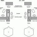
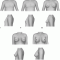
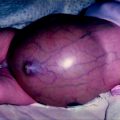
Fig. 5.1
Development of Parathyroid Glands. Superior parathyroid glands originate from fourth pharyngeal pouch and are designated as P-IVs. Their migration path is shorter and final position more reliable than inferior parathyroid glands. Inferior parathyroid glands originate from third pharyngeal pouch and are designated as P IIIs. Their migration path is longer and final position is more variable than superior parathyroid glands. The thymus also originates from the third pharyngeal pouch and the final position of the inferior parathyroids is often adjacent to or even within the thymus
During the descent of the parathyroid glands, the P-IVs are crossed by the P-IIIs and occasionally the superior and inferior glands fuse. The final position of fused (or near fused) glands is usually close to the inferior thyroid artery. Parathyroid gland fusion during development may lead to the two separate glands being mistaken for a single bi-lobed gland which would give a false impression of an unidentified gland during parathyroid exploration. This misinterpretation can be avoided by close inspection of the blood supply to the parathyroid. Each gland will have a unique blood supply so even an enlarged single gland has a single arterial pedicle while a bilobed gland has two separate arterial pedicles.
Supernumerary parathyroid glands can occur in up to 15% of population [1]. They are most often found in the thymus, but they have also been described in the middle mediastinum at the level of the aortopulmonary window and lateral to the jugulo-carotid axis. Intravagal parathyroid tissue has also been reported. Such locations are thought to be due to fragmentation of the foetal superior parathyroid during development.
Human foetal parathyroid glands can synthesize parathyroid hormone (PTH) from as early as 10 weeks of gestation [2]. Foetal parathyroid hormone is essential for foetal calcium homeostasis [3] as intact maternal PTH does not cross the placenta. Maternal serum calcium crosses the placenta and may influence foetal PTH secretion.
The embryological development of the parathyroid gland requires the coordinated expression of many transcription factors including GCMB, SHH, GATA3, TBCE, Six1/4, Eya1, Hoxa3, Tbx1, Sox3, Pax1 and Pax9. Studies in a mouse knockout model demonstrated that loss of GATA3 expression or its target GCM2, a parathyroid-specific transcription factor, resulted in abnormal third and fourth pharyngeal pouch morphology and the absence of parathyroid glands, although some PTH was produced by the thymus gland [4]. Deletion of the homeobox gene Hoxa3 resulted in lack of both thymus and parathyroid glands [5]. Parathyroid hyperplasia or tumours develop in several familial conditions noted in Table 5.1.
Table 5.1
Familial conditions in which parathyroid hyperplasia or tumours develop
Syndrome | Inheritance pattern | Associated gene(s) | Parathyroid abnormality | Other abnormalities |
|---|---|---|---|---|
Multiple endocrine Neoplasia type 1 (MEN-1) | Autosomal dominant | MEN1 | • Multiple gland hyperplasia • Adenomas | • Pancreatic islet cell tumours • Pituitary adenomas |
Multiple endocrine Neoplasia type 2 (MEN2) | Autosomal dominant | RET | • Adenomas | • Medullary carcinoma thyroid (MCT) • Pheochromocytoma |
Hyperparathyroidism jaw Tumour syndrome | Autosomal dominant | HRPT2a, b | • Adenomas • Carcinoma (15%) | • Ossifying jaw tumours • Renal, uterine and pancreatic tumours |
Familial isolated Hyperparathyroidism | Typically autosomal dominant | MEN1, RET or HRPT2 | • Adenomas • Carcinoma • Hyperplasia | • Without additional syndromic features |
Parathyroid Anatomy
As predicted by embryology, most people have four parathyroid glands, two on each side of the neck: a superior parathyroid gland (derived from the fourth pharyngeal pouch) and an inferior parathyroid gland (derived from the third pharyngeal pouch). The superior parathyroid glands (P-IV) are located close to the upper pole of the thyroid gland, at the level with the first tracheal ring. Traditionally, a superior parathyroid gland is said to be found in an area 2 cm in diameter centred 1 cm above the intersection of the inferior thyroid artery and recurrent laryngeal nerve (RLN).
Stay updated, free articles. Join our Telegram channel

Full access? Get Clinical Tree


