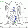
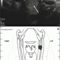
Fig. 10.1
Sonogram of the right side of the neck, sagittal view. (a) A mass (PC) measuring approximately 3.5 × 2.6 × 2.9 cm is seen lying along the inferior and posterior aspect of the right lobe of the thyroid gland (TG). It is heterogeneous with lobulated margins and has internal calcifications. Both lobes of the thyroid are normal in size with normal vascularity. (b) The mass is hypervascular on color Doppler flow imaging
Diagnosis and Assessment
Our patient’s clinical presentation is consistent with hypercalcemia due to primary hyperparathyroidism. Parathyroid carcinoma (PC) is a rare endocrine malignancy, accounting for around 1 % of cases of primary hyperparathyroidism [1]. This infrequent, but potentially life-threatening, clinical entity was first reported by Wilder of Mayo Clinic in 1929 [2]. Conditions that appear to result in an increased risk of PC include multiple endocrine neoplasia type 1 [3], autosomal dominant familial isolated hyperparathyroidism [4], and hyperparathyroidism-jaw tumor syndrome due to mutation of the CDC73 gene [5].
PC presents a diagnostic and therapeutic challenge due to overlapping features with benign primary hyperparathyroidism and the lack of specific differentiating characteristics for malignancy. However, patients with PC typically have a serum PTH greater than five times the upper limit of normal and calcium levels in excess of 14 mg/dL, significantly higher than those with parathyroid adenoma or hyperplasia [6]. Unlike parathyroid adenoma, which has a female preponderance, PC affects both sexes equally, with an earlier median age of diagnosis in the fifth decade [7]. The findings of a palpable neck mass and dysphonia related to recurrent laryngeal nerve palsy as well as the combination of severe renal and bone disease are also suggestive of malignancy [7, 8].
The diagnosis of PC relies on a high index of suspicion, taking into account the patient’s clinical presentation, laboratory studies, imaging, and histopathology. The vast majority of PCs are functioning neoplasms, with clinical features and symptoms resulting from excessive secretion of PTH rather than local infiltration or mass effect from the neoplasm itself. Initial laboratory evaluation should include measurement of total serum calcium, PTH, and serum creatinine and an assessment of overall fluid and electrolyte status.
Ultrasound and sestamibi scintigraphy provide information on localization and the extent of the neoplasm, though neither is specific for carcinoma. Nonetheless, evidence of tumor invasion, irregular margins, and cervical node enlargement on ultrasound may raise suspicion for PC [9]. SPECT-CT with sestamibi, as utilized in this case, provides three-dimensional information with improved sensitivity for the detection of hyperfunctioning parathyroid glands [10]. Computerized tomography and magnetic resonance imaging, although not typically utilized on initial evaluation, may be useful adjuncts to ultrasonography in assessing for distant metastases in the chest or abdomen. Selective venous catheterization and PTH measurement may also be useful if noninvasive tests are equivocal or negative.
Fine needle aspiration (FNA) is not recommended in the setting of suspected PC due to the inability of cytology to differentiate between benign and malignant disease and the potential risk of ectopic tumor seeding [11].
Management
Prior to operative intervention, patients may require urgent management of dehydration, hypercalcemia, and associated metabolic abnormalities. Our patient was admitted for aggressive hydration with saline, initiation of a loop diuretic to promote calcium excretion, and administration of pamidronate. Following this, the decision was made to proceed to the operating room.
The gold standard treatment for PC is en bloc surgical resection [8]. Removal of any contiguous tissues to which the neoplasm adheres is necessary to achieve complete resection. This frequently includes the ipsilateral thyroid lobe and may necessitate resection of the ipsilateral recurrent laryngeal nerve. Ipsilateral compartment VI central neck dissection with removal of all lymphatic tissue should be performed. Care must be taken to avoid rupturing the capsule of the neoplasm, as this increases the likelihood of malignant parathyromatosis [12]. As highest chance of cure is obtainable if en bloc resection is carried out at the initial operation, a high index of suspicion for PC is therefore paramount. Chemotherapy and radiation have generally shown disappointing results [13].
Histopathological diagnosis is challenging, and while early pathologic criteria suggested the presence of a trabecular growth pattern, capsular invasion, vascular invasion, dense fibrous bands, and mitotic figures as suggestive of malignancy [14], later studies have shown that none of these are pathognomonic and may exist in parathyroid adenomas [15, 16]. Thus, the only unequivocal proof of malignancy is the local invasion of contiguous structures noted during surgery or the presence of distant metastasis.
Stay updated, free articles. Join our Telegram channel

Full access? Get Clinical Tree


