Papillary Lesions


Papillary Lesions


Papillary lesions of the breast comprise a heterogeneous group. These lesions have in common a growth pattern characterized by the presence of finger-like projections or fronds of variable length and thickness that are composed of central fibrovascular cores covered by epithelium. Although the histologic identification of a breast lesion as “papillary” is usually straightforward, the distinction among intraductal papilloma, papilloma with atypia (atypical papilloma), papilloma with ductal carcinoma in situ (DCIS), papillary DCIS, or even invasive papillary carcinoma may be a source of diagnostic difficulty.

A few general principles apply to all papillary lesions. First, as will be discussed in more detail, assessment of the presence and distribution of myoepithelial cells in the lesion is one of the most helpful features in arriving at the correct diagnosis (Table 8.1). In some cases, this may require the use of immunostains to myoepithelial cell proteins. Second, the ideal method to examine an excisional biopsy specimen containing a suspected intraductal papillary lesion involves carefully opening the involved duct longitudinally using a pair of fine scissors until the tumor is exposed. Identification of the lesion may be facilitated by the surgeon placing a suture at the end of the involved duct nearest the nipple. Randomly slicing through the excised tissue is not recommended as a small lesion may be missed. Third, if a papillary lesion is suspected on gross examination, a frozen section should not be performed because the distinction of benign from atypical or malignant papillary lesions on frozen sections may be extremely difficult. Moreover, freezing may produce tissue distortion and artifacts that could preclude definitive categorization of the lesion on permanent sections.


TABLE 8.1 Distribution of Myoepithelial Cells in Papillary Lesions


Myoepithelial Cells within Papillae


Myoepithelial Cells at the Periphery of Involved Spaces


Papilloma


Present


Present


Papilloma with atypia/DCIS


Absent in atypical areas; present in areas of residual benign papilloma


Present


Papillary DCIS


Absent


Present


Encapsulated papillary carcinoma


Absent


Absent


Solid papillary carcinoma


Absent


May be present or absent


DCIS, ductal carcinoma in situ.


InTraDuCTal PaPIlloMa

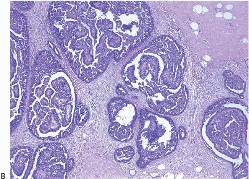
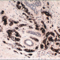
Intraductal papillomas are benign lesions that can be divided into two groups: central papillomas, which involve large ducts and are usually solitary, and peripheral papillomas, which involve the terminal duct lobular units and are usually multiple (Fig. 8.1).


FIGURE 8.1 Intraductal papillomas. A: Solitary, central papilloma. B: Multiple peripheral papillomas.


FIGURE 8.1 (Continued)

Central papillomas are tumors of the major lactiferous ducts and are most frequently observed in women 30 to 50 years of age. Patients usually present with nipple discharge that may be bloody; on occasion, the lesion reaches sufficient size to produce a palpable, subareolar mass. Peripheral papillomas occur in somewhat younger patients and less often present with nipple discharge or a mass.1 They may occasionally present as a mammographic abnormality (microcalcifications or multiple densities).

Central papillomas are generally <1 cm in diameter, but occasionally may be as large as 4 or 5 cm. On gross examination, they appear as tanpink, circumscribed nodules within a dilated duct or cyst. A frankly papillary configuration may be apparent, but more typically the lesion has a bosselated surface. The tumor may be attached to the wall of the involved duct by a stalk or may be sessile. Peripheral papillomas are usually not identifiable on gross examination.

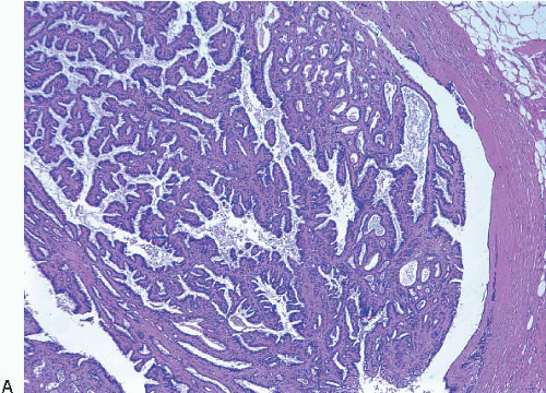
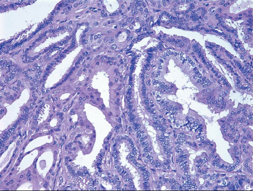
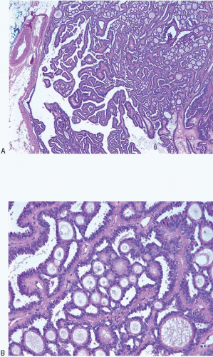
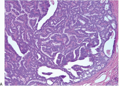
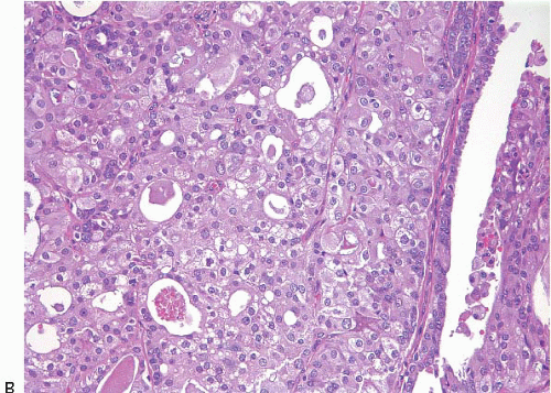
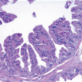
On histologic examination, papillomas are composed of arborizing fronds with well-developed fibrovascular cores (Fig. 8.1, 
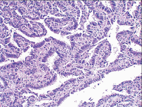
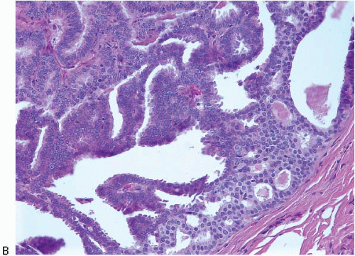
e-Fig. 8.1). The papillary fronds are covered by an inner myoepithelial cell layer and an outer epithelial layer (Fig. 8.2, 
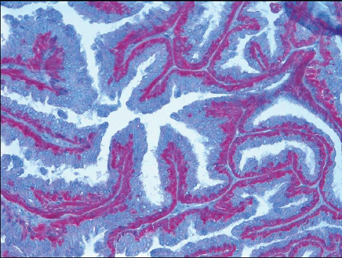
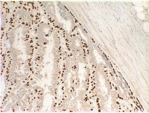
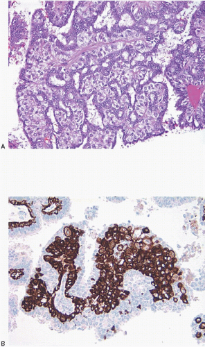
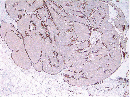
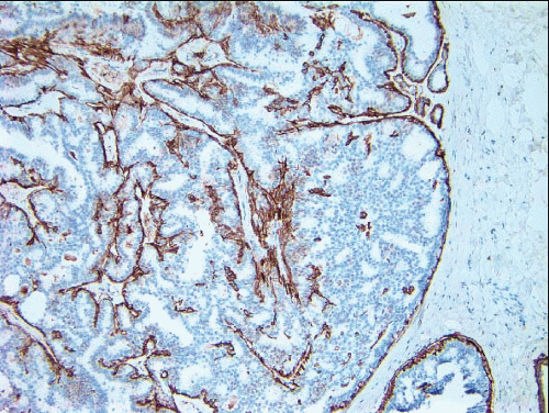
e-Fig. 8.2). The myoepithelial layer is variably prominent, but is always present. In problematic cases, myoepithelial cells can be highlighted by immunostaining for actin, smooth muscle myosin heavy chain, calponin, p63, or other myoepithelial cell markers (Fig. 8.3, e-Fig. 8.3). Myoepithelial cells are also present at the periphery of the involved duct(s) (Fig. 8.4, e-Fig. 8.3) (
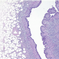
Table 8.1). The epithelium of benign papillomas may consist of one or a few layers of cuboidal to columnar cells or it may show varying degrees of usual ductal hyperplasia (UDH). The epithelial hyperplasia may be extreme and may grow in a contiguous fashion between adjacent papillae (Fig. 8.5, e-Fig. 8.4). In some cases, the epithelial proliferation may in areas have the appearance of atypical ductal hyperplasia (ADH) or DCIS (see subsequent text). Apocrine metaplasia is frequent (Fig. 8.6, e-Fig. 8.5); squamous metaplasia may also be seen.2 Varying degrees of myoepithelial hyperplasia may be observed in some cases (Fig. 8.7). The hyperplastic myoepithelial cells can be epithelioid or spindle-shaped and may have abundant clear cytoplasm. The myoepithelial cell component may be sufficiently prominent that the foci resemble areas of adenomyoepithelioma (
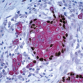
e-Fig. 8.6) (see subsequent text). Collagenous spherulosis is yet another change that may be seen in the epithelium of intraductal papillomas; this should not be misinterpreted as an area of ADH or DCIS (Fig. 8.8, e-Fig. 8.7) (see subsequent text).


FIGURE 8.2 Intraductal papilloma. The papillary fronds consist of fibrovascular cores covered by an inner myoepithelial cell layer and an outer epithelial cell layer.


FIGURE 8.3 Intraductal papilloma. Calponin immunostain highlights the myoepithelial cell layer.


FIGURE 8.4 Intraductal papilloma. p63 immunostain demonstrates myoepithelial cells within the papillae and around the periphery of the involved duct.

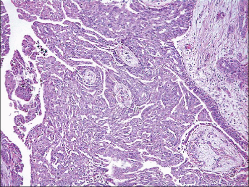
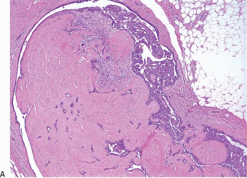
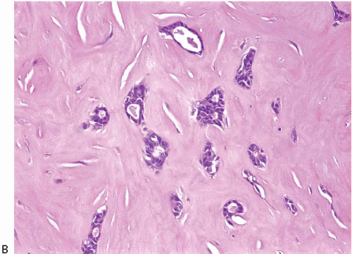
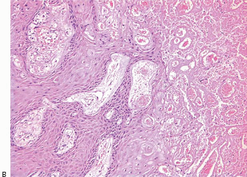
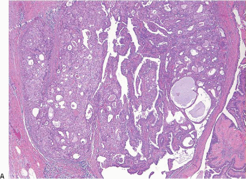
The papillary fronds and/or the surrounding duct wall may show varying degrees of stromal fibrosis and may contain entrapped glands and/or solid epithelial cell nests. On occasion, the fibrosis is so extensive that it distorts or obscures the underlying papillary architecture. In such cases, the term sclerosing papilloma is appropriate. In the most extreme cases, the features overlap with those of a ductal adenoma (see subsequent text) or complex sclerosing lesion. The presence of glands or epithelial nests within a fibrous stroma may produce a worrisome appearance that raises the question of an invasive carcinoma (Fig. 8.9, e-Fig. 8.8). However, in benign intraductal papillomas with sclerosis, myoepithelial cells are discernible around at least some of the entrapped glands and epithelial nests, which is a feature supporting the benign nature of this process. In addition, the stroma of these lesions typically has a more hyalinized, sclerotic appearance than the stroma associated with invasive carcinomas.


FIGURE 8.5 Intraductal papilloma with usual ductal hyperplasia (UDH). This papilloma shows a florid epithelial proliferation that fills the spaces between papillae. The proliferation has the architectural and cytologic features of UDH.


FIGURE 8.6 Apocrine metaplasia in an intraductal papilloma.


FIGURE 8.7 Myoepithelial cell hyperplasia in an intraductal papilloma. A: Numerous myoepithelial cells with clear cytoplasm are apparent. B: Smooth muscle myosin heavy chain immunostain further highlights the myoepithelial cells.


FIGURE 8.8 Intraductal papilloma with collagenous spherulosis. A: Low-power view illustrating prominent collagenous spherulosis (upper right). B: Higher power view illustrating the characteristic features of collagenous spherulosis (see text).


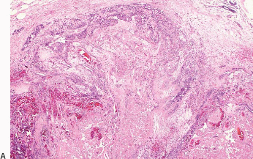
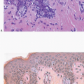
Benign papillomas may undergo infarction, particularly larger central lesions; this may occur spontaneously or may be associated with trauma, such as a needling procedure (fine-needle aspiration or core-needle biopsy). Infarction is frequently associated with entrapment of benign epithelium at the periphery of the lesion. The entrapped epithelium may exhibit reactive cytologic atypia or squamous metaplasia (Fig. 8.10, e-Fig. 8.9).3

Although the histologic features of peripheral papillomas are similar to those of central papillomas, the epithelium of the peripheral lesions more often shows foci of ADH and DCIS than that of solitary, central papillomas.4,5


FIGURE 8.9 Intraductal papilloma with sclerosis (sclerosing papilloma). A: Low-power view illustrates that the majority of this papilloma is replaced by dense collagen obliterating the papillary architecture. B: High-power view illustrating entrapped glandular epithelium within collagen.


FIGURE 8.9 (Continued)

Intraductal papillomas are benign lesions that are adequately treated by excision. The risk of subsequent breast cancer associated with solitary intraductal papillomas is similar to that of women with other proliferative lesions without atypia (relative risk ∽2 compared with the general population). Multiple papillomas appear to be associated with a higher risk of concurrent and subsequent carcinoma than do solitary, central papillomas.1,5, 6 and 7 The key features of intraductal papillomas are summarized in Table 8.2.


FIGURE 8.10 Intraductal papilloma with infarction. A: At low power, necrosis and focal areas of hemorrhage are apparent. B: High-power view of the periphery of the lesion showing necrosis (right side of field) and epithelium with squamous metaplasia.


FIGURE 8.10 (Continued)


TABLE 8.2 Key Features of Intraductal Papilloma


Clinical


Age 30-50 years at presentation


May be central or peripheral


Central lesions most often present as nipple discharge or subareolar mass


Histologic


Variably fibrotic fibrovascular cores covered by epithelial and myoepithelial cells


Epithelium consists of one to several layers of cuboidal to columnar cells, but may exhibit usual ductal hyperplasia, atypical ductal hyperplasia (papilloma with atypia), or DCIS (papilloma with DCIS)


Apocrine metaplasia and/or squamous metaplasia may be seen, the latter most often in association with infarction


DCIS, ductal carcinoma in situ.


PAPILLOMA WITH ATYPIA (ATYPICAL PAPILLOMA) AND PAPILLOMA WITH DCIS

Some intraductal papillomas exhibit monotonous areas of epithelial proliferation that fulfill the combined architectural and cytologic criteria for the diagnosis of ADH or low-grade DCIS (Figs. 8.11 and 8.12, e-Fig. 8.10). These areas may involve the papilloma to varying degrees, but features of a benign papilloma remain evident in part of the lesion. When DCIS is present, it most often exhibits solid and/or cribriform patterns. Small foci of necrosis may be observed (Fig. 8.12). Of note, myoepithelial cells are absent in the foci of ADH or DCIS but remain identifiable in the areas of residual benign papilloma and around the periphery of the involved spaces (Fig 8.13) (Table 8.1). In addition, the epithelial cells comprising the atypical foci lack expression of high-molecular-weight cytokeratins (such as CK5/6) and typically show strong, diffuse expression of estrogen receptor (Fig. 8.14).7 In fact, in problematic cases, the absence of highmolecular-weight cytokeratin staining coupled with the presence of diffuse staining for estrogen receptor is a useful adjunct to help distinguish ADH or DCIS in a papilloma from UDH in a papilloma which typically shows strong expression of high-molecular-weight cytokeratins (often in a mosaic pattern) and patchy, variable expression of estrogen receptor.


FIGURE 8.11 Papilloma with atypia. A: At low power, most of the lesion can be seen to consist of an intraductal papilloma without atypical features. However, in a few areas (seen best in the lower right portion of this photograph), small foci of monomorphic epithelial cells with a cribriform pattern are evident. B: Higher power view illustrates the atypia characteristic of atypical ductal hyperplasia (lower right).


FIGURE 8.11 (Continued)


FIGURE 8.12 Papilloma with DCIS. A: Low-power view illustrates that a large portion of the papilloma contains an epithelial proliferation with the architectural and cytologic features of ductal carcinoma in situ (DCIS) growing in a solid and cribriform pattern (left side). B: Higher power view to illustrate the DCIS. In this case, the DCIS has intermediategrade nuclei.


FIGURE 8.12 (Continued)


FIGURE 8.13 Papilloma with extensive involvement by ductal carcinoma in situ (DCIS). Smooth muscle myosin heavy chain immunostain demonstrating myoepithelial cells lining the fibrovascular cores of residual benign papilloma and at the periphery of the space. Myoepithelial cells are not present within the areas of DCIS.


FIGURE 8.14 Papilloma with DCIS immunostained for cytokeratin (CK) 5/6. The neoplastic epithelial cells are CK5/6 negative; CK5/6 highlights residual myoepithelial cells.

Criteria for distinguishing papilloma with atypia from papilloma with DCIS have been varied. Some have been based on assessment of the size or extent (i.e., number of millimeters) of the atypical proliferation within the papilloma, whereas others have been based on the proportion of the papilloma involved by the atypical proliferation.6,8 The 2011 WHO Working Group recommended that size/extent criteria rather than proportion criteria be utilized to distinguish papilloma with atypia (atypical focus <3 mm in size) from DCIS involving a papilloma (≥3 mm of atypia), an approach to which we subscribe.9 It is important to note that when the atypia within the papilloma is of intermediate or high nuclear grade, a diagnosis of DCIS involving a papilloma should be rendered regardless of extent.

The clinical significance of atypia or DCIS in a papilloma is not well-defined. Some authors have reported a substantially increased risk (7.5-fold) for the subsequent development of breast cancer, predominantly in the ipsilateral breast,6 whereas others have found that the level of breast cancer risk associated with papillomas with atypia was similar to that for patients with ADH elsewhere in the breast (fourfold to fivefold) and that the risk was approximately equal in both breasts.1 Breast cancer risk is reported to be particularly high (sevenfold) among women with multiple papillomas with atypia.1

The risk of subsequent breast cancer and local recurrence does not appear to be related to the extent of atypia or DCIS within the papilloma. In fact, the most important consideration is the presence of atypia or
DCIS in the surrounding breast tissue because this appears to be more closely related to the risk of recurrence than the qualitative features or extent of atypia within the papilloma itself.6,10

Given the foregoing information, papillomas with atypia and papillomas with DCIS are best managed by complete excision with careful follow-up. The surrounding breast tissue should be carefully evaluated for the presence of ADH and DCIS because this should be the major feature influencing management decisions.


PAPILLARY DCIS

As indicated in Chapter 3, some DCIS lesions have a papillary growth pattern, characterized by fibrovascular cores covered by neoplastic epithelium. In our view, these lesions are fundamentally distinct from papillomas with DCIS because they do not exhibit evidence of residual benign papilloma. Features useful in distinguishing papillary DCIS from benign intraductal papillomas are summarized in Table 8.3. In particular, in papillary DCIS, the papillae are usually more delicate and less fibrotic than those of intraductal papilloma. Additionally, the epithelium in papillary DCIS is usually composed of a single cell population with a uniform appearance (Fig. 8.15, e-Fig. 8.11

Only gold members can continue reading. Log In or Register to continue

Stay updated, free articles. Join our Telegram channel

Jun 18, 2016 | Posted by in ONCOLOGY | Comments Off on Papillary Lesions

Full access? Get Clinical Tree

Get Clinical Tree app for offline access