Fig. 26.1
Computed tomography (CT) of a pancreatic neuroendocrine tumor (T) in the body of the pancreas that obstructs the splenic vein with extensive short gastric collaterals and extends into the lumen of the portal vein
The splenic artery was embolized with coils on the night before surgery by interventional radiology. PNET resection required subtotal pancreatectomy with splenectomy, obtaining proximal and distal control of the portal vein, and extraction of tumor thrombi from portal vein (Fig. 26.2) with patch closure of portal vein (Fig. 26.3).
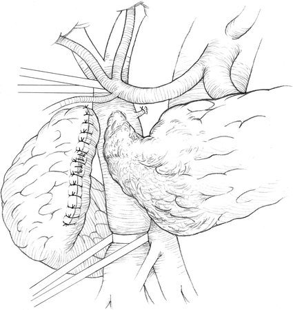
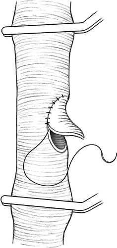
Fig. 26.2
Drawing of the tumor in Fig. 26.1 at surgery just prior to resection. One can see that the pancreas is transected and oversewn to the right of the superior mesenteric vein (SMV)/portal vein confluence. The coronary vein is ligated and divided. Vascular control of the portal vein and SMV proximal and distal to where the tumor extends through the splenic vein into the portal vein is obtained prior to resection of the tumor within the portal vein and splenic vein
Fig. 26.3
Drawing of closure of a portal vein venotomy with a bovine patch. Proximal and distal control of the vein is obtained with vascular clamps. A venotomy is made and the tumor within the portal vein is excised. Following the excision of tumor a pericardial patch is used to reconstruct the vessel and not narrow the lumen
Case #2
A 67-year-old man who presents with pain after eating, and weight loss. CT demonstrates a 3 cm pancreatic mass in the uncinate portion of the head abutting the SMV (Fig. 26.4), with subsequent evidence of invasion into the lumen (Fig. 26.5). EUS was performed and FNA of the pancreatic head tumor shows NET. At surgery he had a Whipple pancreaticoduodenectomy with obtaining proximal and distal control of the SMV, portal vein and splenic vein; given heparin; and removing the tumor thrombus from the portal vein and using a venous patch to reconstruct the SMV at the venotomy (Fig. 26.6).
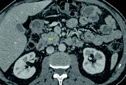
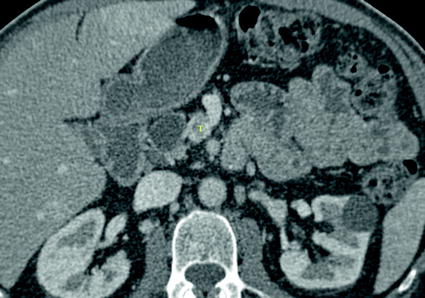
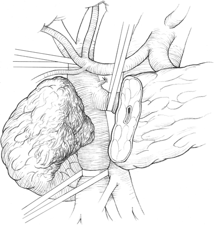
Fig. 26.4
Computed tomography (CT) of a neuroendocrine tumor (NET) in the uncinate portion of the head of the pancreas
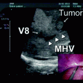
Fig. 26.5
The same PNET as seen in Fig. 26.4 extends through the wall of the superior mesenteric vein into the lumen of the portal vein (T) on superior CT cuts
Fig. 26.6
Drawing of the PNET in Fig. 26.5 that extends into the SMV and is in the process of being removed surgically. Note vascular control of the SMV, portal vein, and coronary vein is obtained prior to excision
Proximal and distal control of the portal vein, splenic vein, inferior mesenteric vein, and the superior mesenteric vein is done with vessel loops and vascular clamps, as seen in Fig. 26.2. Any other smaller branches are ligated and divided. Systemic heparin is given at a dose of 100 Units per Kg IV prior to clamping. We incise the vein and use a spatula to mobilize the thrombus. It may require excision and reconstruction of the vein wall. This can be done with a pericardial patch venoplasty, shown in Fig. 26.3, a superficial femoral vein patch, or interposition graft. After closure of the vein, the heparin is revered with protamine at a dose of 1 mg per 100 units of heparin infused. We start 81 mg per day of aspirin on postoperative day 1. We do not use any other type of anticoagulation. Portal vein thrombectomy is either done with concomitant Whipple pancreaticoduodenectomy or subtotal pancreatectomy with splenectomy.
Clinical Pearls
Proximal and distal control is necessary
Ligate small posterior branches. Make sure that vein is free without any branches
Draw a line on interposition vein to maintain proper orientation
Results
We have published a series of 46 patients with major vascular abutment, involvement, or encasement who underwent surgery to remove all gross neuroendocrine tumor [2]. Our series is contrasted to a more recent series from MD Anderson [1]. They reported on nine patients with PNETs who underwent portal venous tumor thrombectomy. The mean age was 42 and 51, respectively. There were approximately 50% men in both studies. Our study had a much higher percentage of MEN-1 (i.e., 26% compared to 0%). The mean tumor size was similar, approximately 5 cm. Most of their tumors were in the body and tail, while most of ours were in the head. Although not all of the head tumors in our experience required a Whipple pancreaticoduodenectomy, the rate was 23%. Whipple was performed in one-third of patients with pancreatic head tumors. The Ki67 rate was between 1 and 2% for all our tumors because we selected them based on a low malignant potential, which is defined as a positive SRS scan or a Ki67 < 2%, while theirs had a high rate in several patients, and all except one was treated with preoperative chemotherapy. Each of their patients had blood vessel involvement while only 34% of ours had it. Since our patients had less blood vessel involvement; we performed fewer venotomies and vascular reconstructions (Table 26.1 and 26.2).
Table 26.1
Demographics of patients with PNETs and vascular involvement from two series
Study | # patients with pNET + vascular involvement | % Men | Mean age (range) | % functional NET | % MEN1 | NET Size (cm) | % head of pancreas | % vessel involvement |
|---|---|---|---|---|---|---|---|---|
MD Anderson | 9 | 33 | 51 (38-67) | 0 | 0 | 5.4 | 11 | 100 |
Stanford NIH | 46 | 46 | 42 (24-76) | 70 | 26 | 5.8 | 59 | 34 |
Table 26.2
Extent of surgery, complications, disease status, and survival
Study | % Up front chemotherapy | % Whipple procedures | % Vascular resection and reconstruction | % complications (deaths) | % Disease-free | Survival |
|---|---|---|---|---|---|---|
MD Anderson | 67 | 11 | 89 | Not listed | 33% | 78% at 3 years |
Stanford NIH | 0 | 23 | 20 | 27 and no deaths | 30% | 60% at 10 years |
However, the two illustrative cases presented here had tumor thrombus within the vessel that can be excised with a spatula, except where it entered the vessel. We used heparin for our venous procedures, and reversed it with protamine after the procedure on the vessel was completed; however, it is controversial, and some do not use it. We reconstructed the portal or superior mesenteric vein with a vein patch from the femoral vein, or a bovine pericardial patch, while some others use the internal jugular vein, but that is used more for complete replacement of these vessels. We had no cases developing thrombosis of the portal vein, as we have followed the flow through this vessel with Doppler ultrasound. These procedures are done with acceptable morbidity and no operative mortality. The disease-free survival was approximately 30% in both studies, and the long-term survival is between 60 and 70%, suggesting that it is worthwhile to perform this more aggressive surgery [1, 2].
Alternative Approaches
Alternative systemic treatment
Manage bleeding gastric varies with either sclerotherapy or embolization of splenic artery
Discussion
Malignant pancreatic neuroendocrine tumors have a good prognosis [5–9]. Unfortunately, a significant proportion present late, with large tumors that encase or invade adjacent blood vessels [3]. A number of studies have demonstrated that vascular invasion with PNETs is associated with decreased survival [5, 10, 11]. The surgical approach to this group of patients is controversial. Based on analogies to pancreatic adenocarcinoma and limited experience with attempted surgical resection of patients with advanced PETs, for many, involvement of the superior mesenteric vein (SMV), and portal vein (PV) is a contraindication to surgical resection [10, 12]. However, recent studies in standard pancreas cancer surgery question this approach. The operability of pancreatic tumors is usually defined by the results of thin-slice computed tomography with special protocols to enhance visualization of the pancreas and its blood vessels [12–16]. However, these studies may falsely determine operability. For example, in patients with adenocarcinoma of the pancreas, when preoperative imaging studies suggest that the tumor involves the SMV or PV, many surgeons suggest that the tumor is inoperable [10, 12, 13]. However, recent studies dispute this thinking, and suggest that these locally advanced tumors may be resectable for benefit [17–19]. Sarcomas involving blood vessels that were previously thought to be inoperable have been recently excised, with acceptable morbidity and good overall survival [20, 21]. Because PNETs are rare, there have only been two studies of the ability to surgically resect malignant PNETs that invade or involve the major mesenteric veins [1, 2]. Most reports have only a few patients [4, 22–26]. In this review we report the long-term results with PNETs that abut or involve major mesenteric vascular structures, including the PV and SMV. The findings suggest that major vascular involvement on preoperative imaging studies may not be a contraindication to resection of PNET.
Related posts:
 Resection of Centrally Located Cystadenoma/Cystadenocarcinoma
 Debulking of Extensive Neuroendocrine Liver Metastases
 Management of the Gangrenous Gallbladder
 Totally Laparoscopic Right Hepatectomy Combined with En-Bloc Partial Resection of the Inferior Vena Cava
 Gallbladder Cancer with Common Bile Duct Invasion
 Resection of Renal Cell Carcinoma Involving the Liver with Tumor Thrombus Extending into Inferior Vena Cava Requiring Venovenous Bypass
Stay updated, free articles. Join our Telegram channel

Full access? Get Clinical Tree


