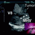
Fig. 27.1
CT scan demonstrating pancreatic tail mass
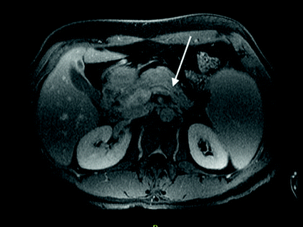
Fig. 27.2
MRI demonstrating splenic vein thrombosis
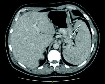
Fig. 27.3
CT scan demonstrating gastric varices secondary to sinistral hypertension
R.D. underwent a distal pancreatectomy and splenectomy for oncologic resection and decompression of his varices. Pathology demonstrated a well-differentiated neuroendocrine tumor with metastasis in 1/27 lymph nodes.
Pancreatic neuroendocrine tumors (PNETs) are rare and account for approximately 2–4% of pancreatic neoplasms. These tumors originate from the pancreatic islet cells and are classified according to the functional syndrome they produce [1]. Insulinoma and gastrinomas account for 70–90% of functional PNETs. Approximately 10–25% of PNETs are nonfunctional, in that they do not cause a syndrome related to hormonal hypersecretion [1]. These tend to be diagnosed at a more advanced stage, given patients do not present until tumors are commonly causing symptoms, such as abdominal pain, from mass effect.
Presentation
Patients with neuroendocrine tumors may present in a variety of ways, and may be subdivided into those producing hormonal syndromes (functioning) and those with nonfunctioning tumors. Functioning tumors tend to present when they are smaller in size, due to hormone hypersecretion resulting in hypoglycemia, peptic ulcer disease, or diarrhea [1]. Nonfunctioning tumors, however, tend to be larger and present later in their course, given they are often only symptomatic once they cause compression or, most commonly, abdominal pain [2]. Patients may also commonly experience symptoms of weight loss and nausea. Those patients who present with symptoms of anemia or GI hemorrhage must be considered for bleeding form gastric varices secondary to sinistral hypertension [3].
Intraoperative Technical Pearls
Ligation of the splenic artery early before manipulation of the splenic vein or venous collaterals is critical, in order to decrease left-sided portal pressure in the venous system.
The splenic vein is divided close to the junction of the mesenteric vein, or 1 cm from the most proximal edge of the lesion.
Ideally, the inferior mesenteric vein is preserved.
Imaging
Given neuroendocrine tumors are hypervascular in comparison to the surrounding pancreatic parenchyma, thin-cut pancreatic protocol CT with IV contrast is the gold standard for preoperative planning. The majority of PNETs will “light up” on the arterial phase of the scan. MRI imaging sensitivity increases with tumor size, and is an alternate mode of imaging that may be used as an adjunct. Additionally, endoscopic ultrasound imaging is utilized to evaluate the extent of disease and perform preoperative biopsies. Immunohistochemical staining with chromogranin A can distinguish neuroendocrine tumors from pancreatic adenocarcinomas [1].
Staging studies include a Chest CT for evaluation of metastatic thoracic disease. Patients may also undergo an octreotide scan, or somatostatin receptor scintigraphy, to localize both primary and metastastic lesions. Octreotide, a synthetic analogue of somatostatin, is radiolabeled and injected into the venous system. A radioactive signal is produced when bound to somatostatin receptors, which are found on primary and metastatic neuroendocrine tumor cells [1].
Operative Planning: Splenic Preservation?
The indication for spleen conserving surgery must be carefully evaluated with each patient. Splenic preservation importantly decreases the risk of overwhelming infection related to encapsulated organisms, as well as provides other immunoprotective capacity. The Warshaw technique, first described in 1988, proposed preservation of the spleen during distal pancreatectomy for tail of the pancreas pathology [4]. This technique divides the splenic artery and vein with the body and tail of the pancreas. The spleen relies on the collateral vasculature via the short gastric and left gastroepiploic vessels to survive. Given the increased flow via these collaterals, patients may develop gastric varices, which theoretically may lead to hemorrhage. After a 23-year experience at MGH, only one-quarter of patients demonstrated asymptomatic perigastric varices on routine imaging, with no resultant clinical consequences [5].
Splenic vein thrombosis complicates pancreatic neoplasms in 7–10% of patients [6]. Splenic vein thrombosis (SVT) can occur secondary to direct tumor invasion or compression of the splenic vein by mass effect. Occlusion of the splenic vein results in sinistral portal hypertension. Greenwald et al. first described the pathophysiology of left-sided portal hypertension in 1939 [7]. Engorgement of the short gastrics and gastroepiploic veins result in gastric varices secondary to back pressure of the left portal venous system [1]. The incidence of gastric bleeding in sinistral portal hypertension varies from 4 to 72% [6].
Related posts:
 Resection of Centrally Located Cystadenoma/Cystadenocarcinoma
 Debulking of Extensive Neuroendocrine Liver Metastases
 Management of the Gangrenous Gallbladder
 Totally Laparoscopic Right Hepatectomy Combined with En-Bloc Partial Resection of the Inferior Vena Cava
 Gallbladder Cancer with Common Bile Duct Invasion
 Resection of Renal Cell Carcinoma Involving the Liver with Tumor Thrombus Extending into Inferior Vena Cava Requiring Venovenous Bypass
Stay updated, free articles. Join our Telegram channel

Full access? Get Clinical Tree


