Stage
Characteristics
1
Pre-adolescent: Elevation of breast papilla/nipple
2
Breast bud stage: Elevation of the breast and papilla as small mound and enlargement of areola
3
Further enlargement of breast and areola, with no separation of contours
4
Projection of nipple and areola to form secondary mound above the level of the breast
5
Mature stage: projection of papilla only due to recession of the areola to the contour of the breast
Premature thelarche may present at any age from the neonatal period (Fig. 28.1), but is commonest before the age of 2, with symmetrical or asymmetrical breast development. It should be noted that physiological breast tissue is palpable in normal children in the first few years of life. Premature breast development is usually an isolated finding but may rarely be associated with hypothyroidism [6]. It is differentiated from precocious puberty by the absence of any other signs of sexual maturation (e.g. pubic hair development) [7] and normal growth. Hormonal investigations include LH (often normal) and FSH (FSH often raised in under 2 year olds [7]). Children who present before age 2 are most likely to undergo regression, with older girls being more likely to have progression of symptoms or persistence of breast tissue [7]. In premature thelarche breast development does not progress beyond Tanner stage 3.
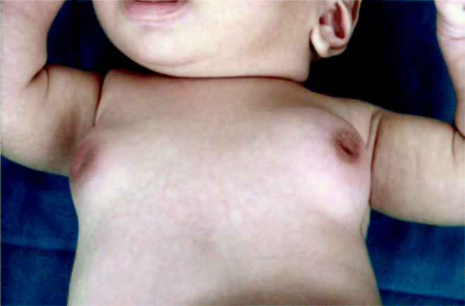

Fig. 28.1
Premature
In normal thelarche, one breast may starts developing first (asynchronous thelarche), and this may be mistaken for a breast lump. On examination, normal breast tissue is palpated and the patient can be reassured.
Precocious puberty (Fig. 28.2) is defined as development of secondary sexual characteristics earlier than would normally be expected [2]. This condition warrants urgent investigation to exclude lesions within the pituitary-endocrine system, as well as the reproductive organs (pituitary, thyroid, adrenal, ovary, testis, etc.). Precocious puberty encompasses breast and pubic hair development, and concerns that children will not reach their full adult height prompt investigation and treatment with GnRH analogues in younger children.
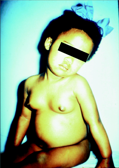

Fig. 28.2
Precocious puberty
Congenital Anomalies
Amastia and Hypomastia
Amastia (congenital absence of the breast) is rare, and may be associated with Poland’s syndrome (hypoplasia or absence of the pectoralis muscle with ipsilateral syndactyly). Amastia may be unilateral or bilateral and may occur in both sexes. It is a condition where the nipple is present but the breast does not develop. Athelia is a rare condition in which breast tissue is present but the nipple is absent [8].
Amastia and hypomastia may be acquired due to iatrogenic damage to the developing breast tissue in a child during central line insertion, chest tube placement, thoracotomy, radiotherapy or burns. Due to nipple complex development only occurring in the eighth month of gestation, premature infants are more susceptible to injury to the developing breast during surgical intervention of central line and thoracic tube insertion.
Lack of breast development may be associated with delayed puberty, which would require investigation of ovarian failure, hypothyroidism or androgen-producing tumours.
Polymastia and Polythelia
Polythelia (accessory nipple) or polymastia (accessory breast) may occur in up to 2% of the population and breast tissue may be found anywhere along the milk line from the axilla to the groyne and very rarely in other anatomical sites [8, 9]. It is common in girls and there is an increased incidence in some geographical populations in America (black, white and Native Americans), Japan and Israel and a decreased incidence in white Europeans (0.2%) [9]. Some cases are familial, with both autosomal dominant and x-linked inheritance described. Accessory breast tissue may be mistaken for other conditions including lipoma, lymphatic disorders and sebaceous cysts [9]. Other congenital anomalies, especially urogenital anomalies are identified in up to 15% of patients. Surgical excision may be indicated for cosmetic reasons or for symptoms including cyclical pain.
Nipple Anomalies
Nipple inversion is a normal variant if present since birth, but should be investigated if it develops in later life as it may be a sign of infection or underlying tumour. It is caused by failure of eversion of the mammary pit in embryonal development [8]. Nipple inversion is susceptible to infection and can be prevented by careful hygiene to the area. Other nipple anomalies include bifid nipples and dysplastic divided nipples. Surgery is not recommended for correction of nipple anomalies in children as invariable damage to the ducts may cause breast feeding problems later in life.
Developmental Anomalies
Neonatal Breast Hypertrophy
Breast tissue is palpable in most babies (75% male infants and 84% female infants [10]) and commonly persists until 10 months of age or longer, especially in breast-fed babies. The palpable nodule is asymmetric in 63% [10], but decreases in size with increasing age. This correlates with the concept of a neonatal ‘mini-puberty’ where endogenous gonadotrophins and sex hormones are produced. Clear or milky fluid nipple discharge may occur. This condition is self-limiting and requires reassurance only.
Breast Asymmetry
Asymmetry is common, in normal development one side usually appears first and may present as a tender lump. Some degree of breast asymmetry is normal in the adult population. Surgery to augment or reduce the breast is not indicated until breast development is complete after puberty.
Macromastia and Breast Hypertrophy
Macromastia (excessively large breasts) may be caused by juvenile hypertrophy, pregnancy, tumours or excess endogenous or exogenous oestrogen or progesterone [11]. Reduction mammoplasty may be indicated after breast development is complete.
Juvenile (virginal) breast hypertrophy is a rare disorder of stromal hyperplasia [12] that may be caused by increased end-organ sensitivity to oestrogen [11]. It is usually symmetrical and may be familial. It usually occurs at menarche and may cause severe mastalgia. Breast growth may be controlled with progesterone or anti-oestrogens. Failure of medical management allows for reduction mammoplasty in teens or young adults when the breasts have achieved maturity. Later in life this condition may affect lactation but there is no risk for breast cancer.
Tubular Breast Deformity
This is a rare condition of skin deficiency at the base of the breast resulting in decreased horizontal and vertical breast growth [13]. The nipple areola complex may become necrosed due to the resulting breast deformity and the overlying skin may be oedematous with venous congestion. Appearance may raise concern for breast malignancy. Therefore, this clinical finding requires careful assessment and reassurance to parents. Reconstructive surgery is indicated after puberty.
Gynaecomastia
Enlargement of the glandular breast tissue in boys is termed gynaecomastia and can present at any age but is commonest in adolescents. A full discussion can be found elsewhere in this book.
Breast Infection
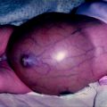
Infection of the breast or nipple may occur at any age and presents with pain, pyrexia, localised tenderness, erythema and induration (cellulitis) progressing to a fluctuant mass (abscess) (Fig. 28.3). The aetiology may be skin infection, nipple abrasion or mammary duct obstruction [14]. Antibiotic therapy may be sufficient, but if an abscess requires drainage, aspiration is recommended initially to prevent damage to the developing breast. If a formal incision and drainage is required, the incision should be as small as possible. Common causative organisms include Staphylococcus Aureus, beta-haemolytic Streptococcus, E.Coli and Pseudomonas Aeruginosa [15].
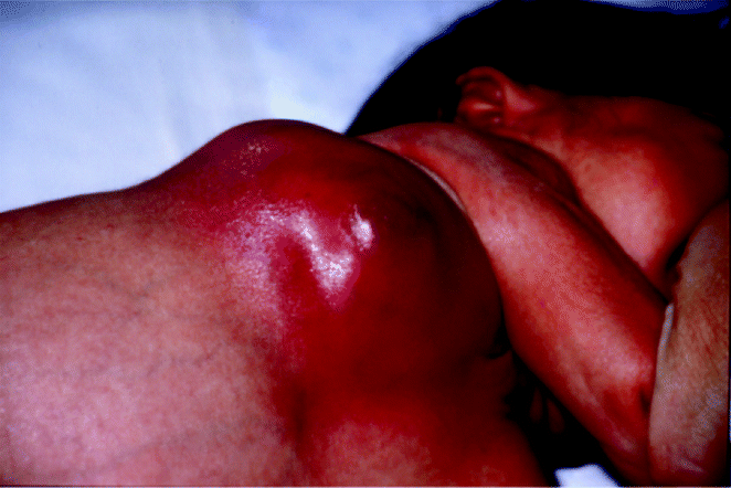

Fig. 28.3
Breast abscess
Mastitis
Neonatal mastitis is uncommon but affects girls more often than boys and commonly proceeds to abscess formation [16]. Certain systemic conditions such as diabetes mellitus, steroid therapy, rheumatoid arthritis could predispose to increased risk of infections. The most common organism is Staphylococcus aureus, less common being Streptococci, gram-negative enteric organisms such as E.coli, anaerobes. Aggressive antibiotic therapy is successful in half of cases and needle aspiration is as effective as formal incision and drainage when required [16].
Periductal mastitis associated with mammary duct ectasia presents with nipple discharge, non-cyclical pain, nipple retraction or a tender lump near the nipple [17]. It is managed with antibiotics and does not usually require surgical duct excision.
Nipple Discharge
Discharge may occur at any age, and is usually associated with benign conditions. It should be investigated if unilateral, persistent and associated with a single duct. Investigations include microbiological culture of the discharge, ultrasound and very occasionally ductogram.
Galactorrhoea
Galactorrhoea is a milky nipple discharge (Fig. 28.4). In the neonate, it is a normal response to the peak of prolactin at birth and will resolve spontaneously. Other causes in children include can be classified as endocrine (hypo or hyperthyroidism), neurogenic (disorders of the chest wall or thorax, including thoracotomy) [6], hypothalamic or pituitary (prolactin-secreting tumours such as prolactinoma), drug-induced (e.g. dopamine receptor blockers) or idiopathic. Investigations include tests of serum prolactin, FSH, LH and thyroid function tests. Treatment of the underlying disorder is required.
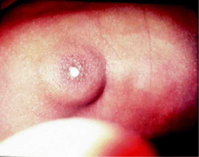

Fig. 28.4
Galactorrhea
Bloody Discharge
A serosanguinous or bloody nipple discharge may be caused by intraductal papilloma—a rare benign condition which can be excised but may recur. Other differential diagnoses include mammary duct ectasia, nipple trauma, foreign body [17] and chronic cystic mastitis. The discharge should be sent for microbiological culture and appropriate antibiotics started.
Mammary duct ectasia is an inflammatory condition characterised by dilatation of the subareolar ducts. Bacterial overgrowth in obstructed ducts may cause abscesses. Ultrasound may confirm tubular anechoic ducts filled with debris [18]. This condition may resolve spontaneously and surgical duct excision is rarely required.
Nipple Adenoma
Nipple adenoma is a very rare benign nipple lesion, usually presenting in middle-aged women (<500 cases reported), with a few case reports in children described. It presents as an enlarging nipple lesion with induration, ulceration or bloody discharge [19]. The recommended treatment is surgical excision, but some recommend that this is deferred in asymptomatic cases until after puberty to preserve developing breast tissue.
Prepubertal Breast Masses
A breast lump in a prepubertal child should be examined carefully and investigated if there are atypical features. Atypical features include hard craggy lump, lump with bloody discharge, inverted nipple, skin changes, ulceration and associated axillary lymph nodes Usually the lump is benign and the child and parents can be reassured, and a policy of watchful waiting is appropriate. Children may present with premature or asynchronous thelarche or breast asymmetry which is mistaken for a mass. Trauma, such as from a seat-belt injury, can result in fat necrosis which may present as a solid mass. Other causes of breast lumps are described below.
Haemangioma
Haemangioma in the breast region may present as a lump. This can be diagnosed clinically and with confirmation on ultrasound or MRI. If the haemangioma grows rapidly it may require treatment with steroids [20]; excision is not usually necessary [14]. There may be associated mammary hypoplasia if the developing breast bud is involved in the haemangioma [14].
Lymphangioma
A lymphangioma is a benign congenital hamartoma or sequestration of the lymphatic system. It most commonly arises in the neck or axilla and is very rare in the breast. A few cases have been described as painless enlarging breast lumps, very rarely in children. Ultrasound may help with diagnosis but usually the definitive diagnosis is made on histological examination of a resected lump, which is curative. In other sites sclerotherapy of large lesions (e.g. with OK432, bleomycin) is used to reduce their size before excision, and this may be useful in the breast to preserve surrounding breast tissue [21].
Stay updated, free articles. Join our Telegram channel

Full access? Get Clinical Tree


