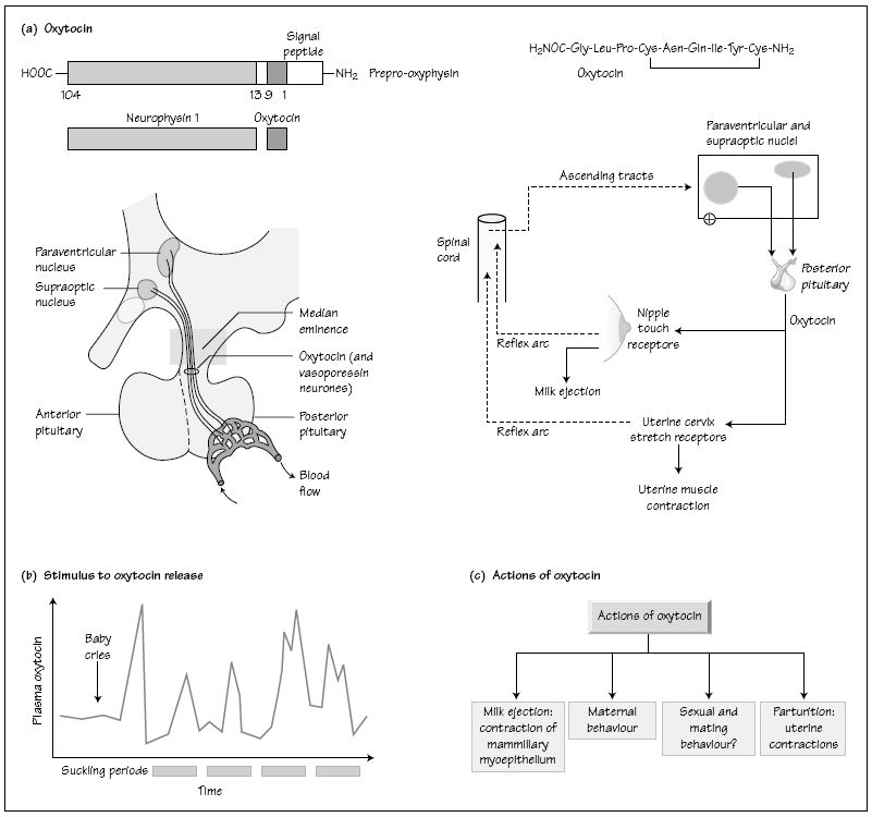
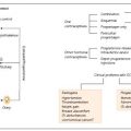
Oxytocin is synthesized in the cell bodies of the magnocellular neurones of the paraventricular and supraoptic nuclei of the hypothalamus (Fig. 34a). Other neurones in the same nuclei produce vasopressin (see Chapter 35). The axons of these neurones pass through the median eminence and terminate in close contact with fenestrated capillaries in the posterior pituitary gland. Both oxytocin and vasopressin are synthesized in the rough endoplasmic reticulum of the cell body, together with proteins called neurophysins. Oxytocin and its neurophysin protein (called neurophysin I) are packaged together in the Golgi apparatus in the same vesicle or secretory granule. The vesicle also contains the enzymes which cleave oxytocin from the neurophysin as the granules migrate along the axon towards the nerve terminal. Neurophysin I is occasionally referred to as the oxytocin transport protein. There is evidence that if the neurophysins fail to be synthesized, then oxytocin and vasopressin do not reach the posterior pituitary. Chemically, oxytocin is a nonapeptide with a disulphide bridge between its two cysteine residues (Fig. 34a).
Oxytocin neurones send axons not only to the posterior pituitary, but also to higher centres in the brain, where the hormone may serve as a neurotransmitter mediating certain forms of behaviour (see below).
Excitatory cholinergic and inhibitory neurones make synaptic contact with the neurosecretory oxytocin neurones in the paraventricular and supraoptic nuclei. Oxytocin is secreted from the nerve terminal by exocytosis, as a result of increased intracellular Ca2+
Related posts:

Stay updated, free articles. Join our Telegram channel

Full access? Get Clinical Tree


