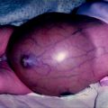
Fig. 25.1
Bimodal age distribution of ovarian torsion
Bilateral ovarian torsion is uncommon and can occur with or without associated underlying adnexal pathology. For example, in one large series of 76 children with ovarian torsion 5% suffered asynchronous bilateral torsion [4] while another large series of 114 patients reported no cases of bilateral ovarian torsion [12]. In the pediatric population, there have been 24 case reports of asynchronous torsion and only three cases of synchronous bilateral torsion [13].
Etiology
Ovarian torsion may occur in a normal ovary or may be associated with an abnormality of the ovary. The normal tube and ovary are mobile and may rotate 90° without any symptoms [14] and can easily twist 360° to 1080° [15]. It is now clear that torsion may occur in normal adnexa without an associated pathologic mass acting as the lead point for torsion. In the two largest case series of ovarian torsion, approximately half of the cases (46 and 48.8%) had no underlying ovarian abnormality [2, 4]. In the series reported by Oltmann et al. of 114 pathologically reviewed torsed specimens, there was no underlying etiology of torsion in one half of cases, but rather, normal tissue with hemorrhage [2]. Likewise, approximately half the cases of ovarian torsion reported in the KID database had no additional ovarian diagnoses [6, 12].
Anatomic Factors in Ovarian Torsion
The absence of a pathologic “lead point” in the setting of torsion suggests that anatomic variants of normal anatomy may predispose to torsion. The increased risk of torsion in the contralateral “normal” ovary in patients who suffer asynchronous torsions also suggests a common anatomic risk factor [16]. Anatomic variants that predispose to ovarian torsion include a long fallopian tube, a long utero-ovarian ligament, such as the mesovarium and mesosalpinx, or an unusually short ovarian hilum [14, 17–19]. All the variants mentioned result in increased mobility in the ovarian suspensory ligaments. A report of familial ovarian torsion with bilateral ovarian loss in the mother and unilateral ovary loss in the daughter suggests that a genetic predisposition to torsion may exist [20]. Other factors that may contribute to hypermobility of the ovary and therefore ovarian torsion are tubal spasm, abrupt changes in intra-abdominal pressure with jarring activity, and adnexal venous congestion seen with constipation, sigmoid distension, and premenarchal or gestational hormonal changes [17, 21, 22].
A predominance of right-sided adnexal torsion has been noted with a 3:2 ratio of right to left sidedness [23]. Even most cases of asynchronous torsion first present on the right side [16]. The explanation of the right side predominance of ovarian torsion may be the associated hypermobility of the cecum and ileum compared to the relative stability of the sigmoid on the left. Alternatively, there may be an ascertainment bias because of the more likely operative intervention for right sided abdominal pain because of concern for appendicitis [14, 24].
Hormonal Factors in Ovarian Torsion
As noted, there are two peaks in the incidence of ovarian torsion—one during infancy and the other around 12 years of age [2]. The bimodal age distribution reflects peak hormonal levels seen in children—the fetal exposure to maternal hormones and the early adolescent response to the maturing reproductive hormonal axis [1]. The association between age and hormonal environment suggests that hormonal effects are a risk factor for ovarian torsion. Potentially, hormonally induced ovarian cysts and adnexal congestion could serve as “lead points” for ovarian torsion.
Ovarian cysts may form in utero in response to maternal hormones and these cysts may persist into infancy, explaining the high incidence of torsion in girls less than 1 year of age [25]. Between ages 1 and 8 years, the hypothalamic-pituitary axis resets to a baseline of low estrogen and FSH levels. As a result, the ovary is quiescent and minimal ovarian cysts are formed in this age group [26]. Around puberty, between 9 and 14 years, the reproductive hormonal axis is activated with high FSH levels long before the onset of menses [27, 28]. The high and fluctuating hormone levels in this age group prime the ovaries for cyst formation. By age 15, hormonal patterns are usually well established, and FSH and LH levels are regulated by the monthly release of estrogen and progesterone by the ovary. Changes in gonadotropin release can result in functional cysts, typically either follicular or corpus luteal cysts, which enlarge throughout the menstrual cycle and then resolve within several months.
Adnexal Abnormalities
As noted, ovaries that undergo torsion in children have no associated abnormality more than 40% of the time, while 33% have underlying cysts, and 23% have benign neoplasms (see Table 25.1). These findings differ from the adults where an underlying cyst is the most common etiology of ovarian torsion [29]. Overall at least 96% of ovaries that undergo torsion in children are normal or have associated benign conditions and only 1.7–3.5% contain a malignancy. (The categorization of serous borderline cysts as benign or malignant accounts for the variance [12].) Summation of all reported pediatric case series of ovarian torsion confirmed an even lower incidence (1.4–1.9%), of an underlying malignancy with the KID database reporting the lowest incidence (0.4%) in over 1232 cases [6, 12]. This incidence of underlying malignancy is markedly lower than the 10% risk of malignancy for all pediatric ovarian masses and is comparable to the 1–2% incidence of malignancy in adults (Table 25.2) [24, 30].
Table 25.1
Patients with ovarian torsion compared to patients with ovarian masses not associated with torsion
Torsion | Nontorsion | P value | |
|---|---|---|---|
Number of cases | 114 (26%) | 324 (74%) | <0.0001 |
Mean age ± SEM | 10.28 years ± 0.53 | 13.23 ± 0.26 | <0.0001 |
Clinical presentation | |||
Abdominal mass | 6% | 23% | <0.0001 |
Abdominal pain | 83% | 56% | <0.0001 |
Prenatal diagnosis | 6% | 1% | 0.0013 |
Othera | 5% | 20% | <0.0001 |
Size by imaging (cm) | 8.1 ± 0.49 (2.9–25) | 9.8 ± 0.60 (0.9–50) | 0.077 |
WBC >12 K | 56% | 28% | 0.0004 |
Pathology | |||
Benign, normal tissue | 46 (40%) | 11 (3.4%) | <0.0001 |
Benign cyst | 38 (33%) | 146 (45%) | 0.0291 |
Benign neoplasm | 26 (24%) | 123 (38%) | 0.0067 |
Malignant neoplasm | 4 (3.5%)b | 44 (13.6%) | 0.0030 |
Table 25.2
Pathology of ovarian torsion—review of literature
Author | Cases ovarian torsion | Malignant neoplasms | Malignant neoplasm pathology | Benign neoplasms | Benign neoplasm pathology | Population |
|---|---|---|---|---|---|---|
Meyer et al. (1995) | 13 | 0 | None | 3 | Cystadenoma (1) Teratoma (2) | Pediatric |
Kokoska et al. (2000) | 51 | 3 | Dysgerminoma (1) Granulosa cell (2) | 26 | Cystadenoma (2) Teratoma (24) | Pediatric |
Cass et al. (2001) | 34 | 0 | None | 15 | Teratoma (15) | Pediatric |
Beaunoyer et al. (2004) | 76 | 0 | None | 21 | Cystadenoma (6) Teratoma (15) | Pediatric |
Anders et al. (2005) | 22 | 1 | Granulosa cell (1) | 5 | Dermoid (5) | Pediatric |
Servaes et al. (2007) | 41 | 0 | None | 11 | Cystadenoma (1) Teratoma (10) | Pediatric |
Rousseau et al. (2008) | 41 | 2 | Dysgerminoma (1) Undifferentiated adenocarcinoma (1) | 26 | Cystadenoma (11) Teratoma (15) | Pediatric |
Oltmann et al. (2010) | 114 | 4 | Dysgerminoma (1) Juvenile granulosa cell (1) Serous borderline (2) | 26 | Cystadenoma (7) Dermoid (2) Fibroma (1) Teratoma (16) | Pediatric |
Houry et al. (2001) | 87 | 1 | Mucinous borderline (1) | 30 | Cystadenoma (11) Teratoma (19) | Adult |
Cohen et al. (2003) | 102 | 0 | None | 13 | Cystadenoma (4) Dermoid (9) | Adult |
White et al. (2005) | 44 | 2 | Granulosa cell (1) Serous borderline (1) | 13 | Cystadenoma (1) Dermoid (7) Fibrothecoma (1) Fibroma (1) Leiomyoma (1) Teratoma (2) | Adult |
Göçmen et al. (2008) | 18 | 0 | None | 11 | Cystadenoma (7) Dermoid (4) | Adult |
Shadinger et al. (2008) | 39 | 0 | None | 12 | Benign Spindle (1) Brenner (1) Cystadenoma (2) Dermoid (7) Fibroma (1) | Adult and pediatric (3–61 years) |
Pediatric totals | 431 | 10 (2%) | Dysgerminoma (3) Granulosa cell (3) Juvenile granulosa cell (1) Serous borderline(2) Undifferentiated adenocarcinoma (1) | 133 (30.8%) | Cystadenoma (28) Dermoid (7) Fibroma (1) Teratoma (97) | Pediatric |
Literature totals | 682 | 13 (1.9%) | Dysgerminoma (3) Granulosa cell (4) Juvenile granulosa cell (1) Mucinous borderline (1) Serous borderline (3) Undifferentiated adenocarcinoma (1) | 212 (31.0%) | Benign Spindle (1) Brenner (1) Cystadenoma (53) Dermoid (34) Fibroma (3) Fibrothecoma (1) Leiomyoma (1) Teratoma (118) | Adult and pediatric |
These findings, based on the pathologic analysis of surgical specimens, show a dramatic, 10–20-fold lower incidence of malignancy in ovarian torsions than found in ovarian masses and clearly refute the traditionally held tenet that an underlying malignant tumor is the most common lead point of torsion. Likewise, fewer benign and malignant tumors are found in torsed specimens compared to nontorsed ovarian masses [12]. Finally, case series show that benign ovarian etiologies cause torsion more commonly than malignant tumors. For example, Sommerville et al. reported the incidence of torsion in benign tumors was 14.3%, which was greater than the 9.7% incidence for borderline tumors and far greater than 1.1% incidence for malignant tumors [31]. The lower rate of torsion seen in malignancies was attributed to their tendency to induce adherence through inflammation and local invasion and thus limit ovarian mobility.
Clinical Manifestations
Symptoms of ovarian torsion in children include lower abdominal pain in almost all cases and associated nausea and vomiting in more than two thirds of girls [1, 8, 32]. However, this classic triad of symptoms is not specific for ovarian torsion and many causes of an acute abdomen present with similar symptoms. Although nearly all children (>97%) with ovarian torsion have abdominal pain, the classic history of an acute onset of severe pain is not common and the variable severity of pain is a potential diagnostic pitfall [3, 33]. There is considerable variation in the intensity, character, location, and duration of pain in patients with ovarian torsion. Often the history of recurrent pain is due to recurrent torsion and detorsion of the adnexa rather than persistent torsion [32, 34]. Torsion in infants is often not accompanied by pain since the torsion may often occur in utero, so the most common clinical presentations of an infant with ovarian torsion are positive prenatal imaging (>50%), presence of a mass (38%), or feeding tolerance (6%) (see Table 25.1) [2].
Stay updated, free articles. Join our Telegram channel

Full access? Get Clinical Tree


