Other Sexually Transmitted Infections Including Genital Ulcers, Pediculosis, Scabies, and Molluscum


Other Sexually Transmitted Infections Including Genital Ulcers, Pediculosis, Scabies, and Molluscum


Wendi G. Ehrman

Mandakini Sadhir

M. Susan Jay


Chancroid, lymphogranuloma venereum (LGV), and granuloma inguinale constitute the classic minor ulcerative sexually transmitted infections (STIs) and should be considered in the differential diagnosis of genital ulcers (see Table 62.1). In the US, the most common causative agent of genital ulcers is the herpes simplex virus (HSV), followed by syphilis. Approximately 3% to 10% of these patients will have more than one infection. There is also an increased risk of human immunodeficiency virus (HIV) infection associated with these ulcerative infections.1 Other potentially sexually transmissible minor infections include scabies, pediculosis, and molluscum contagiosum.


CHANCROID


Etiology

Chancroid is caused by the gram-negative facultative anaerobic coccobacillus, Haemophilus ducreyi.2


Epidemiology

This disease is uncommon in the US. According to Centers for Disease Control and Prevention (CDC), chancroid in the US has fallen dramatically from 4,212 cases in 1990 to only 10 cases in 2013.3 While the reduction in cases is striking, the CDC is concerned that this may in part be related to the difficulty in culturing the causative agent. Worldwide, the highest prevalence of infection is in southern, central, and eastern Africa. In the US, 8 of the 10 cases were in the southern states of California (6), Texas (1), and Alabama (1); 6 cases were in males and 4 in females. Chancroid is a known risk factor for HIV and enhances disease transmission. Co-infection with syphilis or herpes simplex may also occur.4


Clinical Manifestations

The incubation period is generally 3 to 10 days. Classically, chancroid presents as a tender inflammatory papule on the genitalia that becomes pustular and then ulcerates in 1 to 2 days. The characteristic ulcer is painful, soft, friable, and nonindurated with ragged undermined margins, a granulomatous base, and a foul-smelling yellow or gray, necrotic purulent exudate (Fig. 62.1). Males may present with inguinal pain or ulcers located on the prepuce, coronal sulcus, or frenulum. In females, multiple lesions may be present on the vulva, clitoris, cervix, or perianal region. Females may be asymptomatic or present with dysuria, dyspareunia, vaginal discharge, pain with defecation, and rectal bleeding. Rarely, extragenital sites may be involved (breasts, thighs, fingers, and mouth5).

Painful unilateral inguinal lymphadenitis—known as a bubo—develops in as many as 50% of patients and may become suppurative, rupture, and ulcerate. Large inguinal abscesses can occur, leading to significant destruction of skin and soft tissue.5


Other Management Considerations

Clinical improvement should be seen within 3 to 7 days. Failure to improve should raise the possibility of an incorrect diagnosis or co-infection with another STI such as HIV. Large ulcers may require more than 2 weeks to resolve; fluctuant lymphadenopathy heals even more slowly. Adenopathy may progress to fluctuation despite successful therapy and does not represent treatment failure.


LYMPHOGRANULOMA VENEREUM


Etiology

LGV is caused by the obligate intracellular organism Chlamydia trachomatis. Chlamydia has 18 serovars associated with disease; serovars L1, L2, and L3 cause LGV. LGV strains can cause a systemic infection that, if untreated, can lead to colorectal fistulas and
strictures and chronic pain.6,7 These lesions can become superinfected with other STIs or pathogens.


TABLE 62.1 Differential Diagnosis of Genital Ulcers1,7


Infection


Clinical Manifestation


Diagnosis


Chancroid


Cause: H. ducreyi


  • Painful, shallow, friable, nonindurated genital ulcer with ragged undermined margins, granulomatous base, and foul-smelling yellow or gray, necrotic purulent exudate.


  • Painful inguinal adenopathy known as “buboes” present.


  • Culture of lesion (not widely available).


  • CDC criteria for “probable” diagnosis:


    1. ≥1 painful genital ulcer(s)


    2. No evidence of syphilis infection on dark-field examination or serologic test performed 7 days after onset of ulcer


    3. Typical clinical presentation


    4. Negative HSV test of ulcer


Lymphogranuloma venereum


Cause: C. trachomatis


  • Painful inguinal and/or femoral lymphadenopathy. “Groove” sign is pathognomonic.


  • Self-limited genital ulcer or papule at site of inoculation.


Genital lesion swab or lymph node aspirate tested using NAATs, immunofluorescence


Granuloma inguinale


Cause: K. granulomatis


  • Painless, slowly progressive ulcerative lesions on genitals or perineum; bleed easily on contact.


  • Regional lymphadenopathy uncommon.


Identification of Donovan bodies within histiocytes of granulation tissue smears or biopsy specimens


Syphilis


Cause: Treponema pallidum


  • Primary chancre: Painless ulcer with indurated hard raised border and “punched out” appearance.


  • Regional lymphadenopathy may occur.


  • Screen: Nontreponemal tests (RPR, VDRL)


  • Confirm: Treponemal tests (FTA-ABS or TPPA)


  • Dark-field microscopy showing spirochetes


Genital Herpes


Cause: HSV 1 and 2


  • Painful vesicular lesions developing into ulcers.


  • Constitutional symptoms present in primary infection.


Viral culture or PCR for HSV DNA


Nonsexually transmitted genital ulcers


  • Painful, well-demarcated ulcer.


  • Constitutional symptoms may be present if viral in etiology (CMV, EBV).


Negative for HSV or other STIs


RPR, Rapid plasma reagin; VDRL, venereal diseases research laboratory; FTA-ABS, Fluorescent treponemal antibody-absorbed; TPPA, Treponema pallidum particle agglutination; CMV, cytomegalovirus; EBV, Epstein—Barr virus.


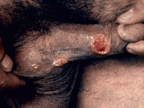
FIGURE 62.1 The lesions in chancroid are painful and more irregular than in syphilis. (From Craft N, Taylor E, Tumeh PC, et al. VisualDx: essential adult dermatology. Philadelphia, PA: Lippincott Williams & Wilkins, 2010.)


Epidemiology

LGV is endemic in parts of Africa, India, South America, and the Caribbean. Outbreaks have been reported in Europe among HIV-positive MSM.8


Clinical Manifestations

The incubation period is 3 to 30 days (usually 7 to 12). Infection occurs in three stages:


  • Primary stage: The initial lesion begins as a small, painless papule or pustule at the site of inoculation that can erode into an asymptomatic herpetiform ulcer that often heals without scarring within a week. Lesions are typically found on the penis, urethral glans, and scrotum in men and on the vulva, vaginal wall, fourchette, and cervix in women. Rectal lesions occur in both sexes from receptive anal intercourse and can be associated with diarrhea, rectal discharge, and tenesmus. Mucopurulent cervicitis and urethritis may also occur. Women usually have primary involvement of the rectum, vagina, and cervix.


  • Secondary or inguinal stage: This stage typically occurs 2 to 6 weeks after the appearance of the primary lesion and involves painful inflammation of the inguinal and femoral lymph nodes. Inguinal adenopathy is unilateral in 70% of cases and is more common in males (Fig. 62.2). The “groove” sign is the result of enlarged inguinal nodes above Poupart’s ligament and the femoral nodes below it and is considered “pathognomonic” for LGV (Fig. 62.3). Nodes can become matted and fluctuant and produce the characteristic bubo.7 Buboes may rupture in one-third of patients or develop into hard, nonsuppurative masses. Most buboes eventually heal, but some will form sinus tracts. Bubonic relapse occurs in 20% of untreated cases. Constitutional symptoms may occur with the inguinal buboes and be associated with systemic spread of chlamydia, leading to arthritis, hepatitis, and pneumonitis.


    TABLE 62.2 Treatment of Chancroid5,18


    CDC Recommended Treatments


    Other Management Considerations


    Follow-Up


    Azithromycin 1 g orally in a single dose or


    Ceftriaxone 250 mg IM in a single dose or


    Ciprofloxacin 500 mg orally twice daily × 3 d or


    Erythromycin base 500 mg orally 3 times daily × 7 d


    HIV-positive patients: May require longer or repeated treatment due to treatment failures and slow healing. Use single-dose therapies only when close follow-up assured.


    Pregnancy/lactation: Ciprofloxacin contraindicated


    Uncircumcised males: Higher treatment failure rates and slower healing especially if ulcers under foreskin


    Sex partners: Examine and treat sex partners who had sexual contact with patient in the 10 days preceding the patient’s onset of symptoms


    Within 3-7 d of start of therapy


    Weekly follow-up until resolution of lesions and symptoms


    Test for HIV at time of diagnosis and 3 mo later along with syphilis if initial test negative


    Buboes (fluctuant adenopathy): Treat by aspiration for symptomatic relief and to prevent rupture or by incision and drainage with wound packing (more definitive). Clinical resolution of fluctuant lymphadenopathy is slower than that of ulcers.


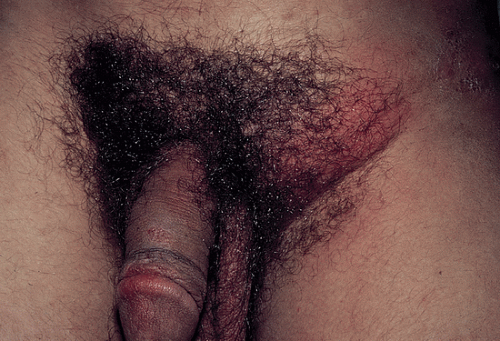
    FIGURE 62.2 Lymphogranuloma venereum. Painful inguinal lymphadenopathy in a man infected with C. trachomatis. (Image from Rubin E, Farber JL. Pathol-ogy. 3rd ed. Philadelphia, PA: Lippincott Williams & Wilkins, 1999.)


  • Tertiary or genito-anorectal syndrome (uncommon): This stage occurs more often in women who were asymptomatic during previous stages and in men who have receptive anal intercourse.9 Patients initially develop symptoms of procto-colitis (anal pruritus, rectal discharge, rectal pain, tenesmus, and fever). Subsequent manifestations include perirectal ab-scesses, rectovaginal and anorectal fistulas, rectal strictures, and rectal stenosis. Chronic untreated LGV can lead to repetitive scarring and fistulous tract formation in the genital region.
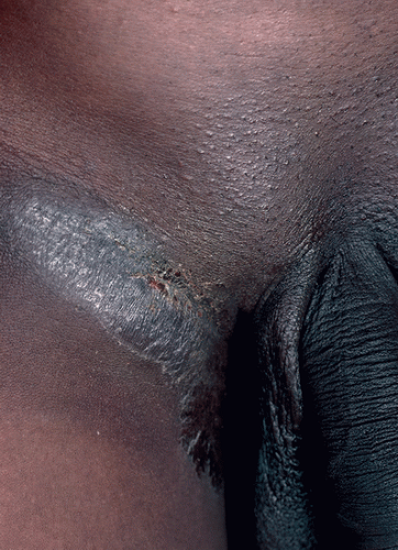


GRANULOMA INGUINALE


Etiology

Granuloma inguinale, or Donovanosis, is caused by Klebsiella granulomatis, an intracellular gram-negative bacillus.2


Epidemiology

Granuloma inguinale is considered endemic in Papua, New Guinea, southeast India, South Africa, central Australia, Brazil, and the Caribbean. It is extremely rare in the US and Western Europe.6,7 It is transmitted primarily through sexual contact; autoinoculation can also lead to spread of the disease.

Only gold members can continue reading. Log In or Register to continue

Stay updated, free articles. Join our Telegram channel

Sep 7, 2016 | Posted by in ONCOLOGY | Comments Off on Other Sexually Transmitted Infections Including Genital Ulcers, Pediculosis, Scabies, and Molluscum

Full access? Get Clinical Tree

Get Clinical Tree app for offline access