Osteomalacia

Rarer causes

These include inadequate alkaline phosphatase activity (hypophosphatasia), inhibition of mineralization by excess fluoride ingestion and abnormal bone matrix, for example in osteogenesis imperfecta or fibrogenesis imperfecta.


Box 26.1 Causes of osteomalacia
Vitamin Ddeficiency
Reduced synthesis, intake or absorption
Inadequate sunlight exposure
Malabsorption: small bowel disease (e.g. coeliac disease, inflammatory bowel disease), extensive bowel surgery, gastrectomy, pancreatic insufficiency
Reduced 25-hydroxylation of vitamin D
Liver disease
Anticonvulsants
Reduced 1α-hydroxylation
Chronic kidney disease
Hypoparathyroidism (reduced stimulation of vitamin D 1α-hydroxylation by PTH)
Vitamin D-dependent rickets type 1 (an autosomal recessive disorder caused by mutations in the gene encoding the enzyme 1α-hydroxylase)
Vitamin D-dependent rickets type 2 (an autosomal recessive disorder caused by mutations in the gene encoding the vitamin D receptor, resulting in target organ resistance to vitamin D)
Primary renal phosphate wasting
Type 2 renal tubular acidosis: Fanconi’s syndrome, multiple myeloma
Hereditary hypophosphataemic rickets (X-linked and autosomal dominant)
Oncogenic osteomalacia
Rarer causes
Abnormal bone matrix: osteogenesis imperfecta, fibrogenesis imperfecta
Hypophosphatasia (associated with periodontal disease)
Mineralization inhibitors: high doses of fluoride (e.g. in certain teas), aluminium, bisphosphonates

Clinical presentations

Osteomalacia may be asymptomatic and present radiologically as osteopenia. Clinically, patients with osteomalacia may present with diffuse bone pain and tenderness, fractures with little or no trauma (typically in the ribs, vertebrae and long bones) and proximal muscle weakness, which may be associated with a waddling gait.

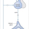
History and physical examination should look for symptoms and signs of the possible underlying cause.

Children with rickets may present with hypotonia, growth retardation and skeletal deformities.

The laying down of uncalcified osteoid at the metaphases leads to widening of the ends of the long bones. This may be seen as ‘rachitic rosary’ in the ribs (enlarged ends of the ribs resembling beads at the costochondral junction) and at the level of the ankle and the wrist. Other skeletal deformities include frontal bossing, pectus carinatum and bowing of the long bones.

Investigations

Radiographic findings

A spine X-ray

Only gold members can continue reading. Log In or Register to continue

Stay updated, free articles. Join our Telegram channel

Jun 4, 2016 | Posted by in ENDOCRINOLOGY | Comments Off on Osteomalacia

Full access? Get Clinical Tree

Get Clinical Tree app for offline access