Abstract
Objectives
To evaluate safety and early and long-term efficacy of CO 2 laser conservative treatments for nonulcerative squamous cell carcinoma (SCC) of the penis.
Material and Methods
Within our institutional database (2002–2022, included), we identified 122 consecutive cT1-2 cN0 cM0 patients with penile SCC who underwent conservative CO 2 laser treatments. Histologically confirmed local relapses were recorded. Local relapses were classified as early recurrences when occurring <2 years and as late new tumor recurrences when occurring >2 years after first laser treatment. Predictors of disease relapse were analyzed with univariable and multivariable Cox regression models (MCRMs).
Results
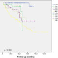
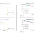
Median follow-up was 36 months [Interquartile range (IQR) 21-73 months]. Median age was 62 years (IQR: 51-69 years). Median largest lesion size was 10 mm (IQR 5-15 mm). 62 patients had penile intraepithelial neoplasia (PeIN) (51.6%), 30 had pT1 m (24.6%), 28 had pT1 (23%) and 1 had pT2 (0.8%), 2 patients were classified as pTx. In case of invasive lesions, tumor Grade was G1 in 37 (60.7%), G2 in 20 (32.8%), G3 in 4 (6.5%). Early and late recurrences occurred in 28 (22%) and in 21 patients (18%), respectively. Proportion of penis preservation was 93.4% and 92.6% at 2 and 5 years. pT1 stage [Hazard Ratio (HR) 13, Confidence Interval (CI) 1.4-73, p-value 0.02] and flat lesions (HR 7.9, CI 1.06-59, 0.04) achieved independent predictor status for late recurrences. No cancer related deaths were recorded after a median follow-up of 36 months.
Conclusions
As far as we know, this is the largest cohort of patients with penile cT1-T2 SCC who underwent conservative CO 2 laser treatment. The vast majority (92%) of these patients preserved their organ at 5 years. Some factors can be of use in preventing or anticipating late recurrences.
1
Introduction
Squamous cell carcinoma (peSCC) of the penis is a very rare entity, accounting for only 1% of male cancers [ ]. The first presentation of the disease is usually a small and superficial penile lesion. The first step in case of these lesions that does not regress is to perform a biopsy. In case of penile cancer diagnosis the subsequent treatment is driven by lesion extent and aggressiveness. Both oncological and aesthetical and functional outcomes should be taken into account in treatment planning.
With these aims, penile-sparing techniques have been proposed along the years and, according to the European Association of Urology (EAU) guidelines, penile-preserving surgery should be, nowadays, the treatment of choice whenever possible [ ]. Given that most of the primary tumors are well to moderately differentiated ( Table 1 ) and in about 80% of cases located on the glans and prepuce, penile amputation represents an over-treatment for some early stage lesions [ ]. On the other side it has to be considered that recurrence rates after penile-sparing surgery vary among the studies, ranging from 4% to 27% [ , ] . Unfortunately, due to rarity, there are no randomized controlled trials (RCTs) or at least observational comparative studies for any of the treatment options available for localized penile cancer [ ]. The aim of our study is to evaluate the oncological efficacy and the ability to preserve organ by the peniscopically controlled CO 2 laser excision of peSCC analyzing of a single high-volume center.
| Early recurrence ( N = 94): Yes | Early recurrence ( N = 27): No | Total ( N = 121) | P -value | |
|---|---|---|---|---|
| Age (Years) , Median [Interquartile range (IQR)] | 61.00 (50.00–67.50) | 64.00 (55.00–73.50) | 62.00 (51.50–69.00) | 0.090 |
| cTstage | 0.974 | |||
| – cT1 | 40 (42.6%) | 11 (40.7%) | 51 (42.1%) | |
| – cT2 | 4 (4.3%) | 1 (3.7%) | 5 (4.1%) | |
| – PEIN | 50 (53.2%) | 15 (55.6%) | 65 (53.7%) | |
| Phimosis | 0.612 | |||
| – No | 78 (82.1%) | 21 (77.8%) | 99 (81.1%) | |
| – Yes | 17 (17.9%) | 6 (22.2%) | 23 (18.9%) | |
| Glans lesion | 0.550 | |||
| – No | 18 (18.9%) | 5 (18.5%) | 23 (18.9%) | |
| – Yes | 73 (76.8%) | 22 (81.5%) | 95 (77.9%) | |
| – Undetermined | 4 (4.2%) | 0 (0.0%) | 4 (3.3%) | |
| Foreskin lesion | 0.506 | |||
| – Yes | 71 (74.7%) | 20 (74.1%) | 91 (74.6%) | |
| – No | 20 (21.1%) | 7 (25.9%) | 27 (22.1%) | |
| – Undetermined | 4 (4.2%) | 0 (0.0%) | 4 (3.3%) | |
| Size (mm), Median (IQR) | 10.00 (5.00–15.00) | 10.00 (6.00–12.50) | 10.00 (5.25–15.00) | 0.973 |
| Lesion type | 0.809 | |||
| – Bumpy | 30 (31.6%) | 7 (25.9%) | 37 (30.3%) | |
| – Plane | 43 (45.3%) | 14 (51.9%) | 57 (46.7%) | |
| – Mixed | 22 (23.2%) | 6 (22.2%) | 28 (23.0%) | |
| HPV-related | 0.255 | |||
| – No | 71 (74.7%) | 23 (85.2%) | 94 (77.0%) | |
| – Yes | 24 (25.3%) | 4 (14.8%) | 28 (23.0%) | |
| Hystotype | 0.335 | |||
| – Basaloid | 6 (6.3%) | 1 (3.7%) | 7 (5.7%) | |
| – Other | 6 (6.3%) | 4 (14.8%) | 10 (8.2%) | |
| – SCC | 83 (87.4%) | 22 (81.5%) | 105 (86.1%) | |
| Grade | 0.795 | |||
| – 1 | 27 (28.4%) | 10 (37.0%) | 37 (30.3%) | |
| – 2 | 16 (16.8%) | 3 (11.1%) | 19 (15.6%) | |
| – 3 | 3 (3.2%) | 1 (3.7%) | 4 (3.3%) | |
| – PEIN | 49 (51.6%) | 13 (48.1%) | 62 (50.8%) | |
| pT stage | 0.879 | |||
| – PEIN | 49 (52.7%) | 13 (48.1%) | 62 (51.7%) | |
| – pT1 | 21 (22.6%) | 6 (22.2%) | 27 (22.5%) | |
| – pT1m | 22 (23.7%) | 8 (29.6%) | 30 (25.0%) | |
| – pT2 | 1 (1.1%) | 0 (0.0%) | 1 (0.8%) | |
| – Unknown | 2 | 0 | 2 |
2
Patients and methods
2.1
Study population
Within our institutional database (2002–2022, included), we identified 122 consecutive cT1-2 cN0 cM0 patients with penile SCC who underwent conservative CO 2 laser treatments for nonulcerative lesions. These early stage tumors were classified according by physical examination as uniformly flat, slightly raised, or exophytic lesions. Exclusion criteria were urethral involvement > 0.5 cm beyond the meatus, regional lymph node involvement and occurrence of other malignancies. All the procedures were performed on a day-surgery setting.
2.2
Surgical technique
The technique of peniscopically controlled CO 2 laser excision is already described by our group since the early series [ ]. Briefly, a Cooper 2502 CO 2 laser instrument with a maximum output power of 35 W, CW, and a peak power of 250 W when used in pulsed mode was used in conjunction with the Zeiss OPMI-6H operating microscope. Timed emission was available for the laser instrument for continuous or pulsed waves, with exposure times of 1/30, 1/15, 118, 112 and 1 s. Particular importance was given to the accurate recognition of the anatomical landmarks of the tissue layers to ensure exact control of the depth of penetration during the peniscopically controlled procedure. Local anesthesia with a 2% solution of lidocaine was used. The anesthetic was injected by a 30-gauge needle, mounted on a syringe, under the lesion area and around the planned incision lines. Laser resection was done at an output power of 10 to 25 W and a spot size of 0.5 to 2.0 mm, depending on the used focal lengths of the microscope (200 or 300 mm.) and laser sources. Consequently, the irradiance (defined as the ratio between laser output power and target area) on the tissue varied from about 500 to 12,700 W/cm 2 . The choice of irradiance of the beam at the focal point varied according to the tissue characteristics at different anatomical sites. Irradiances of greater than 6000 W/cm 2 continuous wave were used to cut the preputial area below the sulcus level, whereas for the more delicate mucosa of the glans and perimeatal area moderate irradiances were used (between 10 and 20 × 102 W/cm 2 ). Also, in these areas pulsed generation of the beam (400 to 600 Hz) and timed emission (1/2 to 1/30 s) of the pulse train with 15 W average power were used to reduce further thermal damage at the incision borders and to control exactly the depth of penetration into the stromal tissue. Tissue removal was obtained by circumscribing the lesion at a distance of 5 mm from the visible borders with a sulcus of the desired depth. Then the tissue was grasped gently with a forceps and retracted to allow the beam to cut the base of the surgical specimen under the lesion. Wet gauze sponges were used intraoperatively to cool resected surfaces and to remove carbonized particles. The width and depth of tissue removal were related mainly to the type and site of the lesion. Whole circumferential surface resections were conducted in 2 half surfaces of the organ. Particular attention was given to overcome the deepest level of the lesion with the orthogonal incidence of the beam, for a thickness more than that of thermal damage, so that histological examination of the surgical specimen would not be affected with thin layers of thermal necrosis. Besides the coagulation, which usually occurs in real time for small vessels during laser cutting, adjunctive coagulation was obtained by defocusing the laser beam (3 to 4 mm. diameter) and using lower levels of irradiance (less than 200 W/cm 2 ).
Concerning postoperative care, an anesthetic-antibiotic cream (Chlortetracycline hydrochloride 3%) and gauze dressing were recommended on the lased area for 2 weeks. A urethral catheter was inserted and maintained for an average of 2 days only when deep perimeatal tissue was involved in the resected area. Information regarding the possible complications was given to all patients.
A follow-up consultation after 2 weeks was planned to evaluate tissue healing and discuss further treatments. Regular follow-up or additional treatments were then provided according to stage and grade.
2.3
Definition of variables for analyses
Patient and disease characteristics as well as diagnostic definition of the disease were recorded along with treatment-related complications and include: previous surgical treatments, clinical stage, size of the lesions, shape of the lesions (bumpy, flat), pathological stage, pathologic grade, histotype (usual SCC, verrucous, warty, basaloid, warty-basaloid, pseudohyperplastic), HPV-related status, nodal staging, surgical techniques, margins status, need of radicalization, recurrence, type of surgery at recurrence, time of follow-up, pathologic progression (progression to higher grade or stage), treatment progression (escalation to a more aggressive treatment: i.e. amputation such as glansectomy, partial and total penectomy), cancer specific survival (CSS), and other cause mortality (OCM) . Moreover, complete information about final pathologic reports, histologically confirmed local relapse, and distant recurrence were collected. Local recurrences were classified as early relapses when occurring <2 years and as late new tumor recurrences when occurring >2 years after first laser treatment.
2.4
Statistical analysis
Predictors of disease relapse were analyzed with univariable and multivariable Cox regression models (MCRM). Analyses were performed using the R software environment for statistical computing and graphics (version 3.6.9) [ ].
3
Results
3.1
Study population
Demographics and clinic-pathological variables of the patients are shown in Tables 1 and 2 . Median follow-up was 36 months [Interquartile range (IQR) 21-73 months]. Median age was 62 years (IQR: 51-69 years). Median lesion size was 10 mm (IQR 5-15 mm). Usual SCC (86.9%) was the most frequent histology followed by basaloid SCC (5.7%) and other SCC (7.4%). 62 patients had penile intraepithelial neoplasia (PeIN) (51.6%), 30 had pT1 m (24.6%), 28 had pT1 (23%) and 1 had pT2 (0.8%), 2 patients were classified as pTx. In case of invasive lesions, tumor grade (G) was G1 in 37 (60.7%), G2 in 20 (32.8%), G3 in 4 (6.5%).
Related posts:
 Is primary retroperitoneal lymph node dissection the way forward for patients with testicular seminoma and limited retroperitoneal metastases?
 Critical care therapy and in-hospital mortality after radical nephroureterectomy for nonmetastatic upper urinary tract carcinoma
 The clinical relevance of cut-off percentage for high-grade urothelial carcinoma within low-grade urothelial carcinoma: A determining factor?
 Cumulative smoking exposure impacts oncologic outcomes of upper tract urothelial carcinoma
Multicenter study of active surveillance for small renal masses: Real world practice pattern
Cumulative smoking exposure impacts oncologic outcomes of upper tract urothelial carcinoma
Multicenter study of active surveillance for small renal masses: Real world practice pattern
 Patient preferences for life expectancy cutoffs for aggressive treatment in clinically localized prostate cancer
Stay updated, free articles. Join our Telegram channel

Full access? Get Clinical Tree


