Oral Toxicities
Shaiba Sandhu
Brittany A. Klein
Sook Bin Woo
Nathaniel Treister
Herve Y. Sroussi
Oral mucosal toxicities are a common finding in patients treated with a range of anticancer therapies and follow several reaction patterns. Cytotoxic chemotherapy-induced mucositis (discussed in detail in Chapter 17) is characterized by irregular ulcerations and erythema, particularly of the nonkeratinized tissues (buccal mucosa, vermillion lips, ventral tongue, floor of the mouth, and soft palatal mucosa), which can interfere with oral alimentation.1
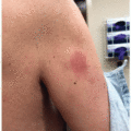
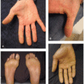
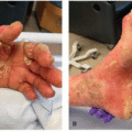
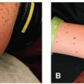
Targeted therapies induce diverse, but at times very specific oral toxicities. Similar to those induced by cytotoxic chemotherapy, irregular oral ulcerations of the nonkeratinized mucosa are observed with VEGF/VEGFR inhibitors as well as EGFR inhibitors.2 Additionally, VEGF/VEGFR inhibitors are associated with the risk of medicationrelated osteonecrosis of the jaw (MRONJ) (Figure 16.1A), although this risk is lower in comparison to bisphosphonates.2 Angiogenesis inhibitors are also implicated in the induction of benign migratory stomatitis/geographic tongue.2 Aphthous-like ulcers (single or multiple well-defined circular or ovoid ulcers with surrounding erythema) affecting primarily nonkeratinized mucosa are a well-known toxicity related to mTOR inhibitors, often referred to as mTOR inhibitor-associated stomatitis, and are also seen in patients treated with PI3K inhibitors and FLT3 inhibitors1,3,4 (Figure 16.1B and C). In patients treated with BRAF inhibitors, asymptomatic focal hyperkeratotic lesions, sometimes with a verrucous/papillomatous appearance (Figure 16.1D), have been observed involving both the keratinized and nonkeratinized oral mucosa.5 Blue-gray hyperpigmentation of the hard-palatal mucosa has been described with first-generation BCR-Abl inhibitor, imatinib5 (Figure 16.1E).
Related posts:

Stay updated, free articles. Join our Telegram channel

Full access? Get Clinical Tree


