Oral Manifestations and Complications of Cancer Therapy


Oral Manifestations and Complications of Cancer Therapy


Eliezer Soto

Jane M. Fall-Dickson

Ann M. Berger


The effectiveness of cancer therapies designed to improve cure rates and/or to extend survival time, including chemotherapy (CT), radiation therapy (RT), and conditioning regimens used in the hematopoietic stem cell transplantation (HSCT) setting, is tempered by side effects that may become intolerable and/or life-threatening. Oral complications are one such side effect and include CT- and RT-related oral mucositis and associated oropharyngeal pain, xerostomia, and oral infection, and oral chronic graft-versus-host disease (cGVHD). The pathogenesis of and management strategies for these oral complications, and future clinical research directions, are presented in this chapter.


ORAL MUCOSITIS

Oral mucositis is an inflammation of the mucous membranes of the oral cavity and oropharynx characterized by tissue erythema, edema, and atrophy, often progressing to ulceration (1). The clinical significance of CT- and RT-related oral mucositis as a dose- and treatment-limiting side effect is well appreciated (2). It is a painful and debilitating side effect that not only interferes with further treatment options but also causes a significant impairment in patient’s quality of life and functional status (3,4).

The frequency and severity of oral mucositis are influenced by numerous patient- and treatment-related risk factors (Table 19.1) (5,6). Risk factors for CT-related oral mucositis are complex, and study results have been conflicting throughout the years. For example, although younger patients are considered at increased risk for oral mucositis, and women have been reported to have more severe oral mucositis more frequently than men, Driezen (7) reported no age or gender risk factors for development of this oral condition. Children are three times more likely than adults to develop oral mucositis because of a higher proliferating fraction of basal cells. Results from a sample of 332 ambulatory CT patients showed no significant differences in CT-induced oral mucositis incidence between outpatients who wore dental appliances, previously had oral lesions, used diverse oral hygiene and oral care practices, and had a history of smoking and those patients who did not (6). Conflicting study results may be related to lack of defined risk factors for subjects entering clinical trials (5).

Although the full spectrum of treatment-related risk factors for oral mucositis is not defined, known risk factors include continuous CT infusion therapy for breast and colon cancer (5-fluorouracil [5-FU] and leucovorin); administration of selected anthracyclines, alkylating agents, taxanes, vinca alkaloids, antimetabolites, and antitumor antibiotics; myeloablative conditioning regimens for HSCT (e.g., high-dose melphalan or carmustine, etoposide, cytarabine, and melphalan [BEAM]) (8,9,10); and RT to the head and neck. Individual drug metabolism affects oral mucositis incidence and severity, as seen with patients who are unable to adequately metabolize certain CT.


CT-INDUCED ORAL MUCOSITIS

Approximately 40% of CT patients develop oral mucositis (11), and approximately half of these patients develop severe, painful lesions requiring parenteral analgesia that may lead to treatment modification (12). Higher oral mucositis incidence rates of 60% are seen in the HSCT setting, with reported incidence rates of up to 78% for ulcerative oral mucositis (13). Oral mucositis is also a risk factor for infections, which may be life-threatening in neutropenic patients. Oral infections, such as herpes simplex virus (HSV) in particular, may increase oral mucositis severity. There is a four times greater relative risk of septicemia in patients with oral mucositis and oral infections when compared with patients without oral mucositis. This greater risk is due to mucosal barrier injury, which allows pathogen entry into the peripheral circulation.

The relationship between severe oral mucositis and clinical outcomes in patients receiving conditioning CT for HSCT has been analyzed in several studies (14,16). McCann et al. (15) performed an observational study in 197 patients with multiple myeloma (MM) or non-Hodgkin’s lymphoma (NHL) undergoing either high-dose melphalan or BEAM CT, respectively. Parameters such as duration of pain score >4, opioid use, dysphagia score >4, total parenteral nutrition (TPN) use, incidence and/or duration of fever and infection, and duration of antibiotic use increased gradiently with maximum grade of oral mucositis. The presence of severe oral mucositis (World Health Organization [WHO] grades 3 and 4) increased the duration of TPN use by 2.7 days, opioids by 4.6 days, and antibiotics by 2.4 days, prolonging hospital stay by 2.3 days in MM patients, but not in NHL patients.

Oral mucositis presents with asymptomatic erythema and progresses from solitary, white, elevated desquamative patches that are slightly painful to large, contiguous, pseudomembranous, painful lesions. Histopathologically, edema of the rete pegs and vascular changes are observed. Typical oral sequelae of CT agents include epithelial hyperplasia,
collagen and glandular degeneration and epithelial dysplasia, atrophy, and localized or diffuse mucosal ulceration. Nonkeratinized mucosal areas are most affected, including the labial, buccal, and soft palate mucosa; the floor of the mouth; and the ventral surface of the tongue. The loss of basement membrane epithelial cells exposes the underlying connective, innervated tissue stroma, which contributes to more severe oropharyngeal pain.


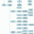
TABLE 19.1 Cancer treatment- and patient-related risk factors for oral mucositis


Patient-Related


Age older than 65 y or younger than 20 y


Gender


Inadequate oral health and hygiene practices


Periodontal diseases


Microbial flora


Chronic low-grade mouth infections


Salivary gland secretory dysfunction


Herpes simplex virus infection


Inborn inability to metabolize chemotherapeutic agents effectively


Inadequate nutritional status


Exposure to oral stressors, including alcohol and smoking


Ill-fitting dental prostheses


Treatment-Related


Radiation therapy: dose; schedule


Chemotherapy: agent; dose; schedule


Myelosuppression


Neutropenia


Immunosuppression


Reduced secretory immunoglobulin A


Inadequate oral care during treatment


Infections of bacterial, viral, fungal origin


Use of antidepressants, opiates, antihypertensives, antihistamines, diuretics, and sedatives


Impairment of renal and/or hepatic function


Protein or calorie malnutrition and dehydration


Xerostomia


Source: Adapted from Barasch A, Peterson DE. Risk factors for ulcerative mucositis in cancer patients: unanswered questions. Oral Oncol. 2003;39:91-100; Dodd MJ, Miaskowski C, Shiba GH, et al. Risk factors for CT-induced oral mucositis: dental appliances, oral hygiene, previous oral lesion, and a history of smoking. Cancer Invest. 1999;17:278-284.


RADIATION-INDUCED STOMATITIS

Oral mucositis is virtually universal when RT targets the oropharyngeal area, with the severity dependent on the type of ionizing radiation, volume of irradiated tissue, daily and cumulative dose, and duration of RT. Oral mucositis is a dose- and rate-limiting toxicity of RT for head and neck cancer and of hyperfractionated RT and CT. Radiation interacts directly with DNA, leading to chromosome and cellular mitotic apparatus damage. Atrophic changes in the oral epithelium usually occur at total doses of 160 to 220 Gy, administered at a rate of 20 Gy/d (17). Doses higher than 600 Gy or concomitant CT place the patient at risk for permanent salivary gland changes (17,18). The addition of total-body irradiation to the HSCT treatment regimen increases oral mucositis severity through both direct mucosal damage and xerostomia.

CT-induced dental effects occur when the glands are within the treatment field and depend more on these effects
than on direct irradiation of the teeth. Teeth in the irradiated field may be desensitized, leading to asymptomatic early caries. Therefore, daily fluoride application is necessary. Health care costs associated with oral mucositis in head and neck cancer patients are significant (19,20). In a prospective, longitudinal, multicenter study with 75 patients with head and neck cancer receiving RT with or without CT (20), 76% reported severe mouth and throat pain necessitating an increased number of visits to health care providers, 51% needed a feeding tube, and 37% were hospitalized with a mean stay of 4.9 days. These complications are directly correlated with a significant increase in resource use and excess costs.


RT-RELATED COMPLICATIONS

Long-term effects of head and neck RT include soft tissue fibrosis, obliterative endoarteritis, trismus, nonhealing or slow-healing mucosal ulcerations, and slow-healing dental extraction sites. RT-induced fibrotic changes may occur in the masticatory muscles and/or the temporal mandibular joint up to 1 year post-therapy, becoming more serious over time. Early phases of fibrogenesis following RT may be viewed through a wound healing model that is characterized by upregulation of tumor necrosis factor-alpha (TNFα) and other proinflammatory cytokines (21). However, as this radiation fibrogenic process continues over time, it functions as a nonhealing wound (21).

Osteoradionecrosis (ORN) is a relatively uncommon condition related to hypocellularity, hypovascularity, and tissue ischemia. Higher incidences are seen after total doses to the bone exceed 65 Gy (22). ORN is usually related to trauma such as dental extraction and may lead to pathologic fracture, infection of surrounding soft tissues, and severe pain. Most studies have reported ORN following tooth extractions that were not timed to allow adequate extraction site healing for 10 to 14 days before the start of RT. Osteonecrosis of the jaw bone has been strongly associated with the use of bisphosphonate therapy that is prescribed to treat malignancy-related hypercalcemia, bone metastasis, and metabolic bone diseases (23,24).

Oral candidiasis is a common acute and long-term oral sequela of head and neck RT. These candida lesions frequently present as angular cheilitis and may appear as white and removable, chronic hyperplastic (nonremovable) or chronic erythematous (diffuse patchy erythema).


CHRONIC GVHD ORAL MANIFESTATIONS

Patients who have undergone allogeneic HSCT frequently develop GVHD, an alloimmune condition derived from an immune attack mediated by donor T cells recognizing antigens expressed on normal tissues. GVHD occurs following allogeneic HSCT because of disparities in minor histocompatibility antigens between donor and recipient, inherited independently of human leukocyte antigen genes (25). Acute GVHD has been classified historically as beginning within the first 100 days after allogeneic HSCT. Chronic GVHD may begin as early as 70 days or as late as 15 months after allogeneic transplant. Increased incidence of cGVHD may be related to the changing patterns of allogeneic HSCT.

The importance of oral cGVHD was recognized by the National Institutes of Health (NIH) Consensus Development Project on Criteria for Clinical Trials in cGVHD (26). Consensus documents have been published, including response criteria guidelines to measure clinical progression over time. The Schubert Oral Mucositis Rating Scale (OMRS) was validated under the auspices of this NIH Consensus Development Project (26). Treister et al. (27) analyzed inter-and intraobserver variability in the component and composite scores using the NIH oral cGVHD Activity Assessment Instrument. Twenty-four clinicians from six major HSCT centers scored high-quality intraoral photographs of 12 patients, followed by a second evaluation 1 week later. Although mean interrater reliability was poor to moderate and unacceptable for the clinical trial setting, greater concordance among the oral medicine experts, high intrarater reliability, and participant feedback suggest that formal training may decrease variability.

Approximately 80% of patients with extensive cGVHD have some type of oral involvement (28) that is a major contributing factor to the morbidity seen with allogeneic HSCT. Although oral lesions are most common in patients with extensive cGVHD, patients may also present with limited disease involving only the oral cavity. Oral cGVHD presents with tissue atrophy and erythema, lichenoid changes (hyperkeratotic striae, patches, plaques, and papules), and pseudomembranous ulcerations occurring typically on buccal and labial mucosa and the lateral tongue, angular stomatitis, and xerostomia (28). Treister et al. (29) correlated the distribution, type, and extent of lesions with patient-reported pain and discomfort. Almost all (93%) of ulcerations, 72% of erythematous lesions, and 76% of reticular lesions occurred on the buccal and labial mucosa and tongue. Ulcerations in the soft palate were uncommon and associated with increased pain. There was a statistically significant inverse relationship between the overall presence of ulceration and time since HSCT. Functional impact was significantly observed as restriction of oral intake due to discomfort. Decreased oral intake related to oral pain leads to serious clinical problems of weight loss and malnutrition.

Although oral cGHVD is one of the major long-term complications after allogenic HSCT, little is known about its pathogenesis. Imanguli et al. (30) have proposed a new pathogenic model of cGVHD in which production of type I interferon by plasmacytoid dendritic cells plays a central role in the initiation and continuation of cGVHD. Fall-Dickson et al. (31) have analyzed the relationship among clinical characteristics of oral cGVHD and related oral pain and dryness, salivary proinflammatory cytokine interleukin 6 (IL-6) and IL-Iα concentrations, and health-related quality of life. Salivary IL-6 was associated with oral cGVHD severity, oral
ulceration, and erythema, suggesting its use as a potential biomarker of active oral cGVHD.


STRATEGIES FOR PREVENTION AND TREATMENT OF ORAL COMPLICATIONS


Pretherapy Dental Evaluation and Intervention

Oral/dental stabilization prior to CT and RT is critically important to avoid serious sequelae and requires an experienced dental team and informed patients working together to provide adequate cleaning, eliminate sites of oral infection and trauma, and promote appropriate oral hygiene (32). Many health care institution-specific policies and preventive approaches exist for oral care for CT and RT patients.

Patients scheduled for CT and/or head and neck RT should receive dental screening at least 2 weeks before therapy starts to allow for proper healing of extraction sites, recovery of soft tissue manipulations, and restoration of teeth. These activities promote optimal mucosal health before, during, and following cancer treatment. Oral hygiene is one of the most important screening areas for all patients, regardless of the type of cancer treatment modality. The initial dental appointment includes examination of the patient’s dentition for carious lesions and defective restorations that may irritate the oral mucosa and necessitate replacement. The periodontium and pulp vitality must be evaluated. Periodontal status assessment includes measurement of pocket depth and assessment of furcation involvement. Denture fit assessment avoids ill-fitting dentures that may cause irritation of irradiated tissue and potential ulceration to underlying bone (33).

A panoramic radiograph combined with intraoral radiographs as needed is necessary to detect periodontal disease, periapical infections, cyst, third-molar pathology, unerupted or partially erupted teeth, and residual root tips. Significant oral/dental problems that should be addressed before cancer treatment begins include inappropriate oral hygiene, periapical pathology, third-molar pathology, periodontal disease, defective restorations, dental caries, orthodontic appliances, and ill-fitting prostheses. Bacterial load should be reduced prior to cancer treatment via root planning, scaling, and prophylaxis, excluding visible tumor located at the site of anticipated dental manipulation. Comprehensive evaluation also includes assessment of the oral mucosa and the alveolar process to prepare for possible future prosthetic intervention and to assess for ulcerations, fibromas, irritation, hyperplasia, bony spicules, and tori. The decision to extract asymptomatic teeth prior to the commencement of RT is related to several important factors, including radiation exposure, type, portal field, fractionization, and total dosage in addition to tumor prognosis, and expediency of control of the cancer (34). Lack of patient motivation regarding appropriate oral hygiene practices should lead to a decision to extract questionable teeth prior to RT. Teeth that are class II or III mobility without use as abutment teeth for prosthetic retention should also be considered for extraction before RT. Extractions of residual root tips and impacted teeth should be performed atraumatically. Alveolectomy and primary wound closure eliminate sharp ridges and bone spicules that could project to the overlying soft tissues. This is important for prosthetic consideration because negligible bone remodeling is predicted after RT.

Communication between the dentist, patient, and radiation therapist is important for successful healthy maintenance of the oral cavity. Patients are very susceptible to dental caries at the cervical areas of all teeth after RT to the head and neck region. Therefore, patient education is important regarding effective daily plaque removal through use of floss, a soft toothbrush, and fluoridated toothpaste at least three to four times a day. Patient and family education and counseling within the context of patient motivation are necessary to promote successful outcomes of preventive strategies. Patients often receive their cancer treatment in the ambulatory setting and need specific written instructions for appropriate use of oral care agents and instruments for effective daily plaque removal, use of prescribed fluoride treatments, and reportable oral observations and symptoms.


Assessment of the Oral Mucosa

Consistent and frequent oral cavity assessment is needed to assess clinical signs before, during, and after the treatment time course. An adequately intense white light is needed to visualize all soft and hard tissues and dentition. All assessors should have an appropriate knowledge base regarding clinical signs and symptoms of oral complications and the predicted negative sequelae. No standard grading system for severity of oral complications of cancer treatment exists. Numerous available oral complications’ grading tools are based on two or more clinical parameters combined with functional status, such as eating ability. One commonly used tool is the National Cancer Institute Common Terminology Criteria for Adverse Events v3.0, which includes both descriptive terminology and a severity grading scale for each reportable adverse event (35). Other frequently used oral mucosal assessment tools are discussed in the subsequent text.


Oral Assessment Guide

The Oral Assessment Guide (OAG) (36) is a concise clinical tool to record oral cavity changes related to cancer therapy using eight assessment categories (voice, swallowing, lips, tongue, saliva, mucous membranes, gingiva, and teeth/dentures), each rated on three levels of descriptors: 1 = normal findings; 2 = mild alterations; and 3 = definitely compromised. The overall oral assessment score is the summation of the subscale score with a possible range of 8 to 24. Content-related validity, construct validity, clinical utility, and a high, trained nurse-nurse interrater reliability (r = 0.912) have been reported (36). The OAG has been used frequently to assess the efficacy of oral care protocols, to compare methods designed to determine the nature and prevalence of stomatitis, and to describe the incidence and severity of stomatitis.


World Health Organization Index

The WHO Index gives an overall rating of stomatitis and has frequently been used as a general comparison index to other oral assessment scales (37). The WHO Index is scaled as follows: grade 0 = no change; grade 1 = soreness, erythema; grade 2 = erythema, ulcers, can eat solids; and grade 3 = ulcers, requires liquid diet only; and grade 4 = alimentation not possible. Limitations of this instrument are the lack of reliability and validity data and also the tool’s inability to capture the variety of oral changes that are observed with cancer treatment (37).


Oral Mucositis Rating Scale

The OMRS was developed as “… a research tool for the comprehensive measurement of a broad range of oral tissue changes associated with cancer therapy” (38). The OMRS was originally tested in 60 patients who were 180 to 500 days post peripheral blood stem cell transplantation (PBSCT), to examine the relationship between oral abnormalities and cGVHD (38). Findings demonstrated that oral manifestations and related sequelae most strongly associated with cGVHD included atrophy and erythema, lichenoid lesions located on the buccal and labial mucosa, and oral pain.

The item pool consists of 91 items covering 13 areas of the mouth that are assessed for several types of changes in 7 anatomic areas: lips; labial and buccal mucosa; tongue; floor of mouth; palate; and attached gingiva. Each site is divided into upper and lower (lips and labial mucosa), right and left (buccal mucosa), dorsal, ventral, and lateral (tongue), and hard and soft (palate). Descriptive categories are atrophy, pseudomembrane, erythema, hyperkeratosis, lichenoid, ulceration, and edema. Erythema, atrophy, hyperkeratosis, lichenoid, and edema are scored using scales of 0 to 3 (0 = normal/no change, 1 = mild change, 2 = moderate change, and 3 = severe change). Ulceration and pseudomembrane are rated by estimated surface area involved (0 = none, 1 = >0 but ≤1 cm2, 2 = >1 cm2 but ≤2 cm2, and 3 = >2 cm2). The total possible score is the sum of all item scores with a possible range of 0 to 273. The OMRS has shown clinical and research utility (38).


Oral Mucositis Index

The Oral Mucositis Index (OMI) was developed from the finalized OMRS. A downsized 20-item version of the OMI (OMI-20) was developed and validated through a consensus panel of bone marrow transplant (BMT) oral complication specialists in the United States (39). The OMI-20 consists of nine items measuring erythema, nine items measuring ulceration, one atrophy item, and one edema item; all scored from 0 = none to 3 = severe, summed for a possible range of 0 to 60. The two sets of nine items measuring erythema and ulceration may be summed to produce subscale scores ranging from 0 to 27. The OMI-20 has demonstrated internal consistency, test-retest, and inter-rater reliability through evaluation in a sample of 133 adult PBSCT/BMT patients (39).


Oral Mucositis Assessment Scale

The Oral Mucositis Assessment Scale (OMAS) was developed as a scoring system for evaluating the anatomic extent and severity of stomatitis in clinical research studies by a team of oral medicine specialists, dentists, dental hygienists, oncologists, and oncology nurses from the United States, Canada, and Europe (40,41). Oral cavity regions assessed are lip (upper and lower), cheek (right and left), right and lateral tongue, left ventral and lateral tongue, floor of mouth, soft palate/fauces, and hard palate (40). Erythema is rated on a scale of 0 to 2 (0 = none, 1 = not severe, and 2 = severe) and ulceration/pseudomembrane is a combined category rated on scores based on the estimated surface area involved (0 = no lesion, 1 = <1 cm2, 2 = 1 to 3 cm2, and 3 = >3 cm2) and summed giving a possible score range of 0 to 45 (34,42). Validity and reliability have been demonstrated for the OMAS through clinical research studies (41,43).


Treatment Strategies

The optimal treatment strategies for oral complications and related sequelae are unknown. Treatment strategies for oral mucositis and related oropharyngeal pain are mainly empirical, and testing is needed in the randomized controlled clinical trial setting. Zlotolow and Berger (44) presented a comprehensive review of clinical research regarding treatment strategies for oral complications of cancer strategies. Conflicting study results may be related to inappropriate design issues, use of limited oral assessment instruments unable to capture variations in oral cavity changes, and incorrect timing and dose of interventions. The only standard forms of care are pretreatment oral/dental stabilization, saline mouthwashes, and oropharyngeal pain management (45).

The need for standardized treatment for oral mucositis was appreciated by the Mucositis Study Section of the Multinational Association of Supportive Care in Cancer and the International Society for Oral Oncology through their formulation of the “Updated Clinical Practice Guidelines for the Prevention and Treatment of Mucositis” (46). The original guidelines published in 2004 were based on a comprehensive review of more than 8,000 English language publications (1966 to 2001). The most recent guidelines published in 2007 included 622 articles (2002 to 2005) (Table 19.2). Publications regarding alimentary tract mucositis were rated using criteria for level of evidence and quality of research design (47).

A standardized approach for the prevention and treatment of CT- and RT-induced oral mucositis is essential. The prophylactic measures usually used for the prevention of oral mucositis include chlorhexidine gluconate, ice-cold water, saline rinses, sodium bicarbonate rinses, acyclovir, and amphotericin B. Regimens used commonly for the treatment of oral mucositis and related pain include a local anesthetic such as lidocaine or dyclonine hydrochloride, magnesium-based antacids, diphenhydramine hydrochloride, nystatin, or sucralfate. These agents are used either alone or in various combinations as a mouthwash formulation. Oral and parenteral opiates are used to relieve oral mucositis-related pain.


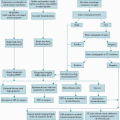
TABLE 19.2 Guidelines for the care of patients with oral mucositis


Foundations of Care


A multidisciplinary development and evaluation of oral care protocols and patient and staff education in the use of such protocols are needed to reduce the severity of oral mucositis from chemotherapy and/or radiation therapy. As part of the protocol, it would be necessary to use a soft toothbrush that is replaced on a regular basis. Elements of good clinical practice should include the use of validated tools to regularly assess oral pain and oral cavity health. The inclusion of dental professionals is vital throughout the treatment and follow-up phases.


Patient-controlled analgesia with morphine is recommended as the treatment of choice for oral mucositis-related pain in patients undergoing HSCT. Regular oral pain assessment using validated instruments for self-reporting is essential.


Radiation Therapy—Prevention


Sucralfate should not be used for the prevention of radiation-induced oral mucositis.


Antimicrobial lozenges should not be used for the prevention of radiation-induced oral mucositis.


The use of midline radiation blocks and three-dimensional radiation treatment was recommended to reduce mucosal injury.a


Benzydamine was recommended for the prevention of radiation-induced mucositis in patients with head and neck cancer receiving moderate-dose radiation therapy.a


Chlorhexidine was not recommended to prevent oral mucositis in patients with solid tumors of the head and neck who are undergoing radiotherapy.a


Standard-Dose Chemotherapy Prevention


Patients receiving bolus 5-FU chemotherapy should undergo 30 min of oral cryotherapy to prevent oral mucositis.a


Acyclovir and its analogs should not be used routinely to prevent mucositis.a


Standard-Dose Chemotherapy Treatment


Chlorhexidine should not be used to treat established oral mucositis.a


High-Dose Chemotherapy with or without Total-Body Irradiation Plus Hematopoietic Cell Transplantation Prevention


In patients with hematological malignancies receiving high-dose chemotherapy and total-body irradiation with autologous stem cell transplant, the panel recommends the use of keratinocyte growth factor-1 (Palifermin) in a dose of 60 µg/kg/d for 3 d prior to conditioning treatment and for 3 d posttransplant for the prevention of oral mucositis.


Cryotherapy should be used to prevent oral mucositis in patients receiving high-dose melphalan.


The use of pentoxifylline was not recommended to prevent mucositis in patients undergoing HSCT.a


Granulocyte-macrophage colony-stimulating factor mouthwashes should not be used for the prevention of oral mucositis in patients undergoing HSCT.


Low-level laser therapy requires expensive equipment and specialized training. Because of interoperator variability, clinical trials are difficult to conduct, and their results are difficult to compare. However, for centers able to support the necessary technology and training, laser therapy should be used to attempt to reduce the incidence of oral mucositis and its associated pain in patients receiving high-dose chemotherapy or chemoradiotherapy before HSCT.a


HSCT, hematopoietic stem cell transplantation; 5-FU, 5-fluorouracil.


a No change from previous guidelines.


Source: Adapted from Keefe DM, Schubert MM, Elting LS, et al. Mucositis Study Section of the Multinational Association of Supportive Care in Cancer and the International Society for Oral Oncology. Updated clinical practice guidelines for the prevention and treatment of mucositis. Cancer. 2007;109:820-831.


BIOLOGICAL RESPONSE MODIFIERS


Epidermal Growth Factors

Studies on epidermal growth factor (EGF) as a potential treatment option for CT- and RT-induced oral mucositis have reported conflicting data. EGF may function as a marker of mucosal damage and could potentially facilitate the healing process (48). In a phase I trial conducted by Girdler et al. (49), EGF mouthwash was used by patients treated with CT, who had a delayed onset and reduction in severity of recurrent ulcerations. However, no statistically significantly difference was seen in resolution of established ulcers. A recent double-blind, placebo-controlled,
prospective phase II study reported a potential benefit from EGF oral spray in the management of oral mucositis in patients undergoing RT for head and neck cancer. In this study, 113 subjects were randomized into one of four arms: EGF treatment groups (10-, 50-, and 100-µg/mL doses twice) and placebo. The 50-µg/mL dose was the most efficacious for the treatment of oral mucositis (50). Further randomized controlled trials are needed to confirm these results.


Hematopoietic Growth Factors

Hematologic growth factors are currently the standard treatment for patients who are treated with high-dose CT because of their well-established efficacy to decrease the duration of CT-induced neutropenia. In vitro studies have demonstrated that EGF is present in saliva and has the ability to affect growth, cell migration, and repair mechanisms (51). The development of increased oral toxicity or mucosal repair may be dependent on the timing of EGF administration in relation to CT treatment (52). Gabrilove et al. (53) reported from a sample of 27 patients with bladder cancer who received escalating doses of granulocyte colony-stimulating factor (G-CSF) during treatment with methotrexate, vinblastine, doxorubicin, and cisplatin. The patients received the G-CSF during the first of two cycles of CT. Although significantly less oral mucositis was seen during the first cycle with G-CSF, the positive results may have been biased related to possible cumulative chemotherapeutic toxicity with resultant increase in oral mucositis severity. Conversely, Bronchud et al. (54) reported from a study of 17 patients with breast or ovarian carcinoma treated with escalating doses of doxorubicin with G-CSF that G-CSF did not prevent severe oral mucositis. A third study was conducted comparing clinical outcomes in a sample of 55 adult patients who received CT for NHL and G-CSF with clinical outcomes in 39 patients who received CT alone. Patients who did not receive G-CSF had neutropenia as the primary cause of treatment delay, when compared with those patients who received G-CSF and experienced oral mucositis as the main cause of treatment delay (55). Granulocyte-macrophage colony-stimulating factor (GM-CSF) has demonstrated conflicting results in patients receiving diverse cancer treatments (56,57,58,59). A randomized controlled phase III trial conducted by Masucci et al. (57) analyzed the efficacy of GM-CSF in head and neck cancer patients with RT-induced oral mucositis. A significant reduction in oral mucositis severity was observed in the GM-CSF treatment group. Conversely, results from a Radiation Therapy Oncology Group-sponsored doubleblind, placebo-controlled, randomized study (N = 121) to analyze efficacy and safety of GM-CSF for reducing severity and duration of oral mucositis and related pain in head and neck cancer patients receiving RT (59) showed that GM-CSF had no significant effect on the severity or duration of oral mucositis. The use of CSFs in the treatment of oral mucositis remains investigational.

Only gold members can continue reading. Log In or Register to continue

Stay updated, free articles. Join our Telegram channel

Aug 25, 2016 | Posted by in ONCOLOGY | Comments Off on Oral Manifestations and Complications of Cancer Therapy

Full access? Get Clinical Tree

Get Clinical Tree app for offline access