Oral Conditions


Oral Conditions


Jude A. Fabiano


CLINICAL PEARLS


  • Oral health is intimately associated with general health and well-being.


  • Poor oral health negatively affects nutrition and systemic health.


  • Dental caries and periodontal disease are preventable.


  • Xerostomia can have devastating effects on the health of the hard and soft tissues of the oral cavity.


  • Root surface dental decay, often a secondary effect of xerostomia, has an increased incidence in older adults.


  • Medications with xerostomic side effects should be avoided when possible.


  • Diagnosis of orofacial conditions in elderly persons may be confounded by coincident medical and dental conditions, multiple medications, and vague histories.


  • The 5-year survival rate of oral cancer has not improved over the past 30 years. Early diagnosis is crucial.


  • Proper oral hygiene in older adults prevents several life-threatening systemic conditions.


  • Follow current recommendations regarding antibiotic prophylaxis and anticoagulant therapy as they relate to dental procedures.

Good oral health has emerged as a critical factor in maintaining general health in geriatric individuals. The oral cavity provides an entrance to the body for every nutrient necessary for life except oxygen. Over the past several decades, the number of older adults who have retained some or all of their natural teeth has dramatically increased. While this has resulted in improved masticatory function and self-image, the risk for acute and chronic oral disease persists later in life. Periodontal disease, dental caries, root surface caries, infections, oral cancer, malocclusion, missing
teeth, and weakness of the orofacial musculature can all inhibit the intake of nutrients and impact the general health of an individual. Compromised oral health has been linked to cardiovascular and cerebrovascular disease, pneumonia, and diabetes. In addition, feelings of social well-being and self-image, quality of life, life satisfaction, and psychological well-being are directly related to an individual’s oral health.

Oral health problems are among the most common chronic conditions found in older people. While only 35% of patients 75 years and older visit a dentist annually, almost 90% of this group see physicians. It is critical, therefore, that physicians be familiar with oral pathology, perform thorough intraoral examinations on their elderly patients and be prepared to manage and/or refer patients for definitive treatment.

This chapter provides basic knowledge of the oral conditions that affect the systemic health and quality of life of older people, and of oral findings related to systemic conditions found in these individuals.


EVALUATION OF THE ORAL CAVITY

Key to identifying and assessing oral conditions is the ability to perform a comprehensive hard and soft tissue oral examination. A complete head and neck examination should precede the intraoral examination, including physical inspection of the head, facial form, skin, eyes, ears, nose, temporomandibular joint, neck, thyroid gland, and cranial nerves. Intraoral physical examination should then proceed as follows:


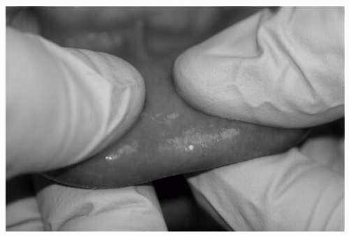
  • Lips. Pale pink, homogeneous in color, well-defined border with skin. Bidigital palpation performed to identify uniform submucosal consistency and thickness (see Figs. 35.1 and 35.2).

    Common Abnormalities: Ulcerations, irregular surface, white thickenings, recurrent herpetic lesions.


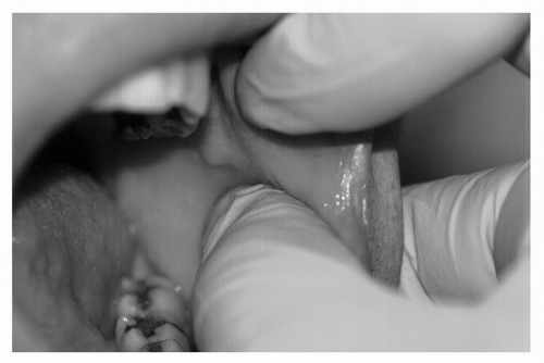
  • Buccal Mucosae. Uniformly pink/red in color; visualize Stensen duct and check for normal salivary flow. Bidigital palpation to rule out submucosal thickenings or tumors (see Fig. 35.3).

    Common Abnormalities: Biteline hyperkeratosis, fibromas, candidiasis.


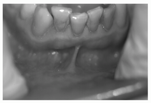
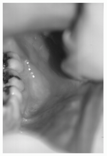
  • Buccal Vestibule. Located at the junction of buccal mucosae and alveolar process. Examine for elevations or depressions. Palpate at height/depth of vestibule to identify tenderness or swelling (see Figs. 35.4 and 35.5).

    Common Abnormalities: Inflammatory lesions associated with dental abscesses.


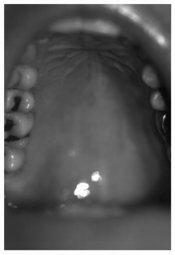
  • Hard Palate. Utilize direct visual inspection. Uniform pink color. Evaluate rugae, palatal raphe, palatine papilla, and maxillary tuberosities (see Fig. 35.6).

    Common Abnormalities: Maxillary torus, candidiasis, papillary hyperplasia.


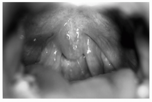
  • Soft Palate:. Utilize direct vision/mouth mirror; depress tongue if necessary. “Ah” for elevation, which should be
    bilaterally uniform. Evaluate uvula for size, color, and texture (see Fig. 35.7).

    Common Abnormalities: Candidiasis, swelling, ulcerations, nicotine stomatitis.


  • Oropharynx. Depress tongue and have patient say “ah.” Evaluate tonsils (usually atrophic in elders) and posterior wall of pharynx (see Fig. 35.8).

    Common Abnormalities: Erythema, exudate, asymmetry.


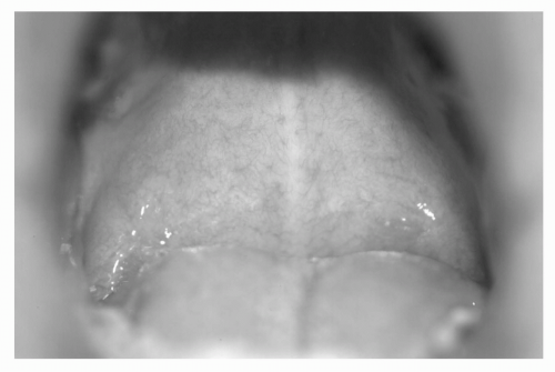
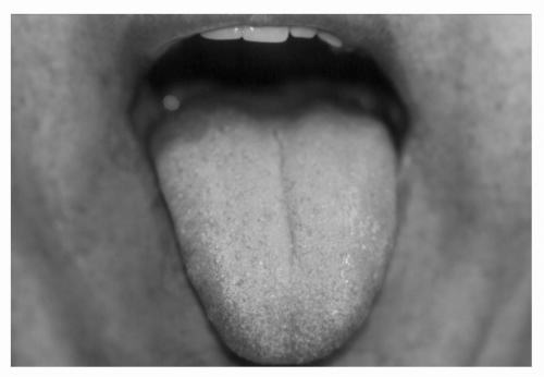
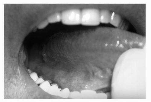
  • Tongue. Have patient extrude tongue and wrap tip with gauze to properly visualize lateral borders. Assess ventral, lateral, and dorsal surfaces, including papillae, lingual frenum, and vasculature (see Figs. 35.9 and 35.10).

    Common Abnormalities: Ulcerations, fibromas, “brown/black hairy tongue” (especially in smokers), geographic tongue. Posterior one third of lateral border is most frequent site of oral cancer.


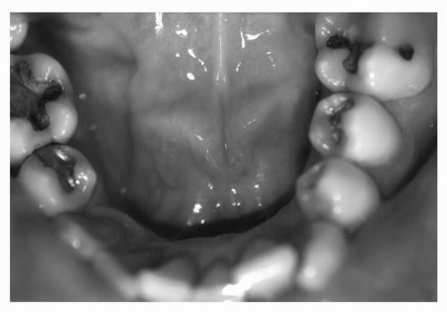
  • Floor of Mouth. Visualize as tongue is elevated. Uniform red color. Evaluate Wharton ducts for salivary flow. Bidigital palpation to evaluate submandibular salivary glands, lymph nodes, symmetry (see Fig. 35.11).

    Common Abnormalities: Ulcerations, varicosities, mucocele.


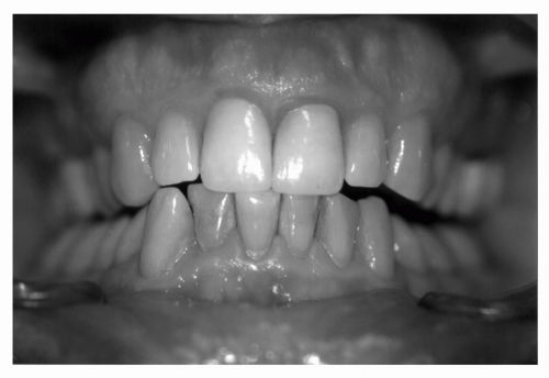
  • Gingivae. Observe color (pink), frenal attachments, and recession.

    Common Abnormalities: Inflammation secondary to periodontal disease, recession, hyperplasia, fistulae (see Fig. 35.12).


  • Teeth. Number present/absent, gross decay, plaque/calculus, mobility, discoloration, and occlusion (Fig. 35.12).

    Common Abnormalities: Decay, mobility, gingival abrasion, fractures, lost/fractured restorations, ill-fitting prosthesis.
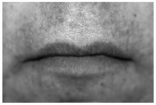


Figure 35.1 Normal appearance of lips. (See color insert.)


Figure 35.2 Bidigital palpation on lower lip. (See color insert.)


Figure 35.3 Bidigital palpation of buccal mucosa. (See color insert.)


Figure 35.4 Visual examination of anterior buccal vestibule, mandibular arch. (See color insert.)


Figure 35.5 Visual examination of posterior buccal vestibule, mandibular arch. (See color insert.)


Figure 35.6 Visual examination of hard palate. (See color insert.)


Figure 35.7 Visual examination of soft palate. (See color insert.)


Figure 35.8 Visual examination of oropharynx. (See color insert.)


Figure 35.9 Visual examination of dorsal surface of tongue. (See color insert.)


Figure 35.10 Visual examination of lateral surface of tongue, including lingual tonsil. (See color insert.)


DENTAL DECAY (CARIES)


Figure 35.11 Visual examination of floor of the mouth, including Wharton ducts. (See color insert.)


Figure 35.12 Visual examination of anterior gingival and teeth. (See color insert.)

Dental caries is the demineralization of the calcified structures of the tooth caused by Streptococcus species and other intraoral bacteria. Caries may occur on the smooth, pit and fissure and root surfaces of the tooth. Smooth and pit and fissure caries occur on the enamel surface and have a higher incidence in children and young adults. Root surface caries typically affect older adults and develop on the dentin surface of the root in areas where gingival recession has occurred. As an increased number of older people maintain their natural teeth, they are predisposed to root surface caries. Other local factors, such as xerostomia, increased sugar intake, acidic foods and drinks, and medications, increase the likelihood that root surface caries will develop.

Dental decay is preventable, with the removal of bacteria-harboring dental plaque. Dental caries occur when three components are present:


  • A tooth surface (enamel and/or dentin)


  • A fermentable substrate (i.e., sugar)


  • Bacteria that metabolize fermentable substrates into acids.
The acids produce a result in the demineralization of the tooth surface and cavitation. Removal of one of the three components will interrupt the decay process and prevent caries from developing. Pit and fissure and smooth surface caries occur on enamel, the hardest substance in the body, and may take months to develop. However, root surfaces are not covered with enamel. Their surface has a thin layer of cementum, which quickly dissolves and exposes dentin. Dentin has a less mineralized composition than enamel, and the decay process on this surface will develop much more rapidly.


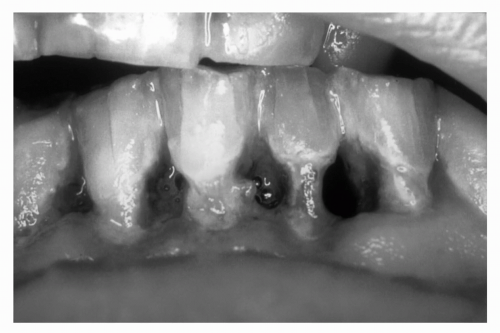
Figure 35.13 Root surface caries. (See color insert.)

Root surface caries is a particularly difficult management issue in the older adult (see Fig. 35.13). Approximately half of all adults 75 years and older have at least one tooth with root surface decay. Individuals who have been caries-free for decades may develop a number of root surface carious lesions in a matter of weeks. Usually this type of episode follows a major change in the oral environment. Medications that cause dry mouth as a side effect are often a contributor. This, coupled with the attempts to counter the dry mouth by frequent use of high-sugar, acidic drinks, and/or candy lozenges, results in the rapid demineralization of the dentin on the root surface and may result in irreparable cavitation and the subsequent loss of the tooth. Care should be taken to avoid medications with a xerostomic effect, and to advise patients to see a dentist for frequent, regular evaluations when their use is unavoidable. Other factors contributing to caries in older adults include history of previous caries, number of existing dental restorations, dietary habits, lack of fluoride exposure, and presence of partial dentures, which often cause food retention and subsequent plaque formation, diminished oral and/or manual motor control, being dependent on others for oral hygiene, systemic disease(s), and infrequent dental visits.

Unless properly treated, dental caries can lead to abscess formation of the hard and soft tissues of the oral cavity, cellulitis (including Ludwig angina), infection of proximal structures (cavernous sinus thrombosis), septicemia, and systemic seeding (subacute bacterial endocarditis). Extraction of the infected tooth may be necessary, affecting dental occlusion, mastication, and nutritional intake.

Prevention of dental caries is focused on:


  • The mechanical removal of dental plaque through proper brushing and flossing;


  • The reinforcement of the composition of enamel through fluoride treatments;


  • Diet modification;


  • The use of antimicrobial rinses;


  • Early attention to salivary dysfunction; and


  • Professional prophylaxis and intervention at appropriate intervals.


Management of dental caries is through removal of the carious lesion and repair of the tooth with the appropriate dental material. Management of dental infections is primarily mechanical, either through root canal therapy, incision, and drainage or extraction. Use of antibiotics should be reserved for the treatment of established abscess formation, and inappropriate prescription of antibiotics should be avoided. Referral to the appropriate dental professional should be the first course of action.

Table 35.1 highlights medications that are useful in the management of oral conditions often found in elderly people.


PERIODONTAL DISEASE


TABLE 35.1 MEDICAL MANAGEMENT OF ORAL CONDITIONS


Indication


Medication/Directions


Strength of Evidence of Effectiveness


Caries prevention


0.2% neutral NaF rinse


A


Disp: 480 mL bottle


Sig: Rinse 10 mL for 1 min and expectorate; do not swallow


Repeat weekly


1.1% neutral NaF dental cream (PreviDent 5,000)


A


Disp: 2 oz tube


Sig: Place 1/2 inch ribbon on toothbrush, then brush for 2-3 min and expectorate; do


not swallow; do not rinse or eat for 30 min following treatment


Perform twice daily


Periodontal disease


Chlorhexidine gluconate 0.12% (Peridex, PerioGard)


A


Disp: 16 oz bottle


Sig: After brushing and flossing teeth, rinse 1/2 oz for 30 s twice daily and expectorate


Xerostomia


Saliva substitutes


C


Sodium carboxymethylcellulose 0.5% aqueous solution (Saliva Substitute, Salivart)


Disp: 8 fl oz


Sig: Rinse as frequently as needed


Salivary stimulants


B


Pilocarpine HCl (Salagen) tablets 5 mg


Disp: 21 tablets


Sig: Take 1-2 tablet(s) 1/2 h prior to meals


Oral mucositis


Topical analgesic rinses


C


Diphenhydramine (Benadryl) elixir 12.5 mg/5 mL and attapulgite (Kaopectate)


C


Disp: Mix equal parts of both liquids (4 oz each) to obtain 8 oz


Sig: Rinse one teaspoon every 2 h for 1 min and expectorate


Aminacrine (Kamillosan liquid) 30 mL


C


Disp: Mix 30 drops in 100 mL of warm water


Sig: Rinse 5-10 mL four times daily for 1 min and expectorate


Dyclonine HCl (Dyclone) 0.5% or 1%


B


Disp: 1 oz bottle


Sig: Rinse one teaspoon for 2 min and expectorate


Intravenous therapy


Palifermin (60 μg/kg/d) intravenously for 3 d immediately before starting high-dose chemotherapy and total-body irradiation (conditioning therapy) and then again for 3 d after stem-cell transplantation.


Oral candidiasis


Candidiasis


Nystatin (Mycostatin, Nilstat) oral suspension 100,000 U/mL


A


Disp: 240 mL


Sig: Rinse 5 mL four times daily for 2 min and swallow until finished


Nystatin lozenge (Mycostatin pastilles) 200,000 U


A


Disp: 70 pastilles


Sig: Dissolve one pastille in mouth five times daily for 14 d; do not chew or swallow whole


Clotrimazole (Mycelex) troches 10 mg


A


Disp: 70 troches


Sig: Dissolve one troche in mouth five times daily for 14 d; do not chew or swallow whole


Angular cheilitis


A


Nystatin-triamcinolone acetonide (Mycolog II, Mytrex) ointment


Disp: 15 mg tube


Sig: Apply to affected areas four times daily for 10-14 d


Clotrimazole-betamethasone dipropionate (Lotrisone) cream


Disp: 15 mg tube


Sig: Apply to affected areas four times daily for 10-14 d


NaF, sodium fluoride.

Only gold members can continue reading. Log In or Register to continue

Stay updated, free articles. Join our Telegram channel

Jul 21, 2016 | Posted by in GERIATRICS | Comments Off on Oral Conditions

Full access? Get Clinical Tree

Get Clinical Tree app for offline access