Imaging plays a central role in the management of patients with suspected or known periampullary masses, including the initial diagnosis, staging, and follow-up to assess treatment response or recurrence. Use of appropriate imaging tools, application of optimal imaging protocols, and knowledge about imaging findings are essential for the diagnosis and accurate staging of these masses. Structured reporting of the imaging studies offers several advantages over freestyle dictations ensuring completeness of the relevant imaging findings, which would in turn help in deciding the best individual treatment strategy for each patient.
Key points
- •
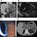
High-quality cross-sectional diagnostic imaging has a central pivotal role in evaluation of patients with known or suspected periampullary masses.
- •
The most commonly used imaging tools include contrast-enhanced multidetector computed tomography and contrast-enhanced MRI with MR cholangiopancreatography.
- •
The role of imaging is to identify periampullary masses, assess the presence or absence of tumor extension to surrounding vessels and organs, and identify distant metastasis.
- •
Accurate and complete reporting of relevant imaging findings should be done through the use of template reporting ensuring proper staging and optimal treatment strategies.
Related posts:
 Role of Endoscopic Ultrasonography and Endoscopic Retrograde Cholangiopancreatography in the Clinical Assessment of Pancreatic Neoplasms
 Minimally Invasive Approaches to Pancreatic Surgery
Adjuvant and Neoadjuvant Therapy for Pancreatic Cancer
Clinical Presentation and Diagnosis of Pancreatic Neuroendocrine Tumors
State-of-the-art Imaging of Pancreatic Neuroendocrine Tumors
Surgical Management of Pancreatic Neuroendocrine Tumors
Minimally Invasive Approaches to Pancreatic Surgery
Adjuvant and Neoadjuvant Therapy for Pancreatic Cancer
Clinical Presentation and Diagnosis of Pancreatic Neuroendocrine Tumors
State-of-the-art Imaging of Pancreatic Neuroendocrine Tumors
Surgical Management of Pancreatic Neuroendocrine Tumors
Stay updated, free articles. Join our Telegram channel

Full access? Get Clinical Tree


