Learning objectives
- •
Vertebral Compression Fractures (VCF) are often silent and referred to as “morphometric vertebral compression fractures.”
- •
The radiological appearances of vertebral deformities and VCF.
- •
In the absence of trauma, and after excluding localized osteolytic lesions, morphometric VCF are diagnostic of osteoporosis and override the densitometric diagnosis.
- •
The importance of opportunistic imaging studies.
The case study
Reasons for seeking medical help
DK has been recently hospitalized because of left lower lobe pneumonia. In addition to the characteristic features of pneumonia, a plain chest X-ray revealed moderate wedge vertebral compression fracture of T7. She denies recent and remote trauma to her back, but admits to experiencing occasional bouts of mild, vague, ill-defined pain in the mid to lower back, especially after standing for prolonged periods of time or carrying her two-year-old granddaughter.
She grades the pain as 3 on a 1 to 10 scale, with 10 being the worst possible pain. The pain is localized, usually readily relieved by lying down, analgesics (Acetaminophen 650 mg), nonsteroidal antiinflammatory drugs (Ibuprofen 200 mg), and local heat application. She uses these medications less frequently than once a month. Osteolytic deposits and other causes of localized bone demineralization have been excluded. She states that she lost about 2 in. in height since early adulthood.
Past medical and surgical history
- •
Surgical menopause: at age of 39 years when she underwent a hysterectomy and bilateral oophorectomy, no hormonal replacement therapy was prescribed.
- •
Menarche at 12 years, regular menstrual periods.
- •
She categorically denies any trauma to the back.
Lifestyle
- •
Sedentary lifestyle.
- •
Daily dietary calcium intake estimated to be about 1300 mg.
- •
No cigarette smoking.
- •
No alcohol abuse: not more than one glass of wine once or twice a week.
- •
No excessive sodium or caffeine intake.
- •
Common causes of low body weight have been investigated and ruled out.
Medication(s)
- •
No regular medications, apart from occasional analgesic and NSAID. Does not use opioids.
Family history
- •
Negative for osteoporosis.
Clinical examination
- •
Weight 131 pounds, steady; height 62″.
- •
Mild kyphosis.
- •
Slight tenderness along mid and lower thoracic vertebrae with adjacent paravertebral muscle spasm.
- •
No other significant clinical findings, no neurologic deficits.
Laboratory result(s)
- •
Complete blood count (CBC) and Comprehensive Metabolic Panel (CMP): no abnormal finding.
- •
Serum 25-hydroxy-vitamin D: within the normal range (46 ng/mL).
- •
Serum and urine protein electrophoresis: no abnormality detected.
- •
Erythrocytic sedimentation rate 3 mm in first hour.
DXA and radiological result(s)
- •
T-scores: right femoral neck −0.5, right total hip −0.3, left femoral neck −0.8, left total hip −0.7, L1–L4 –1.0.
- •
Vertebral Fracture Assessment (VFA): moderate wedge compression fracture of T7 (34% for the T7 antero/posterior ratio of vertebral heights).
- •
FRAX scores: 1.2% and 11% for the 10-year risk of sustaining a hip fracture and major osteoporotic fracture, respectively.
Multiple choice questions
- 1.
In DK’s case, the vertebral compression fracture identified by vertebral fracture assessment (VFA):
- A.
It is not a true fracture, but vertebral deformity.
- B.
It is not relevant because it is essentially asymptomatic.
- C.
It is an incidental benign finding and can be ignored.
- D.
It increases neither mortality nor morbidity risks.
- E.
It is diagnostic of osteoporosis after causes of localized osteolytic lesions have been excluded.
Correct answer: E
Comments:
Although vertebral deformities are common, especially in older patients, they are seldom of the magnitude seen in DK’s case: moderate (34%) wedge compression fracture. Deformities also usually affect several vertebrae. It would be unusual for vertebral deformities to be confined to just one vertebra.
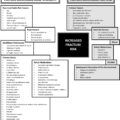
After excluding causes of localized osteoporosis, silent VCFs, also referred to as “morphometric” fragility fractures, are diagnostic of osteoporosis. They are associated with an increased fracture risk of any bone, increased mortality, and significant morbidity, including chronic back pain, loss of height, kyphosis, protuberant abdomen, decreased pulmonary function, sleep disturbances, loss of independence, loss of self-esteem, depression, and cognitive impairment. Nerve entrapment and paraplegia, on the other hand, are rare. The 5-year mortality postvertebral fractures are increased by about 20%. Within 12 months of a VCF, 19% of untreated patients are expected to sustain another vertebral fracture. The presence of these fractures therefore—even though essentially asymptomatic—emphasizes the urgency of addressing the underlying osteoporosis. Abdominal CT scans can be used to identify patients with previously undetected fractures who are at risk of further fractures.
- A.
- 2.
Match the following:
- (a)
Morphometric VCF.
- (b)
Clinical or acute VCF.
- (c)
Both.
- (d)
Neither.
- A.
Usually associated with acute severe localized pain.
- B.
Painless when it occurs.
- C.
Leads to height loss.
- D.
Usually associated with spinal cord compression.
- E.
Usually discovered during imaging studies for an unrelated condition.
- A.
Correct answers: A (b); B (a); C (c); D (d); E (a).
Comments:
Morphometric VCFs are asymptomatic fractures, discovered when imaging studies are conducted for some unrelated medical condition. Clinical VCFs occur spontaneously or after sustaining trauma that ordinarily would not be expected to lead to a fracture. Both are considered fragility fractures and diagnostic of osteoporosis, regardless of the bone density, but after causes of localized bone demineralization have been ruled out. Both lead to height loss. Spinal cord compression may complicate traumatic vertebral fractures, but seldom occurs in patients with osteoporotic VCFs.
Although morphometric VCFs are not associated with the typical acute pain experienced by patients sustaining clinical VCFs, they are often associated with gradually worsening low-grade back pain and kyphosis. The pain is usually at least partly relieved by lying down and analgesics. Heat and gentle massage are useful ancillary aids to relieve pain.
The pain associated with clinical VCFs is acute and severe, usually self-limited, and relieved spontaneously, within 4–6 weeks after the incident. It nevertheless may be replaced by low-grade pain worsened by standing and exertion and relieved by lying down.
The densitometric diagnosis (normal bone density in DK’s case) is overridden by the presence of fragility fractures, including morphometric VCFs. After ruling out common causes of localized bone demineralization, the presence of VCFs in the absence of significant trauma establishes the fracture as a fragility fracture and therefore the final diagnosis is osteoporosis, even if the T-score is not within the osteoporotic range and even if the fracture risk as calculated by the FRAX algorithm does not reach the threshold recommended by the National Osteoporosis Foundation to initiate pharmacologic treatment for osteoporosis.
If the workup of the patient reveals pathologies that may be responsible for the vertebral compression fracture such as, for instance, multiple myeloma or osteolytic deposits, the diagnosis would be “pathological vertebral compression fracture.” Unfortunately, although osteoporosis is a major health issue that affects the day-to-day quality of life, it remains largely undetected, underdiagnosed, and undertreated.
- (a)
- 3.
Morphometric VCFs:
- A.
Most often due to an underlying pathology such as multiple myeloma.
- B.
Vertebral augmentation procedures should be considered.
- C.
Lumbar vertebrae are affected more than thoracic vertebrae.
- D.
All of the above.
- E.
None of the above.
Correct answer: E
Comments:
The commonest cause of morphometric VCFs is osteoporosis. There is no need for vertebral augmentation procedures as the main goal of this procedure is to relieve pain associated with clinical VCF and the results are best when performed soon after the fracture. The short- and especially long-term efficacy of vertebral augmentation procedures has been questioned and is further discussed in the case study on “Acute vertebral compression fractures.”
- A.
- 4.
Vertebral Fracture Assessment:
- A.
The presence of morphometric vertebral compression fractures is diagnostic of osteoporosis and overrides the densitometric diagnosis.
- B.
The exposure to radiation is about the same as a plain X-ray of the thoracolumbar vertebrae.
- C.
Depending on the densitometer used, the patient can remain supine during the VFA imaging study.
- D.
A and C.
- E.
B and C.
Correct answer: D
Comments:
By identifying a VCF in a patient with osteopenia who denies trauma to her back, VFA changes the densitometric diagnosis to osteoporosis, after causes of localized bone demineralization have been excluded. The exposure to radiation resulting from VFA is much less than that resulting from a plain X-ray of the vertebrae. Some densitometers allow for VFA to be done without having to reposition the patient.
- A.
- 5.
Factors predisposing to osteoporosis in DK’s case are:
- A.
Caucasian ethnicity.
- B.
Status postmenopause at a young age, and no hormone replacement therapy.
- C.
Sedentary lifestyle.
- D.
Low body weight.
- E.
A, B, C, and D.
Correct answer: E
Comments:
All listed predispose to osteoporosis.
- A.
- 6.
Clinical signs suggestive of morphometric vertebral compression fractures include:
- A.
Kyphosis.
- B.
Reduced space between lower ribs and pelvic cavity.
- C.
Increased arm span/height ratio.
- D.
Loss of height.
- E.
All of the above.
Correct answer: E
Comments:
All the listed clinical signs are suggestive of multiple vertebral compression fractures, morphometric fractures, if there is no history of back trauma. However, apart from kyphosis and loss of height which can be reliably measured and quantified, the reproducibility of measuring the arm span and the space between the lower ribs and pelvic cavity is such that, unless excessive, it cannot be used reliably for diagnostic purposes.
- A.
- 7.
Loss of height in older patients can be due to:
- A.
The patient assuming a stooped position.
- B.
A reduced volume of the intervertebral disc.
- C.
Vertebral compression fractures.
- D.
Buckling of the knees.
- E.
All of the above.
Correct answer: E
Comments:
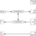
Loss of height is a very common finding in older people and is usually multifactorial. Older people often assume a stooped position to make up for the exaggerated lumbar lordosis and buckling of the knees. In these instances, the patient is able to reduce the thoracic curvature and, at least partly, straighten the kyphotic posture. Old age is often associated with some desiccation and a reduced volume of the intervertebral disc. The intervertebral disc also may invaginate the plates of the adjacent vertebral body: Schmorl’s nodules. It is possible that these represent early stages of vertebral compression fractures.
- A.
- 8.
DK would benefit from the following:
- A.
Estrogen.
- B.
Large daily doses of vitamin D and calcium supplements.
- C.
Vertebroplasty or kyphoplasty.
- D.
A and B.
- E.
None of the above.
Correct answer: E
Comments:
Largely based on the results of Women’s Health Initiative (WHI) study, estrogen is no longer approved for the treatment of osteoporosis in older postmenopausal women even though this study showed that, compared to placebo, hormonal replacement therapy reduced the risk of osteoporosis and the incidence of hip fractures (36%), clinical vertebral fractures (36%), and other osteoporotic fractures (23%). The WHI study, however, also showed that, in older postmenopausal women, HRT is associated with an increased risk of strokes, urinary incontinence, neoplasia, gall bladder diseases, and dementia, including Alzheimer’s disease. At present HRT is approved only for the prevention of osteoporosis, provided it is administered soon after the menopause and in the smallest dose, for the shortest period of time.
It has nevertheless been pointed out that the average age of patients enrolled in the WHI study was about 60 years and therefore the findings of this study may not be applicable to younger postmenopausal women.
DK is getting a good daily amount of calcium through food, and her serum vitamin D level is within the normal range. There is therefore no need for either vitamin D or calcium supplementation. She is also not likely to benefit from vertebral augmentation procedures (vertebroplasty or kyphoplasty) as she is not complaining of severe pain: the main indication for these procedures.
- A.
- 9.
The following are recommended in DK’s case:
- A.
Back brace.
- B.
Heat pads.
- C.
Transcutaneous nerve stimulation.
- D.
All of the above.
- E.
None of the above.
Correct answer: E
Comments:
Although these measures may be useful for the management of pain associated with acute clinical osteoporotic VCFs, they are not indicated for morphometric VCFs, unless the patient is experiencing pain. The pain experienced by DK is, usually, not of a sufficient magnitude to prescribe transcutaneous nerve stimulation. The long-term use of back braces may be counterproductive as it may lead to muscle atrophy. By increasing the local circulation, heat pads may reduce the muscle spasm and hence pain experienced at the level of the affected vertebra(e).
- A.
- 10.
The following exercises are recommended in patients who have sustained vertebral compression fractures:
- A.
Sit-ups.
- B.
Free weightlifting, as high a weight as can be safely tolerated.
- C.
Weighted backpacks.
- D.
All of the above.
- E.
None of the above.
Correct answer: E
Comments:
In healthy premenopausal women, physical exercise, especially a combination of resistive and aerobic high-impact exercises such as running and rope jumping, increases the bone mineral density of the lumbar vertebrae and femoral necks, with the greatest increases in BMD being in the bones undergoing the greatest biomechanical loading.
Physical exercise selectively affects some bones more than others. For instance, aerobic and resistive exercises selectively affect the lumbar vertebrae more than the proximal femur. Walking, on the other hand, has no significant effect on the lumbar vertebrae but significantly increases the BMD at the hips. Several exercise programs, including balance training, gait training, strength training, and Tai Chi, have been shown to be effective at reducing the risk of falls and, hence, the risk of fractures.
It also has been shown that the BMD of patients who have sustained strokes associated with hemiplegia or hemiparesis is lower on the paralyzed or paretic side than the healthy side further emphasizing the exercise/bone density relationship.
Sit-ups are not recommended in patients who have sustained vertebral compression fractures because they increase the mechanical load on the vertebrae and may induce further compression of the osteopenic/osteoporotic vertebra and adjacent vertebrae. Reverse sit-ups, on the other hand, are recommended as they strengthen the paravertebral and abdominal muscles without putting undue stress on the vertebrae.
While performing “reverse sit-ups” the patient lies on a firm surface and uses her arms to sit up. Then, while sitting she crosses her arms on her shoulders and slowly extends her back until she is lying horizontally. When performing reverse sit-ups it is recommended that another person be beside the patient, with one hand under the patient’s head, in case the patient’s muscles give way and she falls back.
Whereas resistive exercises are recommended as part of the physical exercise program to increase muscle bulk and strength as well as bone mineral density, free weightlifting is not recommended because it may be associated with accidents as may happen should the patient feel dizzy and release the free weights. Feeling dizzy while lifting weights is often due to the patient straining which results in an increased intrathoracic pressure, reduced venous return, reduced stroke volume, reduced cardiac output, and reduced cerebral blood flow with the patient feeling dizzy.
Weight machines are much safer than free weights: while exercising on a weight machine, should the patient feel dizzy, she will just let go of the cord fixed to the weights. This may in fact alert the attendees that this person is having problems with the selected weights and a lighter load may be recommended.
Heavy backpacks and jackets laden with weights are not recommended for most patients with osteoporosis as they increase the pressure on the vertebrae and may induce vertebral compression fractures. Heavy handbags may in addition subject the cervical vertebrae to undue physical stress, which may interfere with the vertebrobasilar and cerebral circulation and worsen the extent and degree of dizziness.
Weight bands that can be attached to the wrists and ankles are convenient ways to undertake resistive and aerobic exercises: the patient wears a band on each ankle and each wrist. In order to reduce the risk of putting too much mechanical strain on the rest of the skeleton and especially the vertebral column, light weights, about half a pound each, are recommended. Patients are also more likely to tolerate the half pound on each limb. Many patients wear them all day long, taking them off only at night. In this manner, with every single step they take, they lift 2 pounds. Given that the average person walks 3000–4000 steps a day, the approximate daily weight lifted by having half a pound strapped to each wrist and each ankle is about 6000–8000 pounds each day, performed without needing to go to a gym. After using them for some time most patients get used to wearing these light weights and enjoy being able to alter the weight lifted according to their circumstances that day.
Given the complexity of various exercise programs and the diverse needs and background of patients with osteoporosis, especially older ones, it is advisable to consult a qualified physical therapist or personal trainer, with a special interest in osteoporosis to develop an exercise program tailored to the individual needs of the interested person. Physical exercise programs and related issues are discussed separately.
- A.
Case summary
Analysis of data
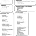
Factors predisposing to bone demineralization/osteoporosis in DK’s case
- •
Status postmenopause, no HRT.
- •
Sedentary lifestyle.
- •
Caucasian ethnicity.
- •
Low body weight.
Factors reducing risk of bone demineralization/osteoporosis in DK’s case
- •
Good daily dietary calcium intake.
- •
No excessive sodium, caffeine intake.
- •
No cigarette smoking.
- •
Negative family history of osteoporosis/fractures.
Factors increasing risk of falls/fractures
- •
Kyphosis, vertebral compression fracture.
- •
Sedentary lifestyle.
Factors reducing risk of falls/fractures
- •
None.
Diagnosis
- •
Osteoporosis, morphometric vertebral compression fracture.
Management
Treatment(s)
- •
Antiresorptive or osteoanabolic medication.
Diagnostic test(s)
- •
Ensure that causes of localized bone demineralization have been excluded.
Lifestyle changes
- •
None recommended.
Rehabilitation
- •
Physical exercises: combination of aerobic and resistive exercises.
Related posts:
 Clinical diagnosis: Fragility fracture long bones
 Clinical diagnosis: Fragility hip fracture
 Clinical diagnosis: Acute vertebral fragility fracture
Osteopenia: Individualizing treatment—Part III: When silent vertebral compression fractures override the densitometric diagnosis
Osteopenia: Individualizing treatment—Part IV
Clinical diagnosis: Acute vertebral fragility fracture
Osteopenia: Individualizing treatment—Part III: When silent vertebral compression fractures override the densitometric diagnosis
Osteopenia: Individualizing treatment—Part IV
 Screening for osteoporosis
Stay updated, free articles. Join our Telegram channel

Full access? Get Clinical Tree


