Fig. 1
Somagram device
Prostate Ultrasound
Over the next decade the use of ultrasound as a safe diagnostic imaging method began to gain acceptance and advances in ultrasound equipment development resulted in reduced size and increased reliability, readability, and usability. In 1963, the first trans-perineal ultrasonic examination of the prostate was published by two Japanese urologists, Takahashi and Ouchi. However, the ultrasound images created with this array were of such poor quality that they had little medical utility [12] (Fig. 2).
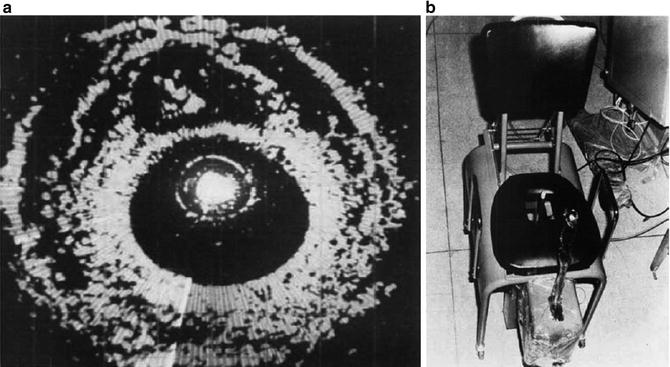
Fig. 2
(a and b) Early TRUS device and image (used with permission)
The first clinically applicable images of the prostate obtained with trans-rectal ultrasound (TRUS) were described in 1967 by Watanabe et al. [13]. Watanabe used a 3.5 MHz transducer, which at that time was a significant improvement upon any previous probe and allowed clinicians to obtain clinically interpretable, real-time images. As ultrasound technology became more developed, the use of TRUS in the evaluation of prostatic disease increased. By the mid 1980s, the 7 MHz ultrasound probe, which more clearly delineated the architecture of the prostate, had become a standard diagnostic instrument of the urologist. Despite adequate visualization of the prostate by TRUS techniques, ultrasound guided biopsies were still performed via the transperineal route. In fact, it was not until 1987 that the first literature appeared describing the use of TRUS with trans-rectal biopsy. Since then, as ultrasound technology has become more refined, this technique has been described as a superior method of performing biopsy of the prostate [14].
Since the initial reports of TRUS of the prostate, substantial technologic advances have improved the diagnostic capabilities of this modality. The current TRUS probe that most urologists consider standard is a 5–8 MHz hand-held, high-resolution probe with multiaxial planar imaging capabilities, allowing for both transverse and sagittal imaging of the prostate in real time. These probes can be fitted with an adapter that accepts the needle of a spring-loaded biopsy gun, thus allowing multiple cores of tissue to be easily obtained. The visualization provided by the new higher resolution transducers, coupled with the ability to direct the biopsy needle into various regions of interest and provide uniform spatial separation of the areas to be sampled, has helped to make TRUS-guided prostate biopsy a standard technique in the diagnosis of prostate cancer.
Related posts:

Stay updated, free articles. Join our Telegram channel

Full access? Get Clinical Tree


