Noninvasive Malignancies and Conditions of Uncertain Malignant Potential
|

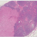
A variety of abnormalities can be identified in the cells lining the terminal duct lobular units short of an invasive malignant appearance penetrating the basement membrane. These include usual type hyperplasia, atypical hyperplasia, and noninvasive (in situ) carcinoma. Ductal and lobular patterns of atypical hyperplasia and in situ cancer can be recognized from the histological pattern of disease and cell type. Ductal carcinoma in situ is the most common form of noninvasive carcinoma (making up 4% of symptomatic and 25% of screen-detected ‘cancers’). It is characterized by distortion, distention, and complete involvement by a homogenous and neoplastic population of cells in adjacent ducts and lobular units. By contrast, lobular carcinoma in situ is rare (<1% of screen-detected ‘cancers’) and exhibits relatively uniform expansion of the whole lobule by regular cells with regular, round or oval nuclei. While each involved lobular unit has a uniform cellular population, the pattern and even cytology may, and often does, vary between units with some intervening units being minimally involved or uninvolved. Some patients present with combined features that should be regarded as having clinical features of both processes.
Criteria have been agreed to distinguish atypical ductal hyperplasia (ADH) from ductal carcinoma in situ (DCIS) and atypical lobular hyperplasia (ALH) from lobular carcinoma in situ (LCIS). In general, lesions that involve only a few membrane bound spaces and that measure less than 2-4 mm in their greatest diameter should be regarded as hyperplastic lesions (with or without atypia) and not in situ carcinoma (Table 5.1). Whether there is progression over time of hyperplasia to in situ disease to invasive carcinoma with increasing genetic mutations within the affected cells analogous to the adenoma/carcinoma sequence of colorectal neoplasia, is not entirely clear.
Difficulty in distinguishing ALH and LCIS and their similar behaviour have led some to combine ALH and LCIS into the single entity of lobular in situ neoplasia.
Table 5.1 Classification of ductal carcinoma in situ | |||||||||||||||||
|---|---|---|---|---|---|---|---|---|---|---|---|---|---|---|---|---|---|
| |||||||||||||||||
Those with atypical hyperplasia and carcinoma in situ are at increased risk of invasive disease, but the timescale is measured in years and it may not occur at all in many individuals. Development of invasive carcinoma can also occur at some distance from the noninvasive disease. Different histological characteristics of in situ disease are recognized which correspond with subsequent risk of progression and recurrence.
Approximately 40% of those with low-grade ductal carcinoma in situ will develop invasive cancer over a 30-year period, with the majority of these developing within the first decade. About 15-20% of women with a diagnosis of lobular carcinoma in situ will develop breast cancer in the same breast, and a further 10-15% will develop an invasive carcinoma in the contralateral breast.
There is an interaction between atypical hyperplasia and family history: women with both atypical hyperplasia and a first-degree relative (mother, daughter, or sister) with breast cancer have an absolute risk of 20-30% of developing breast cancer within the next 15-20 years. Atypical hyperplasia and carcinoma in situ are usually asymptomatic, although a lump is palpable in a small proportion of those with DCIS, and Paget’s disease is often seen in association with DCIS.
Related posts:
Stay updated, free articles. Join our Telegram channel

Full access? Get Clinical Tree


