61 Non-Hodgkin’s Lymphomas
In 2009, approximately 66,000 new cases of NHL were diagnosed in the United States and accounted for almost 20,000 deaths. NHL is the fifth leading cause of cancer among men and women, representing almost 5% of new cancer cases. The incidence of NHL has increased by 150% since 1950. An increase of 50% was documented between 1973 and 1988 (a 3% to 4% increase each year). Over the last 2 decades, this increase has abated, and the fraction of 5-year survivors increased significantly from 48% in 1975-1977 to 65% in 1996-2004.1 Most of the increase in recent years is among patients in their sixth and seventh decades; as a result, many patients with NHL may have significant comorbidities that complicate treatment options, particularly with chemotherapy. Radiotherapy is an attractive option that could reduce the need for aggressive or long chemotherapy regimens in many patients.
Epidemiology
Pertinent epidemiologic facts about NHL include:
Classification
Over the last decades, lymphoma classification has changed multiple times. In the 1980s, the International Working Formulation (IWF) classified NHL into three major categories—low, intermediate, and high grade—based on the morphology and natural history. The Revised European-American Classification of Lymphoid neoplasm (REAL) was developed in 1994. It classified NHL based on the cell of origin (B, T, or NK) and included morphology, immunophenotype, and genetic and clinical features. The currently accepted World Health Organization (WHO) classification is a refinement of the REAL classification.2
The REAL/WHO classification of NHL includes several additional, newly identified entities not recognized by the IWF.2 After consideration of cell of origin (B, T, or NK) the classification subdivides lymphomas into those derived from precursor lymphocytes versus those derived from mature lymphocytes. The classification is further refined based on immunophenotype and genetic features. These considerations have aided in defining active treatment for specific subtypes of lymphoma.
Currently, a comprehensive description of the natural history and clinical features of all NHL diagnoses recognized by the WHO classification does not exist. However, the International Lymphoma Classification Project evaluated 1403 lymphoma cases and identified the 13 most common histologic types responsible for about 90% of cases of NHL in the United States.3 The findings were as follows: diffuse large B-cell (DLBCL), 31%; follicular lymphoma (FL), 22%; small lymphocytic lymphoma/chronic lymphocytic leukemia (SLL/CLL), 6%; mantle cell lymphoma (MCL), 6%; peripheral T-cell lymphoma (PTCL), 6%; and marginal zone B-cell lymphoma (MZL), mucosa-associated lymphoid tissue (MALT) lymphoma, 5%. The remaining subtypes each occurred in less than 2% of cases. Composite lymphomas were not included in these distribution figures. Importantly, in the United States more than 50% of cases of lymphoma are either DLBCL or FL. In a study performed by the International T-cell Lymphoma Project, PTCL–not otherwise specified (PTCL-NOS) was the most common subtype of PTCL (29.3%).4
Cytogenetics and Molecular Biology
Several lymphomas are associated with nonrandom chromosomal abnormalities.5 These abnormalities often correlate with the histologic type, immunophenotype, and clinical behavior. For example, translocation t(14:18) (q32;q21) occurs commonly in follicular lymphoma. It results in the transposition of the bcl-2 oncogene of chromosome 18 to become adjacent to the heavy chain immunoglobulin gene on chromosome 14. The bcl-2 oncogene is essential for apoptosis or programmed cell death. The t(14:18) translocation interferes with normal senescence and death of the follicular center cell and is possibly engaged in its malignant transformation. Most patients with Burkitt’s lymphoma have translocation t(8:14) or one of its variants, and c-myc is the relevant translocated oncogene. The c-myc product is an important transcription factor, and its dysregulation is probably involved in the proliferative process. Translocation t(11:14) involving bcl-1 is characteristic for recognition of mantle cell lymphoma. The gene product of bcl-1, cyclin D, is directly involved in the regulation of cell division. Rearrangement of bcl-6 has been reported in a third of patients with diffuse large-cell lymphoma. In 25% to 40% of patients with MALT lymphoma, t(11:18) has been detected and indicates an unlikely response of MALT lymphoma of the stomach to antibiotic therapy of an associated H. pylori infection.
Pathologic Diagnosis
In Table 61-1, the more common histologies are listed by WHO classification and grouped as low grade (indolent), intermediate grade (aggressive), or high-grade (highly aggressive), a terminology that is still in clinical use.
Table 61-1 Abridged Version of the WHO Lymphoma Classification Organized by Clinical Groups
| Low Grade (Indolent) |
| Intermediate Grade (Aggressive) |
| High Grade (Highly Aggressive) |
Staging and Prognostic Factors
Although histology is the predominant determinant of prognosis in this diverse group of lymphoid malignancies, stage remains important for selection of treatment strategy and predicting prognosis in each histologic category. The Ann Arbor Staging Classification system, developed originally for Hodgkin’s disease, has also been used for NHL. The staging system is described in Chapter 60, Hodgkin’s Disease, and reflects the number of sites of involvement and their relation to the diaphragm, existence of B symptoms, and the presence of extranodal disease. In NHL, most patients are assigned to a stage based on physical examination, imaging studies, and bone marrow biopsy. The Ann Arbor staging system is considered by many to be inadequate for NHL, a shortfall that has led to the incorporation of other prognostic factors to define initial therapy.
The International Prognostic Index
In addition to stage, other important prognostic factors have been identified in patients with NHL.6 They frequently include patient parameters such as age, performance status, and tumor burden indicators such as bulk, number of involved sites, presence of extranodal disease, and lactate dehydrogenase (LDH) level.
An international team developed a prognostic model to predict outcomes of patients with aggressive NHL6 (Table 61-2). The International Prognostic Index (IPI) identified five significant risk factors predictive of overall survival: age (<60 years versus >60 years), serum LDH (normal versus elevated), performance status (0 or 1 versus 2 to 4), stage (I or II versus III or IV), and number of extranodal sites involved (0 or 1 versus 2 to 4). These features were incorporated into a model that identified four groups of patients with significantly different outcomes following treatment. Risk groups were determined by the sum of adverse risk factors: low (0 to 1 factor), low-intermediate (2 factors), high-intermediate (3 factors), and high (4 to 5 factors). The predicted 5-year survival rates for these groups were 73%, 51%, 43%, and 26%, respectively. When only patients younger than 60 were evaluated, stage, performance status, and LDH levels remained significant for poor prognosis (age-adjusted IPI).
Table 61-2 International Prognostic Index for Aggressive NHL
| All Patients |
| IPI Score | 5-Year Survival (%) | |
|---|---|---|
| Low-risk | 0-1 | 73 |
| Low-intermediate | 2 | 51 |
| High-intermediate | 3 | 43 |
| High-risk | 4-5 | 26 |
| Patients ≤ 60 years | ||
| Serum LDH above normal | ||
| ECOG performance > 2 | ||
| Ann Arbor stage III or IV |
| Age-Adjusted IPI Score | 5-Year Survival (%) | |
|---|---|---|
| Low-risk | 0 | 83 |
| Low-intermediate | 1 | 69 |
| High-intermediate | 2 | 46 |
| High-risk | 3 | 32 |
| Patients With Early-Stage (I-II) Disease | ||
|---|---|---|
| Age > 60 | ||
| Serum LDH above normal | ||
| Stage II disease | ||
| ECOG performance status ≤2 |
| Modified IPI Score | 10-Year Survival (%) |
|---|---|
| 0 | 90 |
| 1-2 | 56 |
| 3 | 48 |
For follicular lymphoma, the Follicular Lymphoma International Prognostic Index (FLIPI) has been shown to be a valuable prognostic tool.7 It is depicted in Table 61-3.
Table 61-3 The International Prognostic Index for Follicular Lymphoma4
| Age | ≤60 years |
| Ann Arbor stage | III-IV |
| Hemoglobin level | <12 g/dL |
| Serum LDH level | >ULN (upper limit of normal) |
| Number of nodal sites | >5 |
| Risk Group According to FLIPI Chart | Number of Factors |
| Low | 0-1 |
| Intermediate | 2 |
| High | ≤3 |
FLIPI, Follicular Lymphoma International Prognostic Index.
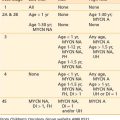
Essential workup studies for staging and IPI based on National Comprehensive Cancer Network (NCCN) guidelines8 are detailed in Table 61-4.
Table 61-4 Essential Workup of Patients With Non-Hodgkin’s Lymphomas