© Springer Nature Singapore Pte Ltd. 2017
Ranu Patni (ed.)Current Concepts in Endometrial Cancer10.1007/978-981-10-3108-3_1010. Newer Perspectives in the Management of Endometrial Cancer
(1)
Department of Gyn Oncology, Malabar Cancer Centre, Thalassery, India
(2)
Department of Medical Oncology, TMH, Mumbai, India
Abstract
CT, MRI, or PET have similar efficacy in detecting extrauterine disease and should be performed when extrauterine disease is suspected in carcinoma endometrium.
Systematic lymphadenectomy is associated with an improvement in overall survival in patients with intermediate- or high-risk endometrial cancer.
Adjuvant RT and chemotherapy in stage I disease with intermediate- or high-risk features prevent recurrence but are not associated with improvement in overall survival.
Introduction
Endometrial cancer is the commonest female malignancy in the western world, but in India, it ranks third after breast and cervical cancer, respectively. Incidence of endometrial cancer in India is 12,335 cases per year, and 4773 persons die because of this malignancy every year [1]. Advances in endometrial cancer at a national level are slow to occur in view of its rarity. However, management of this cancer remains challenging. The aim of this chapter is to comprehensively describe the advances in the management of endometrial cancer.
Methods
A PubMed search was carried out using the following MeSH terms and filters, “endometrial neoplasm’s”[MeSH Terms] OR (“endometrial”[All Fields] AND “neoplasms”[All Fields]) OR “endometrial neoplasms” [All Fields] OR (“endometrial”[All Fields] AND “cancer”[All Fields]) OR “endometrial cancer”[All Fields]) AND ((Clinical Trial[ptyp] OR Comparative Study[ptyp] OR Meta-Analysis[ptyp]) AND “2010/07/19”[PDat]: “2015/07/17”[PDat] AND “humans”[MeSH Terms] AND English[lang]).
Eight hundred and sixty-one articles were available for selection. These articles were manually screened, and relevant articles are compiled under the subheadings of surgical, medical, and radiation oncology.
Recent Perspective in Imaging
Imaging in Carcinoma Endometrium
Detection of extrauterine disease mandates an imaging in carcinoma endometrium. CECT has been traditionally used for this purpose. Whether PET-CT or PET-CECT would improve the rate of detection of extrauterine disease is an open question. A part of this question with regard to nodal involvement was answered by Kitajima et al. [2]. A cohort of 41 patients underwent CECT, PET-CT, and PET-CECT. The sensitivity and specificity of PET-CECT, PET-CT, and CECT were 61.4 % and 98.1 %, 52.3 % and 96.8 %, and 40.9 % and 97.8 %, respectively. The authors concluded that PET-CECT was not significantly superior to PET-CT for nodal staging of uterine cancer. Nodal metastasis cannot be excluded even if PET-CECT findings are negative. A systematic review and meta-analysis were performed by Chang et al. to assess the performances of PET or PET-CT in detecting pelvic and/or para-aortic lymph nodal metastasis [3]. The sensitivity and specificity of PET or PET-CT scans in the detection of nodal metastasis (pelvic and/or para-aortic LN) were 63.0 % and 94.7 %, respectively. Chang et al. concluded that it may help surgeons in selecting appropriate patients for pelvic and/or para-aortic lymphadenectomy.
The value of 18 FDG-PET in preoperative risk determination and prognosis was evaluated in a systematic review by Ghooshkhanei et al. [4]. Pooled mean SUVmax in patients with high-risk factors [grade 3, lymphovascular invasion (LVI), cervical invasion (CI), myometrial invasion (MI) ≥ 50 %] was statistically higher than those in patients without risk factors. Higher preoperative SUVmax was predicted for a lower disease-free survival. However, these findings need large multicentric studies for confirmation.
The current NCCN guidelines suggest that CT, MRI, or PET can be performed as clinically indicated when extrauterine disease is suspected in carcinoma endometrium. On the basis of evidence, it seems that PET-CECT is slightly better in identifying patients with extrauterine nodal disease than CECT.
Recent Perspective in Surgical Oncology
Fertility-Preserving Surgery
Fertility-preserving surgery seems an option for FIGO stage IA endometrial cancer. Laurelli et al. selected patients with age ≤40 years, without Lynch II syndrome, with G1 and ER+/PR+ endometrioid histology, without myometrial invasion, without multifocal tumor, without node metastasis, without ovarian mass, and with normal serum CA 125 for fertility-preserving surgery. They underwent hysteroscopic ablation of the lesion and the myometrial tissue below, followed by either oral megestrol acetate 160 mg/day for 6 months or 52 mg levonorgestrel-medicated intrauterine device for 12 months. Only one patient out of 14 recurred within a median follow-up of 40 months [5]. The Turkish gynecologic oncology group also collected their data on fertility-preserving strategy in early endometrial cancers. They had 43 patients, and with average follow-up of 50 months, 81.4 % patients were disease-free, and 41.9 % patients had conceived [6].
NCCN currently recommends that fertility-preserving treatment can be provided in selected patient cohorts. These patients include those with stage IA G1 endometrioid histology, with endometrial cancer limited to the endometrium, without any myometrial invasion, without Lynch type 2 syndrome or any other genetic syndrome, without any extrauterine disease, and without any medical contraindication for progesterone therapy. These patients need to be counseled that this is not the standard option, and if they still insist, it can be offered. If post 6 months they have a complete response, then they should be encouraged for conception with continuous surveillance.
Extent of Surgery
Prediction of Lymph Nodal Disease
Nomograms
The role of complete prophylactic lymphadenectomy in early stage endometrial cancer is a matter of controversy. Around 5–10 % of early stage endometrial cancers harbor lymph nodal metastasis. Hence, it would be useful to know if we can identify patients who may benefit from lymphadenectomy either preoperatively or intraoperatively. The prediction of nodal disease preoperatively by PET scan has been discussed in the above section. In this section, we would look at other characteristics which have been reported to be important predictors of lymph nodal metastasis. Bendifallah et al. validated two nomograms made and were internally validated by ALHilli et al. [7]. The overall rate of lymphatic spread was 9.9 %. Predictive accuracy was 0.65 (95 % Cl, 0.61–0.69) for the full nomogram and 0.71 (95 % Cl, 0.68–0.74) for the alternative nomogram. The correspondence between recurrence rate and the nomogram prediction suggested only a moderate calibration of the nomograms. The authors concluded that additional parameters are needed to improve upon the accuracy of the nomograms.
In India, patients are commonly seen after incomplete staging surgery, i.e., only TAH-BSO without lymph node dissection. Whether to do para-aortic nodal staging in them is a matter of debate in this situation. Kang et al. addressed this issue and tried to prepare a web-based nomogram which could be utilized to individualize treatment in such cases [8]. Four variables – deep myometrial invasion, non-endometrioid subtype, lymphovascular space invasion, and log-transformed CA-125 levels – were part of the nomogram. It showed good discrimination. The nomogram is available on the website (http://www.kgog.org/nomogram/empa001.html). It can be helpful in individualizing treatment in these patients.
Sentinel Lymph Node
Sentinel lymph node procedure is one of the known ways of predicting lymph node status. It has established itself in breast cancer, melanoma, and carcinoma vulva. The utility of sentinel lymph node dissection in endometrial cancers was studied by Kang et al. in 2011in a meta-analysis [9]. The detection rate and the sensitivity were 78 % (95 % CI=73–84 %) and 93 % (95 % CI=87–100 %), respectively. Paracervical injection technique was associated with the increase in detection rate (P = 0.031). While hysteroscopic injection technique and the subserosal injection technique were associated with decrease in detection rate and decreased sensitivity, respectively, if they were not combined with other injection techniques. The authors concluded that SLN biopsy had shown good diagnostic performance, but this should be interpreted with caution.
Ballester et al. evaluated sentinel lymph node in presumed low- and intermediate-risk endometrial cancers. The detection rates in low and intermediate risk were 61.2 % and 37.1 %, respectively. 21.4 % and 21.2 % of patients in low risk and intermediate risk were upstaged by the procedure. Ultrastaging detected metastases which were undetected by conventional histology in 42.8 % of patients [10].
A repeat systematic review and meta-analysis of sentinel LN sampling by Ansari et al. revealed a detection rate of 77.8 % and sensitivity of 89 % [11]. Similar to Kang’s review, it also observed that paracervical injections were associated with higher detection rates [9]. The authors concluded that sentinel node mapping was feasible in endometrial cancer. Using blue dye, radiotracer, and cervical injection can optimize the sensitivity and detection rate of this technique.
All these reviews had concluded that a large study would be required to confirm these benefits of these techniques. The SENTI-ENDO study was reported by Daraï et al. in 2015 [12]. It was a study evaluating the impact of sentinel lymph node dissection on adjuvant therapy. There was no difference in recurrence-free survival (RFS) whether sentinel LN was detected or not in patients with stage I–II endometrial cancer. Similarly there was no difference in RFS in patients whether the sentinel LN detected was negative or positive. Adjuvant therapies were more frequently administered in patients with a sentinel lymph node-positive status. It seems that these adjuvant therapies may have altered the course of sentinel lymph node-positive cases.
The current NCCN guidelines suggest doing sentinel lymph node dissection as category 3 recommendation.
Role of Lymphadenectomy
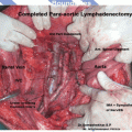
The role of lymphadenectomy has also been debated in endometrial cancers especially after the publication of ASTEC studies. Kim et al. did a systematic review and meta-analysis to address this issue. In all the studies, systematic lymphadenectomy improved overall survival, compared with unsystematic lymphadenectomy (hazard ratio, 0.89; 95 % confidence interval, 0.82–0.97). The systematic lymphadenectomy was associated with an improvement in overall survival in patients with intermediate- or high-risk endometrial cancer (hazard ratio, 0.77; 95 % confidence interval, 0.70–0.86). No such benefit was seen in those with low-risk endometrial cancer (hazard ratio, 1.14; 95 % confidence interval, 0.87–1.49) [13]. The impact of systematic lymphadenectomy was studied by Angioli et al. Lymphadenectomy had no negative influence on global health status [14]. Hence, systemic lymphadenectomy should be performed in patients with intermediate to high risk of lymph nodal metastasis.
The current NCCN guideline suggests doing pelvic and para-aortic lymph node removal for staging in patients with high-risk factors.
Technique of Surgery
Laparoscopic Versus Open Surgery
Lu et al. reported a randomized study comparing the outcomes of open versus laparoscopic surgery in endometrial cancers [15]. Laparoscopic surgery was found to be a safe and reliable alternative to laparotomy, with significantly reduced hospital stay and postoperative complications; however, it did not seem to improve the overall survival and 5-year survival rate. A Cochrane review done on the same topic too had similar conclusions [16]. In early stage carcinoma, laparoscopy was associated with similar overall and disease-free survival. Laparoscopy had reduced operative morbidity and hospital stay. There was no difference in severe postoperative morbidity between the two techniques.
Stay updated, free articles. Join our Telegram channel

Full access? Get Clinical Tree


