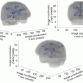
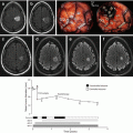
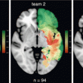
Fig. 28.1
Personalized dynamic multimodal therapeutic strategy in DLGG, with the goal to prevent malignant transformation while preserving quality of life
28.2.1 The Multistage Surgical Attitude
As extensively demonstrated in the chapter by Duffau on “Surgery for DLGG: Oncological Outcomes”, surgical resection is the first option in DLGG—as recommended by the European Guidelines [13] as well as by the National Comprehensive Cancer Network [14]. Indeed, radical surgery has a significant impact on OS by delaying malignant transformation [15–18] while preserving or even improving the QoL thanks to the use of awake mapping [19–22].
Nevertheless, it is worth noting that DLGG patient cannot (yet) be cured by surgery, even in cases of “supratotal” resection. Yordanova et al. [23] and Duffau [24] reported that excision of a margin around the FLAIR-weighted signal abnormalities prevented malignant transformation (with no postoperative adjuvant therapy). Yet, some patients experienced a relapse within the year following supramarginal resection, likely due to isolated tumoral cells still present far beyond the glioma visible on MRI [25]. Indeed, with a mean duration of postoperative follow-up of 132 months (range, 97–198 months), 50% of patients experienced tumor recurrence, with an average time to relapse of 70.3 months (range, 32–105 months)—once again, without malignant progression [24]. This issue should be extensively explained to the patient and his/her family at diagnosis with the goal (1) to inform him/her about the fact that additional treatment(s) will be administrated regularly over the years, and (2) to improve, in parallel, him/her compliance: indeed, this actual “honest information”, very well accepted by the patient, allows build trust that will last throughout the management of this chronic disease.
In other words, following an initial maximal safe surgery, i.e. function-guided resection (see the chapter by Duffau on “Surgery for DLGG: Functional Outcomes”), there is a high risk that DLGG will recur following several years, possibly after (supra)complete resection—and a fortiori in all cases following subtotal resection (i.e. with a residue less than 10–15 cc) [15, 17, 18]. Interestingly, the growth rate of residual glioma, voluntarily left for functional reasons, that is, to preserve QoL, will be similar to its presurgical kinetics [26]. Thus, thanks to the linear progression of the mean diameter of DLGG, one can predict at the individual level when the volume will reach about 15 cc, which represents the threshold for a higher risk of malignant transformation. As a consequence, a second “preventive” treatment can be proposed just before to reach this threshold, but not in the preceding years, in order (1) to not use prematurely (limited) therapeutic options, which will be very useful in the future (2) to preserve QoL of patient by limiting the administration of too much treatment(s)—for example by avoiding early radiotherapy, due the high risk of delayed cognitive disorders preventing to enjoy a normal life (see below) (3) while controlling the tumor by preventing progression towards a higher grade of malignancy, and thus by significantly increasing the OS [5]. Regarding the timing of second (or third, or fourth) surgery, we have reported a series of 19 DLGGs patients who underwent reoperations: 11 tumors had progressed to high-grade glioma in a median time between the two surgeries of 4.1 years. As a consequence, since there was no permanent morbidity associated with re-operations, we have suggested to “over-indicate” an early re-intervention rather than to perform a late surgery when the tumor has already transformed into a malignant glioma [27].
In this spirit, reoperation should be considered as a priority, on the condition nonetheless that at least subtotal resection can be achieved. Such a “multistage surgical approach”—beginning with initial tumor removal up to functional boundaries, followed by a period of several years, and then a second surgery with optimization of the extent of resection while preserving QoL—is possible, even in so-called “eloquent areas”, thanks to mechanisms of brain remapping induced by (1) the first surgery itself (2) the postoperative functional rehabilitation (3) the slow re-growth of the DLGG [28, 29]. Regular neuropsychological examinations as well as serial functional neuroimaging can provide helpful information for predicting the extent of resection during a second (or even third or fourth) surgery, in the philosophy of a recursive surgical attitude [30, 31]. The goal remains to reduce the glioma volume in order to prevent progression to a higher grade while preserving QoL (or even improving it, e.g. by controlling seizures) owing to repeated resections. Thus, the onco-functional balance of surgery can be optimally found for each patient only if the strong relationships between the DLGG course and the cerebral adaptation phenomena are taking into account (see chapter by Duffau on “Interactions Between DLGG, Brain Connectome and Neuroplasticity”).
On the other hand, we should insist again on the fact that a significant oncological benefit of surgery was actually demonstrated only when the resection was at least subtotal, that is, leaving a postoperative residual volume less than 10–15 cc [15, 17, 18]. Consequently, when the glioma is very diffuse, namely with huge invasion of the “minimal common brain” (that is, the cortico-subcortical structures which cannot be resected whatever the patient due to limitations of brain plastic potential, see [32, 33]) and/or with bi-hemispheric infiltration, it is possible to predict using probabilistic atlas before any treatment whether the surgical removal will be only partial—thus with no or only mild oncological impact [34, 35]. In these specific cases, there is no indication to perform surgery first (or reoperation if a subtotal resection was already performed several years before, followed by tumor relapse with a very diffuse pattern)—except (1) in patients with pharmacologically intractable epilepsy, because even partial resection may allow a relief of seizures, especially when the insula and/or mesiotemporal structures are involved [36] (2) in rare cases of intracranial hypertension. In other words, in these invasive DLGG and at this moment, alternative nonsurgical therapies should be envisioned.
28.2.2 The Role of Chemotherapy in a Dynamic Multimodal Therapeutic Approach
As already described (see the chapter by Taillandier and Blonski on “Chemotherapy for DLGG”), whatever the protocol used (PCV versus Temozolomide), chemotherapy may diffuse in the entire brain, i.e. even in critical areas, without inducing functional (neurological and cognitive) deficits [37]. This means that this therapy is perfectly adapted in cases of widely invasive DLGG, typically when (re-)operation is not possible. If one or multiple surgeries have previously been achieved, chemotherapy can be considered when the tumor re-grows with a volume reaching 10–15 cc (the same threshold as discussed regarding re-operation), and when it invades critical structures which cannot be functionally compensated (such as the subcortical white matter connectivity which constitutes the minimal common brain and/or in cases of bilateral extension, see above) [5]. As a consequence, the timing of adjuvant chemotherapy is strongly dependent on the glioma growth rate calculated before and after surgery, based upon regular control MRI performed at least every 3–6 months. The aim is at least to stabilize the DLGG, while preserving QoL, that is, to give chemotherapy before the occurrence of neurological deficits. To this end, Temozolomide is generally preferred, because of fewer adverse effects. In other words, the principle is to control the tumor volume, in order to delay malignant transformation, in patients who should continue to enjoy a normal life [7]. Indeed, QoL does not seem to change over time while patients are receiving Temozolomide. For example, in a recent series, Liu et al., compared of DLGG patients at baseline prior to chemotherapy and through 12 cycles of Temozolomide [38]. Mean change scores at each chemotherapy cycle compared with baseline for all QOL subscales showed either no significant change or were significantly positive (p < 0.01). Authors concluded that DLGG patients on therapy were able to maintain their QOL in all realms. Patients’ QoL may be further improved by addressing their emotional well-being and their loss of independence in terms of driving or working [38]. Interestingly, we have also shown that, in patients with intractable seizures, chemotherapy was able to improve QoL by controlling seizures, thus leading us to give earlier Temozolomide in these specific cases [39].
Furthermore, chemotherapy may also induce a shrinkage of DLGG [37, 40]. In fact, tumor regression with negative growth rate is very common under chemotherapy (see the chapter by Mandonnet on “Dynamics of DLGG and Clinical Implications”). Indeed, over 90% of DLGG patients experienced initial decrease of the mean tumor diameter [41]. It is likely that this rate was underestimated in the traditional literature, because the classical McDonald’s [42] and RANO criteria [43] are not adapted to monitor DLGG, especially under treatment, because they are not sensitive enough. First, it is crucial to achieve objective measurement of 3D volume and not only of two diameters (size) in this kind of invasive tumor (because it is well known that glioma can migrate in one specific direction along white matter pathway) [2]. Secondly, velocity diameter expansion, which is calculated by comparing the evolution of mean diameter (computed from the volume) over time on consecutive MRI scans, is more objective and sensitive at the individual level rather than to wait for the increase or decrease of 25% of the size of the glioma—which is an arbitrary threshold with no demonstrated value. Of note, even in high-grade gliomas, it was shown that, based on the high degree of intra-observer variability, tumor measurements producing an increase in bidimensional products of >25% can routinely be obtained solely by chance [44]. Consequently, the RANO criteria are not sensitive enough to monitor the actual impact of chemotherapy, in comparison with quantitative analyses of glioma volume and kinetics.
In this context, when glioma shrinkage objectively calculated is significant, especially with regression of the tumor invasion within critical structures, chemotherapy may open the door to a subsequent surgery [45]. This original concept of “neoadjuvant chemotherapy” in neurooncology may be envisioned after previous surgical resection(s) when the DLGG recurred with a more invasive pattern, or as the first therapeutic option at the time of diagnosis in very diffuse gliomas—mimicking a gliomatosis [37, 40]. In these particular presentations, a surgical biopsy is recommended to benefit from neuropathological as well as molecular diagnosis. With respect to QoL, in our work concerning patients treated with presurgical chemotherapy, the Karnofsky Performance Scale (KPS) scores ranged from 80 to 100 (median 90) and were globally stable during the whole follow-up period. The global QoL score was preserved after neoadjuvant chemotherapy and subsequent surgery for most patients with a median value of 66.7% (range 33.3–83.3%). Cognitive, emotional, physical and social well-being scores were also relatively preserved (median scores 83.3, 79.2, 100 and 100%, respectively) [37].
Therefore, this dynamic strategy shows that serial multidisciplinary discussions are crucial over years for each patient, because a treatment which seemed impossible (e.g. surgical resection because a too invasive pattern of DLGG) several months or years ago can become possible thanks to a shrinkage elicited by Temozolomide. In other words, it is not reasonable for a tumor board to give a “rigid and definitive” decision regarding the resectability of a DLGG, due to strong links between glioma behavior, neuroplasticity, and treatment(s): such an equilibrium is dynamic and can be modified by administrating the right therapy at the right moment in a given patient, potentially leading to a subsequent surgery initially thought to be impossible [5]. We should nonetheless acknowledge that the question concerning the potential for chemotherapy to induce brain plasticity remains unanswered: such a crucial issue should be investigated in the near future.
When chemotherapy allows (only) stabilization of the glioma volume, without opening the door to a (re-)operation, the duration of Temozolomide is still a matter of controversy (PCV is stopped in all cases after a maximum of 4–6 cycles). Indeed, it is currently very difficult to predict the DLGG course after interruption of chemotherapy, since different patterns have been observed: continuation of shrinkage [46], prolonged stabilization, or rapid re-growth [41]. Several parameters for monitoring should be taken into account in order to solve this problem at the individual level. Firstly, with the goal of preserving QoL, chemotherapy must be stopped if it is (or if it becomes) poorly tolerated. Nevertheless, when the patient has no adverse effects, oncological considerations should be the major criterion. Secondly, radiologically, the tumor volume is again one of the most important prognostic markers of malignant progression free survival and survival [15, 17]. Thus, if the volume is more than 15 cc, the tendency is to give Temozolomide longer, because the risk of malignant transformation is higher—and thus the need to stabilize DLGG at this stage is much more crucial in comparison with a smaller glioma (with a volume less than 10–15 cc). Thirdly, the velocity diameter expansion before administration of chemotherapy is a major criterion. Pallud et al. demonstrated that the growth rate was a significant prognostic marker of OS [8]. Therefore, for DLGG with a higher growth rate (especially more than 8 mm/year), chemotherapy should be administrated earlier and longer. Fourthly, neuropathological parameters can also be taken into account, in particular when micro-foci of malignancy have been detected within a DLGG [47], leading to interrupt Temozolomide later—especially in bigger and rapid-growing DLGG. Even though molecular markers might also been useful [48], it seems today difficult to determine the timing of adjuvant treatment on this sole factor. Indeed, Hartmann et al. considered that no molecular marker was prognostic for “progression-free survival” after surgery alone using multivariate adjustment for histology, age, and extent of resection [49]. In addition, it was not yet demonstrated that molecular biology had a good predictive value of chemosensitivity. Results concerning tumor response according to the molecular status are contradictory in the recent literature (see the chapter by Blonski and Taillandier on “Chemotherapy in DLGG”). Although some teams showed that 1p-19q codeletion, MGMT promoter methylation, and IDH mutation (p = 0.01) were correlated with a higher rate of response to temozolomide [50], others found that tumors were also well controlled by chemotherapy irrespective of molecular profile—especially irrespective of 1p/19q status [51]. In our experience, 1p19q, IDH and MGMT were not predictive of radiological response under chemotherapy [40]. In summary, even though at a population level, there are some preliminary results pleading in favor of possible relationships between molecular status and chemosensitivity, it is nonetheless currently too early to consider the indication of chemotherapy on this sole argument at an individual level. Of note, significant correlations between 1p19q status and delay of relapse after interruption of Temozolomide have been reported, supporting the fact that molecular profile could be a marker of the duration of response and could be useful to decide when to stop Temozolomide [41]. Fifthly, metabolic imaging can also be considered to predict efficiency of chemotherapy on DLGG and to monitor patients under Temozolomide. Indeed, Guillevin et al. [52] found that the proton magnetic resonance spectroscopy profile changes more widely and rapidly than tumor volume during the response and relapse phases, and thus represents an early predictive factor of outcome over 14 months of follow-up.
To sum up, the most important parameters to decide when to administrate chemotherapy are (1) the impossibility to (re)operate DLGG due to an invasion of the white matter connectivity (that is, invasion of the minimal common brain with limitation of plastic potential); (2) QoL of the patient, knowing that intractable epilepsy can lead to propose chemotherapy earlier because it can help to control seizures; (3) the residual tumor volume and its growth rate. In this setting, prospective studies are now mandatory to optimize the management of DLGG under chemotherapy, especially in order to evaluate the possible benefit-to-risk ratio of new protocols alternating periods of 6–12 months with Temozolomide broken by periods of single clinical and radiological follow-up. It is likely that biomathematical modelling for each DLGG could bring precious additional information in the near future (see the chapter by Mandonnet on “Biomathematical Modelling of DLGG Behavior”). In all cases, our decision regarding chemotherapy, as concerning surgical resection(s), should be based upon an evaluation of the onco-functional balance, e.g. the possible efficacy and risk of this treatment at this moment in this patient weighted by the risks of tumor progression at short, medium and long terms [5].
28.2.3 When to Irradiate DLGG?
One prospective randomized trial has strongly demonstrated that early radiotherapy had no impact on overall survival [53]. Although “progression free survival” was significantly increased, one should acknowledge that this issue has no any interest for the patient, for whom the goal is to leave longer and better. In fact, not only the survival was not improved, but in another study by Douw et al., the QoL was worsened due to late cognitive disturbances induced by irradiation [54]. Indeed, at a mean of 12 years after their first diagnosis, long-term survivors of DLGG who did not have radiotherapy had stable radiological and cognitive status. By contrast, of the 65 patients, 32 patients (49%) with DLGG who received radiotherapy showed a progressive decline in attentional functioning, they had poorer executive functioning and lower information processing speed—even those who received fraction doses that are regarded as safe (≤2 Gy). In total, 17 (53%) patients who had radiotherapy developed cognitive disabilities deficits in at least five of 18 neuropsychological test parameters compared with four (27%) patients who were radiotherapy naive. Moreover, white-matter hyperintensities and global cortical atrophy were associated with worse cognitive functioning in several domains [54]. These results suggest that the risk of long-term cognitive and radiological compromise that is associated with radiotherapy should be considered when treatment is planned. Furthermore, conversely to surgery and chemotherapy, radiotherapy cannot be regularly repeated due to potential neurotoxicity. On the basis of these objective data, one could be surprised to see that many DLGG patients have nonetheless continued to be irradiated on an early phase of the disease. Indeed, in the era of “evidence-based medicine”, it is puzzling to note that on one hand, clinicians claim that they would like to benefit from more Class I evidences, but on the other hand, that they do not apply the recommendations when such data have finally been obtained.
For example, in a recent study by Buckner et al. [4] comparing radiotherapy versus radiotherapy plus PCV, that showed that survivals are longer among DLGG patients who received combination chemotherapy in addition to radiation therapy than among those who received radiation, there was no arm with chemotherapy alone (without radiotherapy). This means that, in the design of the study, all patients younger than 40 years of age with incomplete resection and patients who are 40 years of age or over, whatever the extent of resection, were dogmatically irradiated—regardless the results of the two randomized trials mentioned above. In addition, in this series, the extent of resection was not objectively calculated by performing volumetric assessment before and after surgery, but it was based upon the subjectivity of the operating neurosurgeon [4]. In other words, one of the most important (even if not the most important) therapeutic prognostic factor in DLGG was not quantified, introducing a major bias in this trial based upon extent of resection to enrol the patients. Moreover, as previously noticed, radiation can induce subcortical white matter changes, which are associated with behavioral slowing [54]. Yet, in the study by Buckner et al. [4], the QoL was evaluated by using only a mini-mental status examination (MMSE), which was initially dedicated for patients with dementia and not for DLGG patients who enjoy a normal life in the vast majority of cases. Furthermore, none of the MMSE items have time constraints. In addition, longer term follow-up is needed. Indeed, it has been demonstrated that post-irradiation cognitive decline is generally delayed [54]. Nonetheless, this basic cognitive assessment was performed only within the 5 years following radiotherapy (in patients living more than 10 years) [4]. Therefore, due to these major methodological limitations, that resulted in an underestimation of the actual radiation effects, there is no demonstration that radiotherapy is safe. Regarding the efficacy, the sole objective message of this study is that the administration of PCV increases the survival, thus supporting the impact of chemotherapy on the history of DLGG: but it does not prove a possible effect of radiotherapy by itself on the OS. Finally, using the age as a main criterion in the design of this trial is questionable, in particular by differentiating patients under or above the threshold of 40 years. Indeed, in the widest series ever reported on DLGG patients, by the French Glioma Consortium (REG), an independent spontaneous factor of a poor prognosis was an age ≥55 years (and not 40 years) [17]. Moreover, Reuss et al. [55] have recently shown that the analysis of the age impact on survival in three independent series including a total of 1360 adult diffuse astrocytic gliomas with IDH mutation revealed no difference. Stratification of patients in four age groups, 18–29, 30–39, 40–49 and over 50 demonstrated a trend (p = 0.067; log-rank test) for patients over 50 exhibiting a poorer outcome. They concluded that differences in age and survival between A II(WHO2007) and AA III(WHO2007) predominantly depend on the fraction of IDH-non-mutant astrocytomas in the cohort [55].
Stay updated, free articles. Join our Telegram channel

Full access? Get Clinical Tree


