© Mayo Foundation for Medical Education and Research 2016
Ann E. Kearns and Robert A. Wermers (eds.)Hyperparathyroidism10.1007/978-3-319-25880-5_33. Nephrolithiasis in Primary Hyperparathyroidism
(1)
Department of Urology, Mayo College of Medicine, Mayo Clinic, Rochester, MN, USA
(2)
Division of Nephrology and Hypertension, Department of Internal Medicine, Mayo College of Medicine, Mayo Clinic, Rochester, MN, USA
Keywords
HypercalcemiaHyperparathyroidismHypercalciuriaNephrolithiasisUrinary stone diseaseCase Presentation
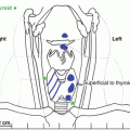
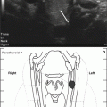
A 73-year-old female is incidentally found to have five right and two left 1–5 mm calyceal kidney stones on an abdominal ultrasound that are confirmed on a subsequent CT scan, both performed for an abdominal pain thought to be biliary in origin. She has no known history of passing kidney stones, but did have an episode of severe crampy abdominal and flank pain some years prior which resolved. Her past medical history is significant for cervical cancer at age 35, hysterectomy, bilateral knee replacement, gastritis, bronchitis, and insomnia. Her two children both have had symptomatic kidney stones. She is not on any lithogenic medications or thiazide diuretics. She is found to have serum calcium of 10.6 mg/dL (nl 8.9–10.1 mg/dL), phosphorus of 3.1 mg/dL (nl 2.5–4.5 mg/dL), and parathyroid hormone of 95 pg/mL (nl 15–65 pg/mL). Her 24-h urinary calcium was modestly elevated at 242 mg/day (normal for females <200 mg/day). Other notable values on the urine collection included a low volume (1200 mL) and a pH of 6.9 with a citrate of 419 mg/day (nl >430 mg/day). A subsequent sestamibi scan revealed an unequivocal left superior parathyroid adenoma.
Assessment and Diagnosis
Primary hyperparathyroidism (PHPT) is a known risk factor for urinary stone disease. Among patients presenting with urinary stone disease, the incidence of PHPT is 2–8 % [1]. In the era before routine serum calcium testing, 40–60 % of PHPT patients developed urinary calculi [2, 3]. In the current era, when more patients are diagnosed with asymptomatic PHPT based on blood calcium and parathyroid hormone (PTH) levels, most studies report a urinary stone incidence of less than 20 % [4]. This compares to a population urinary stone disease incidence of about 11 % [5]. On one extreme of the spectrum, among a cohort of patients with hypercalcemia, high PTH levels, and no secondary cause of hyperparathyroidism, Odvina and colleagues reported in 2007 that 60 % had a urinary stone [6]. In another cohort selected on the basis of surgically proven PHPT, Suh and colleagues reported in 2008 that renal sonograms revealed urinary stones in 7 % of asymptomatic patients [7].
The reason for increased lithogenesis in PHPT is still a matter of some uncertainty. Numerous studies have compared demographic factors as well as serum and urinary chemistries between PHPT patients with stones versus PHPT without stones, as well as controls. The best-established demographic risk factors are male gender and young age [8] (both unlike our current patient).
The physiologic actions of PTH supply some clues regarding possible mechanism(s) whereby PHPT might be associated with urinary stones. Major effects include stimulation of 1-alpha hydroxylation in the kidney, which in turn promotes formation of active 1,25-dihydroxy vitamin D, inhibition of renal phosphate reabsorption, and stimulation of bone resorption to release calcium and phosphate. Increased 1,25-dihydroxy vitamin D also stimulates gastrointestinal absorption of calcium and phosphorus. The net effect is to raise serum calcium but not phosphorus [9].
Although PTH also increases tubular reabsorption of calcium, net renal calcium excretion rises because the filtered load of calcium increases (due to the hypercalcemia) [10]. Since hypercalciuria is a well-defined risk factor for urinary stone disease, it is not surprising that PHPT might increase stone risk.
Nevertheless, the association between hypercalciuria and stone formation in patients with PHPT has been inconsistent in the literature. While some have found an association [6, 11–13], others find none [8, 14–16]. Thus a normal or relatively normal 24-h urine calcium excretion, as was the case for our patient, is often observed. It does seem that calcium metabolism is important, since calcium oxalate and phosphate stones predominate in PHPT, while uric acid stones are proportionally less common than in the general population [6, 10]. In addition, calcium phosphate stones appear to occur somewhat more commonly than in non-PHPT stone formers [10, 17]. Urine parameters other than calcium excretion have not been reproducibly associated with stone formation in PHPT. Polymorphisms in the calcium-sensing receptor gene may be associated with the risk of nephrolithiasis in PHPT [18].
Hypercalcemia is the most important clue to the diagnosis of PHPT in the setting of stone disease, since the vast majority of surgically proven cases will have at least some degree of elevation in serum calcium, often combined with hypophosphatemia [10]. Pooled from several studies, the mean serum calcium level for hyper parathyroid stone formers was 11.2 mg/dL (nl 8.9–10.1 mg/dL). A large portion of patients have values close to 10.5 mg/dL, which is the normal cutoff in many labs. Elevated serum calcium has a high positive prediction of hyperparathyroidism. The added feature of hypophosphatemia decreases the false-positive prediction. As PHPT is one of the few reversible causes of urinary stones, a high level of suspicion for the diagnosis is prudent in a stone former with even mild hypercalcemia [10, 19].
On the other hand, it is not possible to reliably diagnose PHPT in the absence of hypercalcemia, since in many such cases the elevation in PTH represents a physiologically appropriate response [9]. Thus routine screening of stone clinic populations with PTH levels in the absence of hypercalcemia is not recommended [19].
Management
The patient underwent an uneventful left superior parathyroidectomy. Unfortunately, 5 weeks later she suffered a myocardial infarct with somewhat a prolonged recovery. Therefore her asymptomatic renal stones were not addressed surgically at that time. Her serum calcium ranged from 8.7 to 9.7 mg/dL postoperatively.
Outcome
Fourteen and 31 months after the parathyroidectomy, the patient required surgery to remove ureteral stones. Their chemical composition was 80 % calcium oxalate monohydrate and 20 % hydroxyapatite. Prior to the latter event, she spontaneously passed a few other stones. In 8 years since the last stone surgery, she has not had a symptomatic stone event. A CT scan done for another indication 5 years after parathyroidectomy showed no further stones.
Related posts:

Stay updated, free articles. Join our Telegram channel

Full access? Get Clinical Tree


