Chapter Outline
DIAGNOSIS OF HYPERBILIRUBINEMIA
TOXICITY OF UNCONJUGATED HYPERBILIRUBINEMIA
NONPATHOLOGIC UNCONJUGATED HYPERBILIRUBINEMIA
DISORDERS OF PATHOLOGIC UNCONJUGATED HYPERBILIRUBINEMIA
MANAGEMENT AND TREATMENT OF UNCONJUGATED HYPERBILIRUBINEMIA
DISORDERS OF CONJUGATED HYPERBILIRUBINEMIA
Introduction
* URL referenced in this chapter includes: Online Mendelian Inheritance in Man (OMIM), http://www-ncbi-nlm-nih-gov.easyaccess2.lib.cuhk.edu.hk/Omim/ .
Elevation of the serum bilirubin level is a common, if not universal, finding during the first week of life and has been reviewed elsewhere. This can be a transient phenomenon that will resolve spontaneously or can signify a serious or even potentially life-threatening condition. There are many causes of hyperbilirubinemia and related therapeutic and prognostic implications. Independent of the cause, elevated serum bilirubin levels can be potentially toxic to the newborn infant. This chapter will review perinatal bilirubin metabolism and address assessment, etiology, toxicity, and therapy for neonatal jaundice. The diseases in which there is a primary disorder in the metabolism of bilirubin will be reviewed regarding their clinical presentation, pathophysiology, diagnosis, and treatment.Bilirubin Metabolism
Bilirubin Production and Transport
In 1864 Städeler used the term bilirubin, derived from Latin ( bilis, bile; ruber, red), for the red-colored bile pigment. Bilirubin is formed from the degradation of heme-containing compounds ( Fig. 4-1 ), the largest source of which is hemoglobin, and other sources are the cytochromes, catalases, tryptophan pyrrolase, and muscle myoglobin.
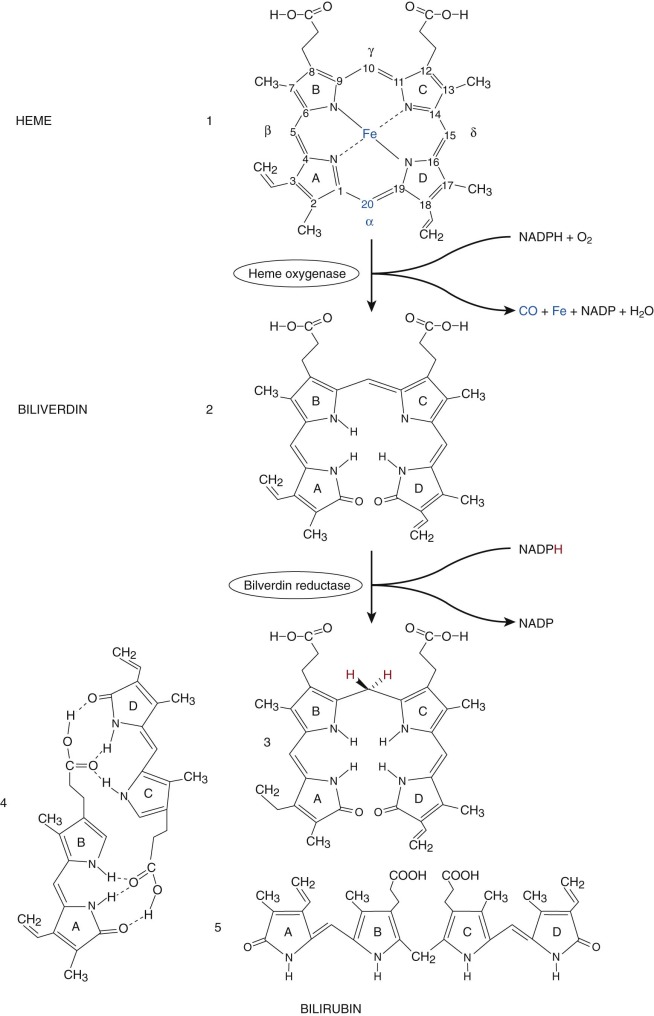
The formation of bilirubin is initiated by cleaving the tetrapyrrole ring of protoheme (protoporphyrin IX), which results in a linear tetrapyrrole (biliverdin). The first enzyme system and rate limiting step in bilirubin synthesis is microsomal heme oxygenase (HO). Two major forms of HO have been identified. HO1, the inducible form, is located in the spleen, liver, and bone marrow. HO2, the constitutive form, is located in testes, central nervous system (CNS), vasculature, liver, kidney, and gut.
HO results in reduction of the porphyrin iron (Fe III to Fe II ) and hydroxylation of the α methine (=C-) carbon. This α carbon is then oxidatively excised from the tetrapyrrole ring, yielding carbon monoxide (CO); this is the only physiologic source of CO. This excision opens the ring structure and is associated with oxygenation of the two carbons adjacent to the site of cleavage. The cleaved α carbon is excreted as CO, which has numerous biological effects, including neurotransmission, vasodilation as well as mediation of apoptosis and anti-inflammatory processes. The iron released by HO can be reutilized by the body. HO produces a linear tetrapyrrole, biliverdin IXα. The IX designation is a result of Fischer’s grouping of the protoporphyrin isomers, group IX being the physiological source of bilirubin. In utero, bilirubin IXβ is the first bile pigment seen and can be found in bile or meconium by 15 weeks’ gestation. Small amounts of bilirubin IXβ are also found in adult human bile. The central (C-10) carbon on biliverdin IXα is then reduced from a methine to a methylene group (-CH 2 -), thus forming bilirubin IXα. This is accomplished by the cytosolic enzyme biliverdin reductase. The proximity of this enzyme results in very little biliverdin ever being present in the circulation.
Bilirubin formation can be assessed by measurement of CO production. Such assessments indicate that the daily production rate of bilirubin is 6 to 8 mg/kg/24 hr in healthy term infants and 3 to 4 mg/kg/24 hr in healthy adults. In mammals, 80% to 85% of bilirubin produced daily originates from hemoglobin. Degradation of hepatic and renal heme appears to account for most of the remainder, reflecting the very rapid turnover of certain of these heme proteins. Although the precise fate of myoglobin heme is unknown, its turnover appears to be so slow as to be relatively insignificant.
Catabolism of hemoglobin occurs very largely from the sequestration of erythrocytes at the end of their life span (120 days is adult humans, 90 days in newborns). A small fraction of newly synthesized hemoglobin is degraded in the bone marrow. This process, termed ineffective erythropoiesis, normally represents less than 3% of daily bilirubin production but may be substantially increased in people with hemoglobinopathies, vitamin deficiencies, and heavy metal intoxication. Infants produce more bilirubin per unit of body weight because of their greater red blood cell mass and shorter red blood cell life span. Additionally, hepatic heme proteins represent a larger fraction of total body weight in infants.
Although bilirubin has long been thought of solely as a waste product, data suggest that some mild degree of hyperbilirubinemia may be helpful because of the anti-oxidant capacity of bilirubin and its potential role as a free-radical scavenger and cytoprotectant.
Bilirubin is poorly soluble in aqueous solvents and thus requires biotransformation to more water-soluble derivatives for excretion from the body. This poor solubility is related to the structure of bilirubin. Rather than being linear (structure 5, Fig. 4-1 ), bilirubin undergoes extensive internal hydrogen bonding (structure 4, Fig. 4-1 ). This shields the polar propionic acid side chains and makes bilirubin very nonpolar and lipophilic. The carbon–carbon double bonds at positions 4-5 and 15-16 can assume two different configurations (similar to cis and trans ) depending on whether the higher priority atoms or groups (based on atomic number) are on the same ( Z, zusammen, German: together ) or opposite ( E, entgegen, opposite ) sides of the double bond. The naturally occurring form of bilirubin, 4Z,15Z-bilirubin IXα, can be represented by any of the three structures (3-5) depicted at the bottom of Figure 4-1 . Knowledge of this stereochemistry is important in understanding phototherapy, which will be discussed later.
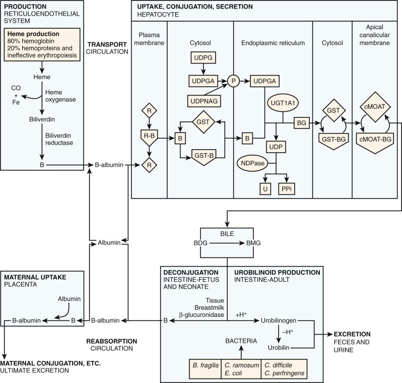
Bilirubin’s poor aqueous solubility necessitates a carrier molecule, albumin, for transport from sites of production in the reticuloendothelial system to the liver for excretion ( Fig. 4-2 ). Each albumin molecule possesses a single high-affinity (K a =7 × 10 7 M -1 ) binding site for one molecule of bilirubin. A binding affinity of this magnitude implies that, at normal serum bilirubin levels, all bilirubin will be transported to the liver bound to albumin, with negligible amounts free to diffuse into other tissues. Secondary binding sites of lesser affinity also exist on albumin. Albumin also serves as a carrier for other compounds such as xenobiotics and fatty acids.
Hepatic Uptake of Bilirubin
The structure of the liver is well suited for the uptake of bilirubin by individual hepatocytes. Cords of hepatocytes are arranged radially so that adjacent sinusoids border all hepatocytes. The flow of blood through the sinusoids is slower than that of other capillary beds because it is generated by portal venous pressure rather than arterial pressure. There is easy passage of albumin-bound bilirubin from the plasma into the tissue fluid space (space of Disse) between the endothelium and the hepatocyte because the sinusoidal endothelium of the liver lacks the basal laminae, which are found in other organ capillary systems. The pores of the endothelium allow direct contact with the plasma membrane of the hepatocyte.
A hepatocyte with a schematic illustration of bilirubin metabolism is shown in Figure 4-2 . In the first step, bilirubin dissociates from its albumin carrier and enters the hepatocyte via a membrane-receptor carrier, which facilitates entry into the hepatocyte. Carrier-mediated transport into the hepatocyte has been demonstrated for several organic anions including bilirubin, bromsulfophthalein (BSP), and indocyanine green (ICG), though bilirubin has also been shown to be able to pass through membranes by simple passive diffusion. Evidence suggests that bilirubin, BSP, and ICG share the same hepatocyte-receptor carrier because they exhibit competitive inhibition when injected simultaneously. This finding cannot be explained by subsequent intrahepatic metabolism, because these anions are handled differently by the hepatocyte: Bilirubin is conjugated with glucuronic acid in the endoplasmic reticulum (ER), BSP is conjugated with glutathione in the cytosol, and ICG is excreted directly without biotransformation. Data from rat hepatocytes suggest that the anion binding–receptor carrier is a dimeric protein with subunit molecular weight of 55,000. Antibody studies confirm the expected location in the plasma membrane and demonstrate blocking of uptake. Organic anion transporting polypeptide 2 (OATP2, recently named OATP1B1 under new nomenclature ) has shown high-affinity uptake of bilirubin in the presence of albumin and is a member of the OATP family, transporter symbol SLC21A.
Carrier-mediated transport of bilirubin into the hepatocyte is necessary because of differences in protein-binding inside and outside the hepatocyte. Outside the hepatocyte, bilirubin is bound to albumin (affinity constant ~10 8 , concentration: 0.6 mM). Inside the hepatocyte, bilirubin is bound to glutathione S-transferase B (GST), historically known as ligandin or the Y protein (affinity constant ~10 6 , concentration 0.04 mM). GST constitutes a family of proteins that exhibit important functions both as enzymes and as intracellular binding proteins for nonsubstrate ligands such as bilirubin. Carrier-mediated uptake helps generate a concentration gradient for bilirubin uptake despite the difference in affinity between albumin and GST. GST is important in the intracellular storage of bilirubin and bilirubin conjugates and reduces efflux from the hepatocyte back into plasma.
Bilirubin Conjugation
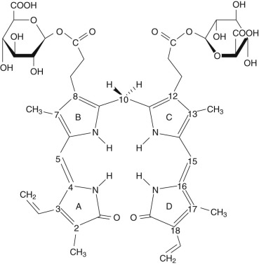
Inside the hepatocyte, bilirubin is conjugated with glucuronic acid within the ER (microsomes). The glucuronic acid donor is uridine diphosphate glucuronic acid (UDPGA). The conjugation results in an ester linkage formed with either or both of the propionic acid side chains on the B and C pyrrole rings of bilirubin ( Fig. 4-3 ). The enzyme responsible for this esterification is bilirubin UDP-glucuronosyltransferase (BUGT; Online Mendelian Inheritance in Man [OMIM] *191740). BUGT is distinct from the other glucuronosyltransferase isoforms which catalyze the conjugation of thyroxine, steroids, bile acids, and xenobiotics. BUGT (which is the same as UGT1A1, see below) is embedded in the lipid environment of the microsomal membrane, and perturbations of this environment greatly affect in vitro measurements of BUGT activity. Since BUGT is located on the interior of the ER, the existence of a permease has been hypothesized to facilitate the transport of UDPGA from the cytosol across the lipid layers of the ER. The permease has been proposed because uridine diphosphate glucose (UDPG) is present in the cytosol in higher concentrations, yet UDPGA serves as the preferred donor for bilirubin conjugation. Uridine diphosphate N-acetyl glucosamine (UDPNAG) is considered to be a natural regulator of BUGT because UDPNAG increases in vitro BUGT activity threefold. The mechanism for this is unknown and could possibly involve facilitation of the permease UDPGA transporter. After providing glucuronic acid for conjugation, UDP is converted to uridine and inorganic pyrophosphate by a nucleoside diphosphatase (NDPase) that is also located on the interior of the ER and prevents the reverse reaction.
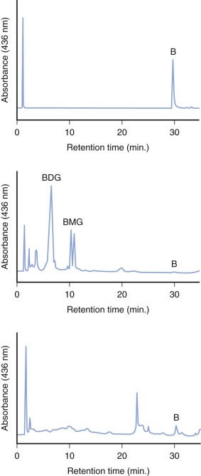
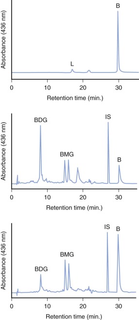
The specific isoform responsible for bilirubin conjugation is UGT1A1 (trivial name HUG-Br1 or BUGT, EC 2.4.1.17). This is part of the UDP glycosyltransferase superfamily of enzymes encoded by the UGT gene complex on chromosome 2 that is involved with metabolism of many xenobiotic and endogenous substances. The UGT1 gene encodes several isoforms and has a complex structure consisting of four common exons (2-5) and 13 variable exons encoding different isoforms ( Fig. 4-4 ). At least 30 different UGT1 mutant alleles have been described that cause Gilbert’s syndrome and Crigler-Najjar (CN) syndromes I and II. UGT1A1 catalyzes the formation of both bilirubin mono- and diglucuronides but also metabolizes other hormones and drugs, so mutations could be involved in carcinogenesis and adverse drug reactions. In normal adult humans the majority of bilirubin conjugates are excreted in the bile as bilirubin diglucuronides (approximately 80%) ( Fig. 4-5 , middle panel). Lesser amounts of bilirubin monoglucuronides (approximately 15%) are also excreted along with very small amounts of unconjugated bilirubin (UBIL) and other bilirubin conjugates (e.g., glucose, xylose, and mixed diesters). In infants, because there is lower UGT1A1 activity, bile contains less bilirubin diglucuronide and more bilirubin monoglucuronide than the adult ( Fig. 4-6 , middle panel).

Bilirubin Excretion
Following conjugation, bilirubin conjugates are excreted against a concentration gradient from the hepatocyte through the canalicular membrane into the bile. The ATP-dependent transporter responsible for bilirubin glucuronide passage from the hepatocyte through the canalicular membrane is canalicular multispecific organic anion transporter (cMOAT). cMOAT is a member of the ATP-binding cassette (ABC) transporter superfamily and is homologous to the multidrug resistance–associated protein (MRP2). It is also known as ABCC2 because it is encoded by the ABCC2 gene. cMOAT/MRP2/ABCC2 is involved with ATP-dependent transport across the apical canalicular membrane of a variety of endogenous compounds and xenobiotics including both bilirubin mono- and diglucuronide. cMOAT has previously been described as the nonbile acid organic anion transporter, the glutathione S-conjugate export pump, or the leukotriene export pump. Genetic mutations which alter these ABC transporters cause diseases which include cystic fibrosis, hyperinsulinemia, adrenoleukodystrophy, multidrug resistance, and, as will be discussed later in this chapter, Dubin-Johnson syndrome. This mechanism can be saturated with increasing amounts of bilirubin or bilirubin conjugates. Many other organic anions (e.g., BSP, ICG), share this same canalicular membrane excretion mechanism. Simultaneous infusions of BSP and ICG will decrease the maximal canalicular excretion of bilirubin and vice versa. The canalicular excretion mechanism for bilirubin and BSP are different from that of bile salts. Biliary excretion of conjugated bilirubin and BSP is decreased in individuals with the Dubin-Johnson syndrome, though bile salt excretion in not impaired. However, bile salt and bilirubin conjugate excretion by the canalicular membrane are not completely independent since infusion of bile salts does increase the maximal excretion of bilirubin conjugates. A similar effect in seen with phenobarbital. Conversely the maximal excretion of bilirubin conjugates can be decreased by cholestatic agents like estrogens and anabolic steroids.
Under normal conditions there is evidence that bilirubin conjugates equilibrate across the sinusoidal membrane of hepatocytes resulting in small amounts of bilirubin conjugates being present in the systemic circulation. If there is diminished hepatic glucuronidation of bilirubin (e.g., in the neonate), there will be a decreased amount of bilirubin conjugates present in the serum. Data show that in full-term newborns there is an increase in the serum level of bilirubin diconjugates (0.55 ± 0.25% on days 2 to 4 to 1.62 ± 0.99% on days 9 to 13) which are consistent with the maturation of bilirubin glucuronidation. In contrast, in premature infants less than 33 weeks’ gestation, bilirubin diconjugates were very low and remained so, suggesting a more severe immaturity of the glucuronidation process.
In many pathologic circumstances, bilirubin mono- and diglucuronides are not excreted from the hepatocyte fast enough to prevent significant reflux back into the circulation. The resulting elevation of serum bilirubin conjugate levels results in the transesterification of bilirubin glucuronide with an amino group on albumin, producing a covalent bond between albumin and bilirubin. This product is formed spontaneously and is known as delta bilirubin or bilirubin-albumin. Similar nonenzymatic reactions have been demonstrated between albumin and various drugs. Delta bilirubin is not formed in hyperbilirubinemic conditions unless there is elevation of the conjugated bilirubin fraction. Both delta bilirubin and bilirubin conjugates are direct reacting, which explains a situation that has long confounded clinicians. The direct bilirubin may continue to be elevated in patients who otherwise are recovering from a hepatic insult because the delta bilirubin formed lingers due to the long (~20 day ) half-life of albumin.
Enterohepatic Circulation of Bilirubin
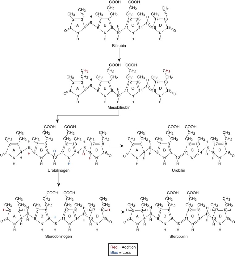
When bilirubin conjugates enter the intestinal lumen (see Fig. 4-2 ), several possibilities for further metabolism arise. In adults, the normal bacterial flora hydrogenate various carbon double bonds in bilirubin to produce assorted urobilinogens ( Fig. 4-7 ). Subsequent oxidation of the middle (C-10) carbon produces the related urobilins. Because there are a large number of unsaturated bonds in bilirubin, there are many compounds formed by reduction and oxidation of these bonds. This large family of related reduction–oxidation products of bilirubin is known as urobilinoids and excreted in the feces and urine. Bacteria capable of producing urobilinoids include Clostridium ramosum, Escherichia coli, Bacteroides fragilis, Clostridium perfringens, and Clostridium difficile. The conversion of bilirubin conjugates to urobilinoids is important because it blocks the intestinal absorption of bilirubin known as the enterohepatic circulation. Neonates lack an intestinal bacterial flora and are more likely to absorb bilirubin from the intestine. This difference in bile pigment excretion between adults and neonates is demonstrated by comparing Figures 4-5 and 4-6 (lower panels).
Bilirubin conjugates in the intestine can also act as substrate for either bacterial or endogenous tissue β-glucuronidase. This enzyme hydrolyses glucuronic acid from bilirubin glucuronides. The UBIL produced is more rapidly absorbed from the intestine. In the fetus, tissue β-glucuronidase is detectable by 12 weeks’ gestation and is believed to play an important role in facilitating intestinal bilirubin absorption that enables bilirubin to be cleared via the placenta. Following birth, increased intestinal β-glucuronidase can increase the neonate’s likelihood of experiencing higher serum bilirubin levels. The ability of endogenous tissue β-glucuronidase to deconjugate bilirubin glucuronides has been demonstrated in germ-free animals. Breastmilk can contain high levels of β-glucuronidase, and it has been suggested that this is one factor related to the higher jaundice levels seen in breastfed infants. Feeding specific nutritional ingredients that inhibit β-glucuronidase, such as hydrolyzed casein or L-aspartic acid have been shown to result in increased fecal bilirubin excretion and lower levels of jaundice.
Diagnosis of Hyperbilirubinemia
Jaundice and icterus both refer to the yellow discoloration of the tissues (skin, sclerae, etc.) caused by deposition of bilirubin. Jaundice, from the French jaune means yellow. Icterus is derived from the Greek word for jaundice (ikteros). Jaundice is a sign that hyperbilirubinemia exists (i.e., total serum bilirubin greater than approximately 1.4 mg/dL after 6 months of age, 1 mg/dL = 17 µmol/L). The degree of jaundice is directly related to the level of serum bilirubin and the amount of bilirubin deposited in the extravascular tissues. Hypercarotenemia, occasionally mistaken for jaundice, can impart a yellow hue to the skin, but the sclerae remain white. There are many conditions associated with neonatal jaundice. Some of these states are so commonly recognized as to be termed physiologic. Alternatively jaundice can be a sign of severe disease.
Total Serum Bilirubin Measurements
Measurement of the total serum bilirubin concentration allows quantitation of jaundice. Such measurements are very common in the newborn nursery and in one study were made at least once in 61% of term newborn infants. Two components of total serum bilirubin can be routinely measured in the clinical laboratory: conjugated bilirubin (“direct” reacting, because in Van den Bergh’s test, color development takes place directly without adding methanol), and UBIL (“indirect” fraction). Although the terms direct and indirect are often used equivalently with conjugated and UBIL, this is not quantitatively correct because the direct fraction includes both conjugated bilirubin and albumin-bound delta bilirubin. Elevation of either of these fractions can result in jaundice. There is a long history of undesirable variability in the measurement of serum bilirubin fractions. One problem is the ditaurobilirubin (DTB) content of standards set by the College of American Pathologists (CAP). The DTB influences the result variably because of protein matrix differences related to the specific bilirubin measurement used. This has prompted the suggestion that standards consist of human serum enriched solely with UBIL rather than bovine serum containing a mixture of UBIL and DTB. The automated laboratory methods now used to measure serum bilirubin have been reviewed elsewhere. The Jendrassik-Grof procedure is the method of choice for total bilirubin measurement, though this method also has problems. When the total serum bilirubin level is high, factitious elevation of the direct fraction has been reported. Three newer methods have been developed which can more accurately determine the various bilirubin fractions (unconjugated, monoconjugated, diconjugated, and albumin-bound or delta): high-performance liquid chromatography (HPLC), multilayered slides, and use of bilirubin oxidase. HPLC analysis is superior and used as the gold standard, but it is too expensive and time consuming for the clinical laboratory. HPLC analysis of serum from normal human neonates in the first 4 days of life showed unconjugated and conjugated bilirubin levels rose in parallel with the conjugated fraction, making up only 1.2% to 1.6% of total pigment (3.6% in adults). Although the absolute concentration of conjugates was 2 to 6 times higher in neonates, only 20% were diconjugates (54% in adults). These sensitive HPLC data are consistent with the increased bilirubin production and relatively deficient glucuronidation seen in the neonate. Analysis with automated multilayered slide technology (VITROS, Johnson & Johnson) used in many clinical laboratories allows measurement of specific conjugated and UBIL fractions without inclusion of delta bilirubin. The conjugated bilirubin measurement is an earlier indicator of relief from biliary cholestasis than is direct bilirubin because of the long half-life of delta bilirubin. A comparison of the bilirubin oxidase method concluded that determinations of total bilirubin in neonatal serum were not advanced by this method.
Newer methods of total bilirubin measurement (Twin Beam, Ginevri, Rome, Italy; ABL 735, Radiometrer, Copenhagen, Denmark; Roche OMNI S, Roche Diagnostics, Graz, Austria) using nonenzymatic photochemical analysis offer the convenience of bilirubin quantitation outside of a core lab setting (i.e., blood gas analyzer in the nursery) using a smaller blood sample than traditional serum analysis. However, these measurements must be interpreted with caution because the instruments tend to underestimate the total bilirubin concentration at greater than 15 mg/dL.
There are conflicting data regarding the accuracy of capillary versus venous serum bilirubin levels. However, as Maisels has pointed out, the literature regarding kernicterus, phototherapy, and exchange transfusion is based on bilirubin measurements in capillary samples.
Transcutaneous Bilirubinometry
Noninvasive methods to measure jaundice levels have been shown to be useful in neonates. Current commercially available methods include the BiliChek (Respironics, Pittsburgh, Pennsylvania) and the Minolta/Hill-Rom Air-Shields Transcutaneous Jaundice Meter 103 (Air-Shields, Hatboro, Pennsylvania). The technique utilizes principles of reflectance spectrophotometry, has been validated against both HPLC and clinical laboratory measures, and is advocated by the American Academy of Pediatrics (AAP). The device is touched to the skin in a painless manner with the immediate resulting point-of-care measurement of transcutaneous bilirubin (TcB). Significant TcB levels (e.g., greater than 13 mg/dL) should prompt measurement of a serum or plasma bilirubin level. Some suggest that the yellow color of the skin is a better risk indicator of bilirubin-dependent brain damage than is the serum bilirubin level. A less expensive method useful in assessing jaundice utilizes a plexiglass color chart pressed against the baby’s nose (Ingram icterometer, Thos. A. Ingram and Co. Ltd, Birmingham, England).
Toxicity of Unconjugated Hyperbilirubinemia
Kernicterus
Reviews of neonatal bilirubin toxicity have been published elsewhere. Kernicterus (German: kern : nucleus ; Latin: icterus : yellow) is the neuropathologic finding associated with severe unconjugated hyperbilirubinemia and is named for the yellow staining of certain regions of the brain, particularly the basal ganglia, hippocampus, cerebellum, and nuclei of the floor of the fourth ventricle. Acute clinical findings associated with kernicterus, termed acute bilirubin encephalopathy, include sluggish Moro reflex, opisthotonos, retrocollis, hypotonia, vomiting, high-pitched cry, hyperpyrexia, seizures, paresis of gaze (“setting sun sign”), oculogyric crisis, and death. Long-term findings (chronic bilirubin encephalopathy) include spasticity, choreoathetosis, dental enamel dysplasia of the desiduous teeth, and sensorineural hearing loss. Milder forms of bilirubin encephalopathy include cognitive dysfunction and learning disabilities. In 17-year-old males, the risk of having an IQ score less than 85 was found to be significantly higher among full-term subjects with neonatal serum bilirubin levels above 20 mg/dL. The mechanisms of bilirubin cytotoxicity are complex and have been reviewed elsewhere. Though the neonatal period is the most common time for bilirubin-related brain damage, the neurotoxicity of bilirubin has also been documented in older children and adults with CN syndrome, type I.
The absolute level of serum bilirubin has not been a good predictor of the risk of bilirubin encephalopathy. However, it has long been known that kernicterus is likely with serum UBIL levels greater than 30 mg/dL and unlikely with levels less than 20 mg/dL. In one study, 90% of the patients who had a bilirubin level greater than 35 mg/dL either died or had cerebral palsy or physical retardation. Alternatively, no developmental retardation was found in 129 infants with bilirubin levels less than 20 mg/dL. Albumin concentration is an important variable because of the high affinity binding with bilirubin, and the ratio of bilirubin to albumin is a risk factor that has been included in the most recent AAP guidelines. Drugs and organic anions also bind to albumin and can displace bilirubin, thereby increasing the free bilirubin which can diffuse into cells and cause toxicity, such as when sulfisoxazole was given to premature infants. Walker has reviewed neonatal bilirubin toxicity and drug-induced bilirubin displacement. In recent years there has been a reemergence of kernicterus. While it has been acknowledged that total serum bilirubin may not be the most important factor related to risk, there are, at present, no other generally accepted tests (e.g., albumin saturation, reserve bilirubin binding capacity, or free bilirubin concentration) which are more helpful in identifying infants at risk for bilirubin encephalopathy. Recently the basics of bilirubin–albumin binding and neonatal jaundice have been reviewed by Ahlfors and Wennberg. Additionally, Ahlfors and colleagues have recently described how to use zone fluidics (the precisely controlled physical, chemical, and fluid-dynamic manipulation of zones of miscible and immiscible fluids and suspended solids in narrow bore conduits to accomplish sample conditioning and chemical analysis) to measure free bilirubin and overcome some of the existing problems with unbound (free) bilirubin measurement. Wennberg and colleagues speculate that enhanced sensitivity and specificity of newer methods of free bilirubin measurement will provide for the establishing of improved risk threshold for kernicterus and therefore reduce unnecessary aggressive intervention (i.e., exchange transfusion) and its associated cost and morbidity.
Another approach aimed at measuring early changes in the CNS caused by bilirubin has assessed brainstem auditory evoked potentials (BAEP). Abnormalities in the BAEP have been demonstrated in jaundiced infants and shown to improve after exchange transfusion. Data have shown that even moderate hyperbilirubinemia (mean ± SD: 14.3 ± 2.8 mg/dL) affects the BAEP, specific components of the Brazelton Neonatal Behavioral Assessment Scale, and cry characteristics.
Another reported toxicity of neonatal jaundice relates not to the CNS of the jaundiced infant, but rather to the attitude of that infant’s parents. Mothers of jaundiced infants more frequently exhibit behavior suggesting the “vulnerable-child syndrome,” including inappropriate visits to the physician because of unrealistic perceptions of illness, separation difficulties, and early termination of breastfeeding. Another study showed, however, that hyperbilirubinemia and/or phototherapy during the neonatal period are not associated with impaired mother–child attachment after the first year of life.
Nonpathologic Unconjugated Hyperbilirubinemia
Physiologic Jaundice
The term physiologic jaundice has been used to describe the frequently observed jaundice in otherwise completely normal neonates. However, physiologic jaundice is merely the result of a number of factors involving increased bilirubin production and decreased excretion.
Jaundice should always be considered to be a sign of a possible disease and not routinely explained as physiologic. Specific characteristics of neonatal jaundice to be considered abnormal until proven otherwise include: (1) development before 36 hours of age; (2) persistence beyond 10 days of age; (3) serum bilirubin greater than 12 mg/dL at any time; (4) elevation of the direct bilirubin (direct bilirubin greater than 1 mg/dL if the total bilirubin is less than 5 mg/dL, or direct bilirubin greater than 20% of the total bilirubin if the total bilirubin is greater than 5 mg/dL ). The physiologic immaturity of premature infants is associated with both higher peak serum bilirubin levels and pathologic conditions.
In general, infants are not jaundiced at the moment of birth, due to the impressive ability of the placenta to clear bilirubin from the fetal circulation. However, within the next few days most if not all infants will develop elevated serum bilirubin levels (greater than 1.4 mg/dL), with clinical jaundice evident when the total bilirubin level exceeds 2 to 5 mg/dL. As the serum bilirubin rises, the skin becomes more jaundiced in a cephalocaudal manner. Kramer found the following serum indirect bilirubin levels as jaundice progressed: head and neck: 4 to 8 mg/dL; upper trunk: 5 to 12 mg/dL; lower trunk and thighs: 8 to 16 mg/dL; arms and lower legs: 11 to 18 mg/dL; palms and soles: greater than 15 mg/dL. Hence when the bilirubin was greater than 15 mg/dL, the entire body was icteric. However, darker skin tones can make jaundice difficult to estimate visually. Jaundice is best observed by blanching the skin with gentle digital pressure under well-illuminated (white light) conditions. At least one third of infants develop visible jaundice. A combined analysis of several large studies involving thousands of infants during the first week of life showed that moderate jaundice (bilirubin greater than 12 mg/dL) occurs in at least 12% of breastfed infants and 4% of formula-fed infants, while severe jaundice (greater than 15 mg/dL) occurs in 2% and 0.3% of these respective feeding groups.
There are a number of epidemiologic risk factors related to neonatal jaundice that have been reviewed elsewhere. Risk factors include male gender, low birth weight, prematurity, polycythemia, certain ethnicities (Asian, Native American, Greek), maternal medications (e.g., oxytocin, promethazine hydrochloride), premature rupture of the membranes, increased weight loss after birth, delayed meconium passage, breastfeeding, and neonatal infection. Delivery with the vacuum extractor increases the risk of cephalohematoma and neonatal jaundice. Data suggest pancuronium is associated with an increased risk of hyperbilirubinemia. There is a significant correlation between umbilical cord serum bilirubin level and subsequent hyperbilirubinemia. Maternal serum bilirubin level at the time of delivery and transplacental bilirubin gradient also correlate positively with neonatal serum bilirubin concentrations. Factors associated with decreased neonatal bilirubin levels include African race, exclusive formula feeding, gestational age 41 weeks, maternal smoking, and certain drugs given to the mother (e.g., phenobarbital).
Breastfeeding and Jaundice
Breastfeeding has been clearly identified as a factor related to neonatal jaundice, and this subject has been reviewed elsewhere. Breastfed infants have been shown to have significantly higher serum bilirubin levels than formula-fed infants on each of the first 5 days of life, and this unconjugated hyperbilirubinemia can persist for weeks to months. Jaundice during the first week of life is sometimes described as breastfeeding jaundice in order to differentiate it from breastmilk jaundice syndrome, which occurs after the first week of life. The former is frequently associated with inadequate breastmilk intake, whereas the latter generally occurs in otherwise thriving infants. There is probably overlap between these conditions and physiologic jaundice. Early reports linking breastmilk and neonatal jaundice with a steroid (pregnane-3(α),20(β)-diol) in some milk samples have not been confirmed by more recent larger studies utilizing more sensitive methods. There are also conflicting data regarding efforts to attribute this jaundice to increased lipase activity in the breastmilk, resulting in elevated levels of free fatty acids, which could inhibit hepatic glucuronosyltransferase. It has been suggested that the enterohepatic circulation of bilirubin can be facilitated by the presence of β-glucuronidase or some other substance in human milk. Other factors possibly related to jaundice in breastfed infants include caloric intake, fluid intake, weight loss, delayed meconium passage, intestinal bacterial flora, and inhibition of bilirubin glucuronosyltransferase by an unidentified factor in the milk. Lascari suggests that a healthy breastfed infant with unconjugated hyperbilirubinemia, normal hemoglobin concentration, reticulocyte count, blood smear, no blood group incompatibility, and no other abnormalities on physical examination may be presumed to have early breastfeeding jaundice. Because there is no specific laboratory test to confirm a diagnosis of breastmilk jaundice, it is important to rule out treatable causes of jaundice before ascribing the hyperbilirubinemia to breastmilk. Some infants with presumed breastmilk jaundice exhibit elevated serum bile acid levels suggesting mild hepatic dysfunction or cholestasis, though in general this is not the case. Breastfed infants who are fed specific nutritional ingredients, such as L-aspartic acid, that inhibit β-glucuronidase excrete more fecal bilirubin and have lower levels of jaundice than breastfed infants who receive no supplements. No commercial preparations of these specific ingredients are presently available.
Disorders of Pathologic Unconjugated Hyperbilirubinemia
Disorders of Production
Isoimmunization
The most common cause of severe early jaundice is fetal–maternal blood group incompatibility with resulting isoimmunization (see Chapter 3 ). Maternal immunization develops when erythrocytes leak from fetal to maternal circulation. Fetal erythrocytes carrying different antigens are recognized as foreign by the maternal immune system that forms antibodies against them (maternal sensitization). These antibodies (IgG immunoglobulins) cross the placental barrier into the fetal circulation and bind to fetal erythrocytes. In Rh incompatibility, sequestration and destruction of the antibody-coated erythrocytes takes place in the reticuloendothelial system of the fetus. In ABO incompatibility, hemolysis is intravascular, complement mediated, and usually not as severe as in Rh disease. Significant hemolysis can also result from incompatibilities between minor blood group antigens (e.g., Kell). Although hemolysis is predominantly associated with elevation of UBIL, the conjugated fraction can also be elevated. Any type of isoimmune hemolytic disease is a risk factor for kernicterus.
Rh incompatibility problems do not usually develop until the second pregnancy, thus prenatal blood typing and serial testing of Rh-negative mothers for the development of Rh antibodies provide important information to guide possible intrauterine care. If maternal Rh antibodies develop during pregnancy, potentially helpful measures include serial amniocentesis (with bilirubin measurement), ultrasound assessment of the fetus, intrauterine transfusion, and premature delivery. The prophylactic administration of anti-D gamma globulin has been most helpful in preventing Rh sensitization. The newborn infant with Rh incompatibility presents with pallor, hepatosplenomegaly, and a rapidly developing jaundice in the first hours of life. In severe cases the infant may be born with generalized edema (fetal hydrops). Laboratory findings in the neonate’s blood include reticulocytosis, anemia, a positive direct Coombs’ test, and a rapidly rising serum bilirubin level. Conjugated hyperbilirubinemia is occasionally observed in Rh erythroblastosis, particularly when intrauterine transfusions have been used. This may be the result of exceeding the hepatic excretory capacity. Alternatively, cholestasis may also result from hepatic congestion from extramedullary erythropoiesis and heart failure, or tissue hypoxia resulting from reduced hepatic blood flow and anemia. Cholestasis in infants with erythroblastosis fetalis may also be exacerbated by secondary complications, including biliary obstruction, sepsis, or hepatic failure.
Therapeutic intervention with phototherapy and consideration of exchange transfusion should be initiated at lower serum bilirubin concentrations in infants with isoimmune hemolysis, and intravenous gamma-globulin has been shown to reduce the need for exchange transfusions in Rh and ABO hemolytic disease. Serum bilirubin measurements should be obtained as frequently as every 4 hours to establish the rate of increase and to assess the effect of therapy. Exchange transfusions may be performed as emergency procedures in severe cases almost immediately after delivery in order to treat anemia and heart failure.
In contrast to Rh incompatibility, ABO incompatibility usually presents clinically with the first pregnancy. ABO hemolytic disease is largely limited to blood group A or B infants born to group O mothers. ABO hemolytic disease is relatively rare in type A or B mothers. Development of jaundice is not as rapid as with Rh disease, and a serum bilirubin greater than 12 mg/dL on day 3 of life would be typical. Laboratory abnormalities include reticulocytosis (greater than 10%) and a weakly positive direct Coombs’ test, though this is sometimes negative. Anti-A or anti-B antibodies may be seen in the serum of the newborn if examined within the first few days of life, before they rapidly disappear. Spherocytes are the most prominent feature seen in the peripheral blood smear with ABO incompatibility.
Erythrocyte Enzymatic and Structural Defects
A number of specific abnormalities of the red blood cell can result in neonatal jaundice, including hemoglobinopathies and red blood cell membrane and enzyme defects. Hereditary spherocytosis (see Chapter 16 ) can be a neonatal problem with significant hemolysis and present with a rising bilirubin level and a falling hematocrit. A family history of spherocytosis, anemia or early gallstone disease (before age 40) is helpful in suggesting this diagnosis. The characteristic spherocytes seen in the peripheral blood smear may be impossible to distinguish from those seen with ABO hemolytic disease. Other erythrocyte conditions associated with neonatal jaundice include drug-induced hemolysis, deficiencies of the erythrocyte enzymes (glucose 6-phosphate dehydrogenase [G6PD] deficiency [see Chapter 17 ], pyruvate kinase deficiency, and others) (see Chapter 18 ), and hemolysis induced by vitamin K or bacteria. Recent studies of G6PD activity in African-American neonates suggest that hemolysis is neither the predominate factor in the pathogenesis of hyperbilirubinemia, nor is it alone predictive of hyperbilirubinemia, though African-American male neonates may be at higher risk for hyperbilirubinemia, particularly if breastfed and premature. This emphasizes the important relationship between bilirubin excretion and serum bilirubin concentration. α-thalassemia (see Chapter 21 ) can result in severe hemolysis and lethal hydrops fetalis. β-thalassemia (see Chapter 21 ) may also present with hemolysis and severe neonatal hyperbilirubinemia. There are a wide variety of clinical findings associated with the thalassemias, extending from profound intrauterine hydrops and death, to mild neonatal jaundice and anemia, to no jaundice or anemia. Southeast Asian ovalocytosis (see Chapter 16 ) has been associated with severe hyperbilirubinemia. These red blood cell abnormalities are more likely to result in hyperbilirubinemia in the presence of Gilbert’s syndrome as described later in this chapter.
Drugs or other substances responsible for hemolysis can be passed to the fetus across the placenta or to the neonate via breastmilk. Induction of labor with oxytocin has been shown to be associated with neonatal jaundice. There is a significant association between hyponatremia and jaundice in infants of mothers who received oxytocin to induce labor. The vasopressin-like action of oxytocin prompts electrolyte and water transport, and it is suggested that the erythrocyte swells, and increased osmotic fragility and hyperbilirubinemia can result. Steroid administration at the initiation of oxytocin and 4 hours later may be helpful in preventing this hyperbilirubinemia.
Infection
Bacterial sepsis increases bilirubin production through initiation of erythrocyte hemolysis or via endotoxin-mediated reduction of canalicular bile secretion, resulting in a combination of conjugated and unconjugated hyperbilirubinemia. However, hyperbilirubinemia is rarely the only manifestation of bacteremia or sepsis, and some data suggest sepsis may result in lower peak levels of UBIL through consumption of bilirubin as an antioxidant in response to oxidant production by phagocytic cells.
Sequestration
Extravascular blood within the body can be rapidly metabolized to bilirubin by tissue macrophages. Examples of this increased bilirubin production include cephalohematoma, ecchymoses, petechiae, and hemorrhage. Although the diagnosis can often be made on physical examination, occult intracranial, intestinal, adrenal, or pulmonary hemorrhage can also produce hyperbilirubinemia. Similarly, swallowed blood can be converted to bilirubin by the HO of intestinal epithelium. From a red sample of bloody gastric contents, meconium, or stool, the Apt test can be used to distinguish blood of maternal or infant origin because of differences in alkali-resistance between fetal and adult hemoglobin; however, this test has low sensitivity and poor reproducibility, and an HPLC method has been shown to be superior.
Polycythemia
Polycythemia can cause hyperbilirubinemia because the absolute increase in red cell mass results in elevated bilirubin production through normal rates of erythrocyte breakdown. A number of mechanisms may result in neonatal polycythemia (usually defined by venipuncture hematocrits greater than 65%) as reviewed by Werner. Maternal–fetal transfusion during peripartum placental separation, a delay in umbilical cord clamping at birth, and twin–twin transfusion syndrome are all commonly encountered causes of polycythemia. Similarly, intrauterine hypoxia and maternal diseases such as diabetes mellitus can result in neonatal polycythemia. Therapy for symptomatic polycythemia is partial exchange transfusion, though therapy for asymptomatic polycythemia remains controversial.
Disorders of Conjugation
The disorders described in this section, summarized in Tables 4-1 and 4-2 , are those in which there is a primary abnormality in bilirubin metabolism without other liver disease. The disorders can best be understood in the context of the normal pathway in which bilirubin is cleared from the circulation (see Fig. 4-2 ). Defects in these metabolic steps are responsible for the disorders to be described in the final section of this chapter.
| Gilbert’s Syndrome | Crigler-Najjar Type I | Crigler-Najjar Type II | ||
|---|---|---|---|---|
| Prevalence | 3% | Rare | Rare | |
| Inheritance | Autosomal dominant or recessive | Autosomal recessive | Autosomal recessive, rarely dominant | |
| Genetic Defect | UGT1A1 gene | UGT1A1 gene | UGT1A1 gene | |
| Hepatocyte Defect Site | Microsomes +/− plasma membrane | Microsomes | Microsomes | |
| Deficient Hepatocyte Function | Glucuronidation +/− uptake | Glucuronidation | Glucuronidation | |
| BUGT Activity | 5%-53% of controls | Severely decreased | 2%-23% of controls | |
| Hepatocyte Uptake | Decreased in 20%-30% | Normal | Normal | |
| Serum Total Bilirubin Level (mg/dL) | 0.8-4.3 | 15-45 | 8-25 | |
| Serum Bilirubin Decrease with Phenobarbital (%) | 70 | 0 | 77 | |
| HPLC Serum Bilirubin Composition | ||||
| (Normal %) | ||||
| Unconjugated | (92.6) | 98.8 | ~100 | 99.1 |
| Diglucuronide | (6.2) | 1.1 | 0 | 0.6 |
| Monoglucuronide | (0.5) | 0 | 0 | 0 |
| Bile Bilirubin Conjugates | ||||
| (Normal %) | ||||
| Diglucuronide | (~80) | 60 | 0 to trace | 5-10 |
| Monoglucuronide | (~15) | 30 | Predominant if measurable | 90-95 |
| Other Routine Liver Function Tests | Normal | Normal | Normal | |
| Prognosis | Benign | Kernicterus common | Occasional kernicterus | |
| Rotor’s Syndrome | Dubin-Johnson Syndrome | |
|---|---|---|
| Prevalence | Rare | Rare |
| Inheritance | Autosomal recessive | Autosomal recessive |
| Genetic Defect | SLCO1B1/SLCO1B3 gene | cMOAT/MRP2/ABCC2 gene |
| Hepatocyte Defect Site | GST | Apical canalicular membrane |
| Deficient Hepatocyte Function | Intracellular binding of bilirubin and conjugates | Cannalicular secretion of bilirubin conjugates |
| Brown-Black Liver | No | Yes |
| Serum Total Bilirubin Level (mg/dL) | 2-7 | 1.5-6 |
| Serum Conjugated Bilirubin (%) | >50 | >50 |
| Other Routine Liver Function Tests | Normal | Normal |
| Oral Cholecystogram | Usually visualizes | Usually does not visualize |
| 99m Tc- HIDA Cholescintigraphy: Liver Gallbladder | Poor to no visualization Poor to no visualization | Intense, prolonged visualization Delayed or no visualization |
| BSP Clearance Test | Serum BSP levels elevated (delayed clearance) | Serum BSP levels normal at 45 min, but elevated at 90-120 min |
| ICG Clearance Test | Delayed clearance | Normal |
| Response to Estrogens or Pregnancy | No change | Increased jaundice |
| Total Urinary Coproporphyrin Excretion (Isomers I + III) | 2.5-5 times increased | Normal |
| Urinary Coproporphyrin Isomer I Composition (%) (Normal = 25%) | Usually <80% of total | >80% of total |
| Prognosis | Benign (asymptomatic) | Benign (occasional abdominal complaints; probably incidental) |
Gilbert’s Syndrome
Clinical Presentation.
Gilbert’s syndrome (GS) (OMIM #143500) was first described in 1901 by Gilbert and Lereboullet. It is characterized by a hereditary chronic or recurrent, mild unconjugated hyperbilirubinemia with otherwise normal liver function tests (for reviews, see ). The serum UBIL elevation is variable and usually ranges from 1 to 4 mg/dL (17 µmol/L = 1 mg/dL). Frequently patients are first identified when an elevated serum bilirubin is found on screening blood chemistry or mild jaundice (perhaps only scleral icterus) is noted during a period of fasting associated with a nonspecific viral illness or religious activities. Icteric plasma from a blood donor may suggest GS. Alternatively, hyperbilirubinemia post liver transplant may be a sign that the donor had GS. GS in normal adults is generally associated with no negative implications for health or longevity; however, a large variety of common symptoms have been reported by patients with GS. These symptoms include vertigo headache, fatigue, abdominal pain, nausea, diarrhea, constipation, and loss of appetite. The possible relationship of these symptoms to GS was evaluated in a group of 2395 Swedish subjects. The only symptom that was more common in GS was diarrhea in males aged 57 to 67 years. The authors suggested that this was most likely type I error because of the large number of comparisons made and concluded that there was no higher prevalence of symptoms associated with GS. There are limited reports suggesting that GS is a risk factor for chronic fatigue syndrome.
In two large surveys of normal individuals, approximately 3% of the population had serum bilirubin levels greater than 1 mg/dL. If the normal upper limit of serum bilirubin is defined as 1.4 mg/dL, then there is a strong male predominance (approximately 4 : 1). This finding might be related to the observation that females clear bilirubin better than males. GS may be inherited in either an autosomal dominant or recessive fashion.
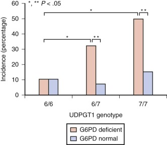
Although GS is a congenital disorder, it rarely becomes clinically apparent until after puberty. The reasons for this are unknown but have been suggested to be related to the hormonal changes of puberty. Steroid hormones can suppress hepatic bilirubin clearance. During pregnancy, increased estrogen levels are associated with impaired clearance of exogenous bilirubin. Gonadectomy has been shown to alter BUGT activity. Odell speculated that some infants with nonhemolytic neonatal jaundice are manifesting GS. Use of genetic markers (see below) has allowed investigation of the role GS plays in neonatal jaundice. Individuals carrying such markers have been shown to have a more rapid rise in their jaundice levels during the first 2 days of life, a predisposition to prolonged or severe neonatal hyperbilirubinemia, variably increased jaundice when the GS polymorphism occurs with pyloric stenosis or is coinherited with hematologic abnormalities such as G6PD deficiency, β-thalassemia, or hereditary spherocytosis. Thus studies from several different parts of the world indicate that GS, as detected by UGT1A1 analysis, does play some role in neonatal jaundice. Kaplan et al noted that, in their study, neither G6PD deficiency nor the GS type UDPGT1 promoter polymorphism (also known as UGT1A1*28 ) alone increased the incidence of hyperbilirubinemia, but both in combination did ( Fig. 4-8 ). They speculated that this gene interaction may serve as a paradigm of the interaction of benign genetic polymorphisms in the causation of disease, in other words, it may take two genetic abnormalities to produce disease symptoms. Numerous studies indicate the increased risk of cholelithisis in patients with GS.
Pathophysiology.
GS is a heterogeneous group of disorders all of which share at least a 50% decrease in hepatic BUGT activity. Based on plasma clearance of other organic anions (BSP and ICG) that share the same hepatocyte uptake receptor carrier, there appear to be at least four subtypes of GS. In GS type I, clearance of BSP and ICG are normal. In GS type II there is delayed BSP clearance, but ICG clearance is normal. Because BSP uptake is normal in type II, delayed clearance must be related to subsequent intrahepatic metabolism or canalicular excretion. In GS type III, clearance of both BSP and ICG are delayed. The delay in the initial rate of disappearance from the plasma suggests a defect in uptake at the hepatocyte plasma membrane. Those with GS type IV have delayed uptake of ICG but not BSP. Thus some individuals with GS have delayed uptake of bilirubin into the hepatocyte, others have delayed biotransformation, and others demonstrate both abnormalities. Immunohistochemical staining for UGT shows a clear reduction throughout the hepatic lobule in specimens from individuals with GS, when compared to normals.
The elucidation of the structure of the UGT1 gene, which encodes human bilirubin, phenol and other UDP-glucuronosyltransferase isozymes led to the discovery of UGT1A1 mutations or polymorphisms associated with GS. In Caucasian populations, the homozygous finding of an additional TA repeat in the promoter region, or so called TATA box (i.e., [TA] 7 TAA, rather than [TA] 6 TAA) of the UGT1A1 gene has been shown to be a necessary, though not sufficient, condition for GS. Individuals who are heterozygous for seven TA repeats have significantly higher serum bilirubin levels than the homozygous wild type six repeats. In Asian populations, the (TA) 7 TAA mutation is relatively rare, but several different UGT1A1 mutations have been associated with GS. These Asian mutations involve exon one of the UGT1A1 gene, rather than the TATA promoter region. One of the most common mutations in Asians, a Gly71Arg mutation in exon 1, has also linked GS and severe neonatal hyperbilirubinemia. It has been reported that although within Caucasians promoter TA repeat number and bilirubin level are strongly positively correlated, in other ethnic groups (e.g., Africans, where two other variants, [TA] 5 and [TA] 8 , have been identified) there is a negative correlation. Rarely the (TA) 8 variant has been reported in Caucasians, thus the ethnic implications of these genetic polymorphisms of the UGT1A1 gene require further analysis.
Although decreased hepatic BUGT activity is universal in GS, there is poor correlation between measured enzyme activity in liver and serum bilirubin level. This may be explained by the increased bilirubin production associated with the decreased red cell half-life seen in as many as 40% of patients with GS. While early authors concluded that there was little or no induction of BUGT following administration of phenobarbital, more recent work has revealed that, in fact, phenobarbital does increase UGT1A1 activity via a phenobarbital-responsive enhancer module (PBREM) of the UGT1A1 promoter region and that such polymorphisms could represent an additional risk factor for GS.
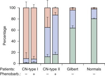
Related to the decreased BUGT activity is the observation that duodenal bile from individuals with GS contains a decreased amount of bilirubin diglucuronides and an increased amount of bilirubin monoglucuronides compared to normals ( Fig. 4-9 ). This distribution is similar to that seen in infants. Administration of phenobarbital has been shown to normalize the bile pigment profile in duodenal fluid, lower plasma bilirubin levels, and increase hepatic clearance of bilirubin. Clofibrate and glutethimide also normalize serum bilirubin concentrations but do not normalize the duodenal bile pigment profile. In animals clofibrate is an inducer of BUGT but does not effect BSP uptake or GST. An additional unexplained phenomenon in GS is the exaggerated rise in serum bilirubin associated with fasting. Although normal individuals can double their serum bilirubin level in response to fasting, in GS a more pronounced rise occurs. This is not related to decreased hepatic blood flow because ICG clearance is not affected. Although fasting can reduce BUGT activity and increase HO (the enzyme responsible for bilirubin production) activity, neither of these effects are believed to explain the fasting hyperbilirubinemia of GS. Enhanced enterohepatic circulation of bilirubin is suggested to be a major factor in the pathogenesis of fasting-induced hyperbilirubinemia. Intraluminal noncalorie food-bulk can blunt the bilirubin rise.
Diagnosis and Treatment.
Generally, a diagnosis of GS can be made when there is a mild, fluctuating unconjugated hyperbilirubinemia, the rest of the liver function tests are normal, and there is no hemolysis. Hemolysis can add confusion because it can result in similar findings and it is not unusual in GS. Hence other tests are sometimes used to aid in diagnosis, including the intravenous administration of nicotinic acid (niacin) or one oral dose or rifampin with assessment of the subsequent rise in serum bilirubin concentration.
Another test suggested to aid in the diagnosis of GS involves fractionation of the total serum bilirubin using alkaline methanolysis and thin-layer chromatography. This allows precise measurement of the conjugated and UBIL levels. This approach has shown that in GS ~6% of the total serum bilirubin was conjugated compared to approximately 17% in normals and those with chronic hemolysis. Individuals with chronic persistent hepatitis had 28% of their total bilirubin present as conjugates. Fasting did not change the percentage of conjugates in GS, despite the rise in total serum bilirubin concentration. An overlap of only three individuals was seen among the 77 with GS and 60 normal subjects. Other studies support these findings. In patients with GS, fractionation of the total serum bilirubin by HPLC showed significantly decreased bilirubin monoglucuronides (1.1% vs. 6.2% in normals) and increased UBIL (98.8 vs. 92.6 in normals).
Currently genetic testing is easily available to diagnose GS. Monaghan et al. have suggested GS genetic screening for the UGT1A1 TA repeat as a simple, useful additional test in the investigation of very prolonged neonatal jaundice in North American, African, and European populations; and for the Gly71Arg mutation in Asians. However, the value of such a genetic test cannot be fully determined until accurate data regarding the prevalence and penetrance of the GS genotype are known. Thus genetic testing for GS cannot be routinely recommended.
GS has no significant negative implications regarding morbidity or mortality. In general, drug metabolism studies have revealed no major dangers, although there appears to be an increased incidence of slow acetylators, and lorazepam clearance is 20% to 40% decreased. Concurrent genetic deficiencies in other xenobiotic pathways may put individuals with GS at increased risk of drug toxicity to such compounds as acetaminophen, cancer chemotherapeutic agents CPT-11 (irinotecan) or TAS-103, or the viral protease inhibitor, indinavir. For this reason, Bosma suggests that screening for GS is of clinical importance. No specific treatment is necessary for GS, though phenobarbital has been shown to lower serum bilirubin levels in these patients. If the well-documented antioxidant effect of bilirubin provides a biologic advantage, then the mild hyperbilirubinemia of GS might actually be a significant benefit against such things as vascular disease, in which free radicals are involved in pathogenesis. However, Bosma concludes that a selective advantage in GS because of the antioxidant capacity of bilirubin seems unlikely.
Crigler-Najjar Syndrome Types I and II
In 1952, Crigler and Najjar described seven infants with congenital familial nonhemolytic jaundice who developed severe unconjugated hyperbilirubinemia shortly after birth and died from kernicterus within months. These infants were from three related families. The serum bilirubin concentration reached 25 to 35 mg/dL despite a lack of hemolytic disease. Other liver function tests were normal. Liver histology was normal except for the deposition of bile pigments. Subsequent reports document that the main risk for patients with the CN is kernicterus. An excellent review of the neurologic perspectives of CN has been published. Although some patients survive into the second decade with normal development, the possibility of developing late kernicterus is always a concern, even in adulthood. Serum bilirubin levels vary from approximately 15 to 45 mg/dL.
In 1969, Arias and colleagues described a second, more frequent type of severe nonhemolytic hyperbilirubinemia. The previous syndrome was termed CN type I (CNI, OMIM #218800), while the new findings were termed CN type II or Arias’ syndrome (CNII, OMIM #606785). Hyperbilirubinemia is less severe in type II patients and varies from approximately 8 to 25 mg/dL. Hence these individuals have a much lower incidence of kernicterus, though such damage occurs.
Pathophysiology.
Both CNI and CNII are generally inherited in an autosomal recessive manner, though one case of autosomal dominant inheritance of CNII has been reported. CNI and CNII result from mutations to the UGT gene complex. Patients with one normal allele demonstrate normal metabolism of bilirubin. The genetic details determine the severity of clinical disease. In CNI there is a complete absence of functional UGT1A1, while in CNII, UGT1A1 activity is markedly reduced. In CNI, 18 of 23 described mutations of the UGT1 gene are found in the common exons 2 to 5 (see Fig. 4-4 ) and thus affect many UGT1 enzymes. Intronic mutations causing CNI have also been reported. However, in CNII four out of nine known mutations are found in exon 1A1. There is some overlap in classification of mild CNII and GS (e.g., Gly71Arg) which relate to differences in definitions based on serum bilirubin levels. The TATA box TA 7 repeat mutation seen in GS can be seen along with other mutations resulting in either CNI or CNII. In both CNI and CNII, assays of liver tissue from affected patients demonstrate negligible or very low BUGT activity. Hence patients with these disorders experience a profound block in bilirubin excretion, because they lack the ability to conjugate bilirubin with UDPGA. Thus liver biopsy is not helpful in differentiating CNI and CNII. Study of the resected livers from four patients with CNI undergoing liver transplantation showed that there was heterogeneous glucuronidation of various substrates other than bilirubin. Hence several in the family of glucuronosyltransferase isoenzymes can be affected in the same patient. There is considerable overlap of hepatic BUGT activity between CNII and GS (see Table 4-2 ).
In family studies of CNI patients, partial deficiencies have been found in the glucuronidation of salicylate and menthol among siblings, parents, and grandparents. Hence it has generally been accepted that this represents an autosomal recessive inheritance. In family studies of CNII patients, the original report by Arias and colleagues found abnormalities of glucuronidation (menthol) in only one parent, suggesting autosomal dominant inheritance. Subsequent studies of siblings or parents often found elevated serum bilirubin levels (1.2 to 4 mg/dL), delayed bilirubin clearance tests, and decreased hepatic BUGT activity, as would be seen in GS. These findings in both parents suggested that CNII may represent homozygous GS. However, if CNII were truly the homozygous form of GS, one would expect many more affected individuals because GS occurs in at least 3% of the population. Pertinent genetic data have been reviewed supporting the inheritance mode of CNII as autosomal recessive.
The major differentiating characteristic between CNI and CNII is the response to drugs that stimulate hyperplasia of the ER. When CNII patients received phenobarbital or diphenylhydantoin there was a significant decline in the serum bilirubin level, increased hepatic clearance of radiolabeled bilirubin, and increased biliary levels of bilirubin diglucuronides (see Fig. 4-9 ). In a study of five CNII patients, the magnitude of the phenobarbital induced decrease in serum bilirubin ranged from 2.1 to 12.1 mg/dL (27% to 72%) with pre- and postphenobarbital serum bilirubin levels ranging from 7.8 to 16.9 and 4.7 to 10.1 mg/dL, respectively. Summarizing data from seven earlier studies regarding the response of CNII patients to oral phenobarbital treatment revealed the following: 11 females and 13 males had a total serum bilirubin of 15.7 ± 13.8 (mean ± SD) prior to phenobarbital. After doses ranging from 90 to 390 mg/day, or alternatively 4 mg/kg/day, the serum bilirubin decreased 12 ± 4 mg/dL (77% ± 13%). The lowest total serum bilirubin following phenobarbital therapy was 5.9 mg/dL. In contrast, CNI patients show neither decrease in serum bilirubin nor significantly increased biliary bilirubin conjugates in response to drugs (see Fig. 4-9 ). The response to phenobarbital is the criterion used to differentiate between these two disorders. Bile analysis has also been suggested as another method to differentiate CNI and CNII. In CNI, bile contains insignificant bilirubin conjugates (less than 10%), and UBIL predominates. In CNII, bile contains small amounts of bilirubin conjugates, and those present are predominantly bilirubin monoglucuronides (greater than 60%).
Two cousins with CN have been described with unique features that raise the possibility of a new variant of this syndrome (type III). This new variant resembled CNI in that there was no biliary excretion of bilirubin di- or monoglucuronide. However, the type III patients did excrete mono- and diglucoside conjugates of bilirubin. It has been speculated that type III patients lack the long proposed permease that has been hypothesized to transport UDP-glucuronic acid to the luminal side of the ER, where glucuronosyltransferase is located. This absence is suggested to force utilization of a very inefficient substrate for conjugation to bilirubin, UDP-glucose.
Diagnosis and Treatment.
Although CN can be diagnosed during the prenatal period, evaluation of infants with CN more typically begins during the first days of life when serum bilirubin levels exceed 20 mg/dL. The conjugated fraction will not be elevated except possibly for the factitious elevation sometimes seen when the total serum bilirubin level is very high. Evaluation of such infants will eliminate hemolysis, hypothyroidism, infection, and other more common causes of jaundice. Formula feedings will help identify those infants with jaundice related to human milk. During this period of testing, the magnitude of the serum bilirubin elevation will prompt use of phototherapy to avoid kernicterus. Exchange transfusion may be necessary. Yet despite these efforts, CN patients will have persistent jaundice. There is currently no widely available simple clinical test to confirm a diagnosis of CN. CN can be excluded by finding significant amounts of bilirubin conjugates in neonatal stools, if collected prior to establishment of sufficient intestinal bacteria that convert bilirubin conjugates to urobilinoids. High pressure liquid chromatographic (HPLC) analysis of duodenal bile will show that in CNI, there are negligible bilirubin di- or monoglucuronides, while in CNII these conjugates are present but in low concentration. An easy method to collect such fluid for analysis utilizes the pediatric Entero-Test capsule. This approach to diagnosis is much less invasive than performing a liver biopsy to confirm negligible BUGT activity with in vitro assay. The ratio of serum bilirubin conjugates (as determined by alkaline methanolysis with thin-layer chromatography) to total bilirubin, although abnormally low, does not allow differentiation of CN patients from those with GS. Similar overlap occurs with HPLC fractionation of serum bilirubin conjugates. DNA analysis can be very helpful in establishing the correct diagnosis, and in the future it is expected that DNA array technology will allow rapid screening for known mutation.
A world registry of patients with CNI aimed at developing management guidelines has been published. Phenobarbital (4 mg/kg/day in infants) should be used when there is concern about deficiency of BUGT. Within 48 hours CNII patients can demonstrate a significant decrease in serum bilirubin levels (as detailed above) and an increased biliary excretion of bilirubin di- and monoglucuronide, while the CNI patients will show no significant response. Occasionally CNII patients do not respond to the first trial of phenobarbital therapy, but subsequent trials months later will demonstrate the significant decrease in serum bilirubin level. However, despite the decrease in serum bilirubin in response to phenobarbital, CNII patients will usually continue to manifest a significant hyperbilirubinemia (approximately 5 to 15 mg/dL). Phototherapy for 6 to 12 hours daily has been the primary modality to keep serum bilirubin levels below 20 mg/dL during the first several months of life because CNI patients can excrete all bilirubin photoisomers. CNI patients will require lifelong treatment with phototherapy until more definitive therapy such as liver transplant. Phototherapy has been found to be least intrusive when given at night, and improvements have been made in effectiveness and comfort. Although phototherapy is very helpful in infancy, in adolescence social inconvenience and compliance problems can bring increased risk of kernicterus.
Other therapeutic considerations involve the oral administration of binding agents such as agar or cholestyramine or calcium phosphate. These agents bind to bilirubin in the intestinal lumen due to phototherapy or through direct intestinal permeation. They prevent the enterohepatic circulation of bilirubin. Problems associated with the use of cholestyramine include cost, taste, and concern about bile salt depletion and fat malabsorption. Problems regarding agar include significant variation in bilirubin binding affinity among various preparations and batches. During acute episodes of severe hyperbilirubinemia after the first year of life, plasmapheresis has been shown to rapidly decrease serum bilirubin levels. Peritoneal dialysis and exchange transfusion have not been helpful in this setting. Repeated intramuscular injections of tin protoporphyrin, an HO inhibitor that blocks bilirubin formation, have been used in one CNI patient with data suggesting a decreased need for phototherapy. Two patients with CNI were treated with tin mesoporphyrin to block bilirubin formation and also received daily phototherapy and intermittent plasmapheresis over a 400-day period. They developed an iron deficiency anemia believed to be due to the porphyrin therapy, but tin mesoporphyrin (stannsoporfin) (2 to 4 µmol/kg) is suggested to offer a promising, though still experimental, additional therapy for controlling episodes of acute, severe jaundice. Drugs which bind to albumin and can potentially displace bilirubin should be avoided at all times.
Because patients with CN syndrome have good hepatic function other than conjugating bilirubin, they are ideal candidates for auxiliary liver transplantation, wherein the patient’s own liver remains within the body while another whole or partial liver is transplanted just beneath or adjacent to the recipient’s. This option has recently become clinically available. More commonly, orthotopic liver transplantation has been performed, and this represents the only true cure for the hyperbilirubinemia of CNI. Ideally the timing of transplantation would precede irreversible neurologic injury. Transplantation of other BUGT-containing tissues (e.g., segments of small intestine, kidneys, or hepatocytes ) remain experimental. Successful cloning of the gene responsible for bilirubin glucuronosyltransferase activity offers the hope of future gene therapy to correct this deficiency based on studies in Gunn rats, the congenitally jaundiced model for CNI.
Disorders of Enterohepatic Circulation
Increased enterohepatic circulation of bilirubin is believed to be an important factor in neonatal jaundice. As previously reviewed, neonates are at risk for the intestinal absorption of bilirubin because (1) their bile contains increased levels of bilirubin monoglucuronide, which allows easier conversion to bilirubin; (2) they have significant amounts of β-glucuronidase within the intestinal lumen, which hydrolyses bilirubin conjugates to bilirubin, which is more easily absorbed from the intestine; (3) they lack an intestinal flora to convert bilirubin conjugates to urobilinoids; and (4) meconium, the intestinal contents accumulated during gestation, contains significant amounts of bilirubin. Conditions that prolong meconium passage (e.g., Hirschsprung’s disease, meconium ileus, meconium plug syndrome) are associated with hyperbilirubinemia. Earlier passage of meconium has been shown to be associated with lower serum bilirubin levels. This may be facilitated by rectal temperature measurement during the neonatal period. The enterohepatic circulation of bilirubin can be blocked by the enteral administration of compounds which bind bilirubin, such as agar, charcoal, and cholestyramine.
Other Causes of Unconjugated Hyperbilirubinemia
Various hormones may cause development of neonatal unconjugated hyperbilirubinemia. Congenital hypothyroidism can present with serum bilirubin greater than 12 mg/dL prior to the development of other clinical findings. Prolonged jaundice is seen in one third of infants with congenital hypothyroidism. Similarly hypopituitarism and anencephaly may be associated with jaundice due to inadequate thyroxine, which is necessary for hepatic clearance of bilirubin.
Certain drugs may affect the metabolism of bilirubin and result in hyperbilirubinemia or displacement of bilirubin from albumin. Such displacement increases the risk of kernicterus and can be caused by sulfonamides, moxalactam, and ceftriaxone (independent of its sludge-producing effect ). The popular Chinese herb, Chuen-Lin, given to 28% to 51% of Chinese newborn infants, has been shown to have a significant effect in displacing bilirubin from albumin. Pancuronium bromide and chloral hydrate have been suggested as causes of neonatal hyperbilirubinemia.
Infants of diabetic mothers have higher peak bilirubin levels and a greater frequency of hyperbilirubinemia compared with normal neonates. These patients have shown a positive correlation between total bilirubin and hematocrit, thus implicating polycythemia as one possible mechanism. Other potential reasons for this hyperbilirubinemia include prematurity, substrate deficiency for glucuronidation (secondary to hypoglycemia), and poor hepatic perfusion (secondary to either respiratory distress, persistent fetal circulation, or cardiomyopathy).
The Lucey-Driscoll syndrome consists of neonatal hyperbilirubinemia within families in whom there is in vitro inhibition of glucuronosyltransferase by both maternal and infant serum. It is presumed that this is caused by gestational hormones.
Prematurity is frequently associated with unconjugated hyperbilirubinemia in the neonatal period. Hepatic UDP-glucuronosyltransferase activity is markedly decreased in premature infants and rises steadily from 30 weeks’ gestation until reaching adult levels 14 weeks after birth. In addition there may be deficiencies for both uptake and secretion. Bilirubin clearance improves rapidly following birth.
Hepatic hypoperfusion can result in neonatal jaundice. Inadequate perfusion of the hepatic sinusoids may not allow sufficient hepatocyte uptake and metabolism of bilirubin. Causes might include patent ductus venosus, congestive heart failure, and portal vein thrombosis. Other specific liver diseases, as listed in Box 4-1 and described elsewhere in this text, can result in neonatal jaundice.
Increased Production of Bilirubin
Fetal–maternal blood group incompatibilities
Extravascular blood in body tissues
Polycythemia
Red blood cell abnormalities (hemoglobinopathies, membrane, and enzyme defects)
Induction of labor
Decreased Excretion of Bilirubin
Increased enterohepatic circulation of bilirubin
Breastfeeding
Inborn errors of metabolism
Hormones and drugs
Prematurity
Hepatic hypoperfusion
Cholestatic syndromes
Obstruction of the biliary tree
Combined Increased Production and Decreased Excretion of Bilirubin
Sepsis
Intrauterine infection
Congenital cirrhosis
Management and Treatment of Unconjugated Hyperbilirubinemia
In recent years changes in perinatal care have made severe neonatal jaundice a larger problem and there has been a reemergence of kernicterus. Possible reasons for this include (1) early hospital discharge (before extent of jaundice is known and signs of impending brain damage have appeared); (2) lack of adequate concern for the risks of severe jaundice in healthy term and near newborns; (3) an increase in breastfeeding incidence; (4) medical care cost constraints; (5) paucity of educational materials to enable parents to participate in safeguarding their newborns; (6) limitations within in health care systems to monitor the outpatient progression of jaundice; (7) difficulty in estimating the degree of jaundice, particularly in dark-skinned infants; and (8) demonstration of bilirubin being an antioxidant. Newborn infants are now being described who are discharged early, develop severe hyperbilirubinemia (30 to 40 mg/dL) at home, and go on to develop classic signs of kernicterus. This has been reported in otherwise healthy breastfed infants with no other identified etiology for their jaundice. Although early postpartum discharge has advantages, one disadvantage is the risk associated with delayed diagnosis of severe hyperbilirubinemia. The AAP has recommended that infants discharged at less than 48 hours of age be seen in follow-up within 48 hours of discharge. Many physicians do not follow these recommendations, despite the serious impact that short hospital stay has on the jaundiced newborn. The AAP updated their 1994 guidelines for the management of hyperbilirubinemia in newborn infants in 2004.
Jaundice can be caused by increased bilirubin production, decreased bilirubin excretion, or combinations of these mechanisms (see Box 1 for specific examples). An approach to management of jaundice in the newborn nursery has been published elsewhere. Although Newman and colleagues found that obtaining a direct bilirubin measurement was seldom helpful because of low yield and poor specificity, others advocate measurement of an early conjugated bilirubin as a population screening test, which could lead to the earlier diagnosis of neonatal liver disease. Bhutani and colleagues advocated universal bilirubin measurement prior to hospital discharge in order to identify infants at risk for severe neonatal hyperbilirubinemia on the basis of a predischarge hour specific total serum bilirubin measurement ( Fig. 4-10 ). Such universal predischarge screening is now recommended by the AAP and has been shown in one recent study to reduce the incidence of significant hyperbilirubinemia and the rate of hospital readmission of infants for the treatment of jaundice. Despite the large number of etiologies for neonatal jaundice, no cause for the jaundice could be identified in nearly half of the infants evaluated during one study of 447 infants and in one third of the infants in a kernicterus registry.
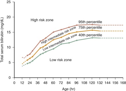
Hyperbilirubinemia is the most frequent reason that infants are readmitted to the hospital in the first weeks of life. The management of neonatal jaundice has been reviewed elsewhere ( Figs. 4-11 and 4-12 ). The most important step in treatment of jaundice is determination of the primary etiology. However, independent of the etiology of the jaundice, elevation of the serum UBIL fraction prompts concern about possible kernicterus. As previously reviewed, when the UBIL fraction is elevated, care must be given to avoid administration of agents which bind to albumin and displace bilirubin, thus promoting kernicterus. Although historically, sulphonamides are the most well known bilirubin displacing agents, drugs such as ceftriaxone and ibuprofen are also strong bilirubin displacers with a potential for inducing bilirubin encephalopathy. Therapeutic options to lower UBIL levels include phototherapy, exchange transfusion, interruption of the enterohepatic circulation, enzyme induction, and alteration of breastfeeding. Research into these and other modalities continues actively. The outcome of rational therapeutic guidelines for unconjugated hyperbilirubinemia during the first 7 days of life have been published elsewhere; phototherapy can reduce the incidence of exchange transfusion and the risk of bilirubin-induced neurologic disease.
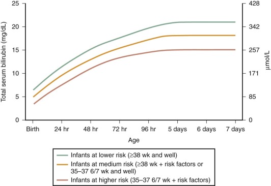

Stay updated, free articles. Join our Telegram channel

Full access? Get Clinical Tree


