Nasopharyngeal Carcinoma


Nasopharyngeal Carcinoma


Brigette B. Y. Ma

Michael K. M. Kam

Anthony T. C. Chan


BACKGROUND


Introduction

Nasopharyngeal carcinoma (NPC) has a unique epidemiology and pathogenesis, and the treatment of NPC has undergone several paradigm shifts over the last 30 years. Advances in the planning and delivery of radiotherapy (RT) and the adjunctive use of chemotherapy over the decades have all contributed to the improved prognosis of NPC. In Hong Kong where NPC is among the top 10 most common cancers with an ageadjusted mortality rate of 3.9 per 100,000 persons, the 5-year overall survival (OS) of patients with stage I and II NPC now approaches 90%.1 However, worldwide, up to 65,000 people still died of NPC annually in recent times.2,3 This is mainly due to the fact that 20% of patients with locoregionally advanced (i.e., stage III and nonmetastatic stage IV) NPC will invariably develop distant metastases despite modern treatment. In Hong Kong, the 5-year OS for nonmetastatic stage III and IV is ˜60% in patients who were treated with 2-dimensional (2D) RT.1,4 For patients with metastatic NPC, the median OS (12 to 18 months) is still well short of that of other cancers such as metastatic colorectal cancer (over 20 months) following modern drug therapy. Although concurrent chemoradiotherapy has significantly improved the treatment outcome for patients with stage III and nonmetastatic stage IV NPC, this approach also increases the incidence of certain acute and late toxicities. Thus, newer biomarkers and more accurate staging are needed to identify those patients who are most likely to benefit from multimodal therapy.

On a global scale, NPC is a rare cancer with ˜80,000 new cases reported per year and accounts for 0.7% of all cancers.2 In nonendemic areas like North America and Europe, the incidence rate is <1 case per 100,000 populations. This is in marked contrast with the endemic or “high-risk” areas such as Hong Kong and Southern China, where the annual agestandardized incidence rates are as high as 20 to 30 cases per 100,000 population in men and 8 to 15 cases per 100,000 population in women.5,6 Recent studies have suggested a fall in the incidence of NPC in the last 30 years in China. However, other reports have found a stable to rising incidence in certain parts of Southern China.5,7 According to the Hong Kong Cancer Registry,3 the age-standardized incidence rates of NPC in 2010 have decreased from 12.8 cases per 100,000 population in 2001 to 8.7 cases per 100,000 population for both sexes. Several studies have speculated on the reasons for this declining trend in Hong Kong. One report found that only the incidence rate of the keratinizing subtype of NPC has fallen, whereas the rate of the more common nonkeratinizing subtype has remained relatively stable.8 Other studies have attributed the falling incidence to improvements in socioeconomic status and standard of living in the local population, as well as an improvement in the standards of medical care in Hong Kong.1,9

NPC is classified into different histologic subtypes: type 1 (I) squamous cell carcinoma, type 2a (II) keratinizing undifferentiated carcinoma, and type 2b (III) nonkeratinizing undifferentiated carcinoma. The World Health Organization (WHO) III subtype is the most common form of NPC in endemic areas and is ubiquitously associated with the Epstein-Barr virus (EBV). It also differs from WHO I NPC with regard to its sensitivity to chemotherapy and RT. The staging of NPC is based on the depth of invasion of the soft tissue, cranial nerves, and bony structures at and near the nasopharynx by the primary tumor, the involvement of local and regional lymph nodes of the head and neck, and the presence of distant metastases. In Hong Kong, the distribution of stage groups at presentation is as follows: stage II 7%, stage IIA to B 41%, stage III 25%, and stage IVA to B 28%. This means that nearly half of all patients with NPC in Hong Kong present at an advanced stage; hence, much of the clinical research in NPC has been focused on improving the treatment of advanced NPC.


Staging of Nasopharyngeal Carcinoma with Radiologic Imaging

The 7th edition of the International Union Against Cancer (UICC) and the American Joint Committee on Cancer (AJCC) TNM classification for NPC is currently the most commonly used staging system internationally (Table 11.1). It is based purely on the anatomical spread of both the primary tumor and the metastatic nodes, without taking into account the size of the tumor or its histologic grade. Significant stage migration has been observed over time with the advances in imaging technology and treatment modalities. Magnetic resonance imaging (MRI) has generally replaced computerized tomography (CT) for local tumor staging because it provides better soft tissue resolution. MRI also plays an important role in RT treatment planning because it provides more accurate delineation of tumor target. For the staging of M stage, studies do not support the routine use of CT thorax, bone scan, or abdominal ultrasonography in average-risk patients because of the low positive detection rate for metastases, and such imaging is usually reserved for patients at high risk of distant metastasis.10,11,12,13

18F-fluoro-2-deoxy-D-glucose (18F-FDG) positron emission tomography (PET)-CT has become increasingly popular in staging and as a tool to detect tumor persistence or recurrence. Several studies have compared the accuracy of PET-CT and MRI in detecting primary tumor, retropharyngeal nodes, cervical nodes, and distant metastasis, but their results are inconsistent.14,15,16,17 In the larger study by Ng et al.,17 MRI appears to
be superior for assessing the primary tumor and retropharyngeal nodes, whereas PET-CT is more accurate than MRI for detecting cervical nodal metastasis and more accurate than CT and bone scans for detecting distant metastasis. A recent metaanalysis of eight series has confirmed the reliable performance of PET or PET-CT in the evaluation of distant metastasis, with a pooled sensitivity of 83% and specificity of 97%.18


Table 11.1 The 7th Edition of the International Union Against Cancer (UIC) and the American Joint Committee on Cancer (AJCC) TNM Classification for NPC


T Stage


N Stage


T1: Nasopharynx, oropharynx, or nasal fossa


T2: Parapharyngeal extension


T3: Bony structures, paranasal sinuses


T4: Intracranial extension, cranial nerve, hypopharynx, orbit, infratemporal fossa(masticatory space)


N0: None


N1: Unilateral cervical or retropharyngeal (irrespective of laterality), <6 cm, above supraclavicular fossa


N2: Bilateral cervical node, <6 cm, above supraclavicular fossa


N3a: >6 cm


N3b: In supraclavicular fossa


M Stage


Overall Stage


M0: No distant metastasis


M1: Distant metastasis is present.


I: T1 N0 M0


II: T1 N1 M0, T2 N0-1 M0


III: T1/2 N2 M0, T3 N0-2 M0


IVA: T4 N0-2 M0


IVB: Any T, N3, M0


IVC: Any T, any N, M1


Used with the permission of the American Joint Committee on Cancer (AJCC), Chicago, Illinois. The original source for this material is the AJCC Cancer Staging Manual, Seventh Edition (2010) published by Springer Science and Business Media LLC, www.springer.com.


Early detection of local recurrence is crucial to successful salvage. However, it is notoriously difficult to diagnose submucosal or deep-seated recurrence by endoscopy alone, and MRI or CT is unable to distinguish post-RT scarring or inflammation from genuine recurrence.19 Controversy remains as to the superiority of PET-CT over MRI on this aspect. In a systematic review of 21 articles, Liu et al.20 suggested that PET was the best modality for diagnosis of local residual or recurrent NPC. In the foreseeable future, PET-CT is going to play a more important role in the staging of NPC at diagnosis and at recurrence and in the monitoring of treatment response.


PRIMARY TREATMENT FOR NONMETASTATIC NASOPHARYNGEAL CARCINOMA


Two-Dimensional Radiotherapy

External RT has been the mainstay treatment for nonmetastatic NPC since the introduction of megavoltage machines in the mid-1960s, when the OS was only 25% at 5 years. With improvement in simple two- to three-field arrangements delivering 60 to 70 Gy of two-dimensional RT (2DRT) to the nasopharynx and its regional lymphatics, the OS rate was typically in the order of 50% during the 1970s to 1980s, with the rate of locoregional failure over 25%.21,22,23 The major drawback of 2DRT is the unnecessary irradiation of abundant normal tissues, and any attempt at vital organ sparing would inevitably compromise target coverage.24 Further improvement of treatment outcome was seen in the 1990s, and this occurred as a result of advances in diagnostic imaging and the use of more aggressive strategies such as RT dose escalation and chemotherapy. In a retrospective analysis on 2,687 patients treated during 1996 to 2000s, the OS rate was 75% and local failurefree rate was 85% at 5 years.1


Three-Dimensional Conformal Radiotherapy

The transition from 2DRT to three-dimensional conformal radiotherapy (3DCRT) marked a great advance in RT development in the late 1990s, and the development of computer planning system and multileaf collimator is a representative achievements during this period. The integration of CT or MRI images into the 3D treatment planning system provides more accurate spatial information on the tumor target and normal organs, which in turn enables a more flexible adjustment of the beam directions. The use of multileaf collimator allows better shaping of beam aperture that conforms to the shape of the target and avoids vital organs in the vicinity. The dosimetric advantage of 3DRT has been translated into significant improvement in patient survival as well as reduction in serious toxicities.4


Intensity-Modulated Radiotherapy

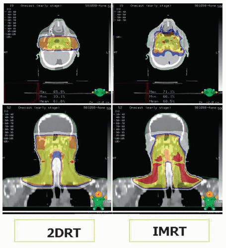
The use of 3DCRT was rapidly overtaken by the introduction of intensity-modulated radiotherapy (IMRT), in the late 1990s. IMRT is an advanced form of 3DCRT with additional capacity to modulate beam intensity pixel by pixel across the treatment field. Working in conjunction with the inverse planning computer optimization algorithm, an optimized fluence can be obtained according to the dose-volume constraints set by the physician. It is particularly useful in generating a concaveshaped dose distribution with steep dose-gradient around the brainstem, spinal cord, and optic pathway. The principal benefit of IMRT in early-stage NPC is the sparing of the parotid glands. In locoregionally advanced NPC, IMRT offers better tumor coverage and protection of critical neurologic organs and allows room for dose escalation.25,26,27 Moreover, IMRT permits the delivery of different dose intensities to different targets according to their clinical risks and enables biologic enhancement through the concept of simultaneous integrated boost (SIB) technique. A comparison of dose distribution between 2DRT and IMRT is demonstrated in Figure 11.1.

This dosimetric advantage has been translated into better local control rate as reported in many retrospective series28,29,30,31,32,33,34,35,36,37,38,39,40,41,42,43,44,45 (Table 11.2). In addition, three randomized studies comparing IMRT versus 2DRT have been reported thus far. In early-stage NPC, Kam et al. and Pow et al. both confirmed that IMRT results

in significantly better recovery of parotid salivary function than 2DRT.49,50 Peng et al.51 randomized patients to 2DRT or IMRT and found a significant improvement in 5-year local control rate (91% vs. 84%, p = 0.046) and OS (80% vs. 67%, p = 0.001) and significant reduction in late toxicities including cranial neuropathy, temporal lobe necrosis, xerostomia, trismus, and fibrosis of the neck. However, several controversies in IMRT remain unresolved. It is uncertain whether IMRT is superior to 3DCRT in terms of patient’s outcome. Fang et al.37 and Lee et al.4 did not observe significant differences in locoregional control, OS, or toxicities between IMRT and 3DCRT. The suboptimal local control rate in locally advanced NPC patients, and the tangible figures on some severe RT complications are really too significant to be ignored.29 The optimal total dose level and time-dose-fractionation schedule are still unclear, because several RT acceleration or dose escalation strategies (such as SIB technique, or sequential boost) have not been formally compared in randomized studies. Another outstanding issue is the need to validate and standardize the definition of clinical target volume (CTV). This is important because the therapeutic window of RT in NPC is narrow, and tailoring of treatment volume is essential to avoiding inadequate or futile treatment margins. Despite all these uncertainties as mentioned, IMRT is rapidly becoming a contemporary standard of care for the treatment of NPC worldwide.


Figure 11.1. Comparison of isodose distribution between 2DRT and IMRT.


Table 11.2 Results of Studies on the Treatment of NPC with IMRT


Study


N


Stage T3-T4 (%)


Median Follow-Up (mo)


Total Dose (Gy)


Dose/Fraction (Gy)


Time Point (y)


Local Control (%)


Nodal Control (%)


Distant Control (%)


Overall Survival (%)


Lee39


118


41


30


70


2.12


4


96


98


72


74


Kam46


63


51


29


66


2


3


92


98


79


90


Wolden47


74


53


35


70.2


2.34


3


91


93


78


83


Kwong43


50


0


14


70


2


3


100


92.3


100


100


Kwong42


50


100


25


76


2.17


2


96a


NR


94


92


Lin44


326


61


33


62.6-69.75


2.2-2.25


3


95


98


90


90


Lee45


20


40


27


72


2.4


2


88a


NR


90


NR


Koom30


24


29


26


64.8


2.4


3


93


87


88


96


Fang37


110


24.5


40


72


2.4


3


84.2a


NR


82.6


85.4


Tham36


195


NR


37


70


2.12


3


89.6a


NR


89.2


94.3


RTOG 022548


68


34


31


70


2.12


2


92.6


90.8


84.7


80.2


Wong38


175


35


34


70


2.12


3


93.6


93.3


86.6


87.2


Ng35


193


61


30


70


2-2.12


2


95


96


90


92


Bakst34


25


28


33


70.2


2.34


3


91


91


91


89


Xiao33


81


100


54


68


2.27


5


94.9


NR


NR


74.5


Lai32


512


52


52.8


NR


2.27


5


93


97


84


NR


a Locoregional control.


NR, not reported; y, years; mo, months.


Dose Escalation

The dose-response effect in NPC has been well established. Lee et al.52 showed that risk of local failure was found to be decreased by a factor of 9% per additional Gy. The value of dose escalation after a basic course of 66 to 70 Gy large-field 2DRT has also been demonstrated by a retrospective review from the Hong Kong NPC Study Group on 2,462 patients, of whom 65% received brachytherapy, 2DRT, 3DRT, or stereotactic RT (SRT) boost.53 The use of RT boost in patients with T1 to T2a and T3 to T4 disease was a significant determinant of local control. The commonly agreed standard total dose should be more than or equal to 70 Gy at 2 Gy fraction per day. The achievable total dose is limited by the tolerance of the surrounding normal critical organs, in particular, the temporal lobe.54 Several techniques have been studied to achieve a total dose beyond 66 to 70 Gy, which include brachytherapy boost or 3DRT/IMRT/SRT boost after the conventional fractionation RT and the SIB technique in the IMRT era.


Brachytherapy Boost

Studies from Hong Kong have shown that the use of two to three fractions of brachytherapy could significantly enhance local control in early T-stage disease.55,56 The indication of the brachytherapy boost is currently confined to the treatment of early T-stage disease due to its limited dose penetration. Because distant failure is the dominant mode of failure in more advanced disease, the role of brachytherapy boost is questionable. Levendag et al.57 reviewed 411 patients with advanced NPC (T1 or T2 with nodal metastases, or T3 or T4 and nodenegative disease), of whom 50% of patients received brachytherapy boost of 11Gy. A significant reduction in local failure was observed in the early T-stage subgroup (0% vs. 14%, p = 0.023), but not in the advanced T-stage group.57 Rosenblatt et al.58 randomized 274 patients with stage III and nonmetastatic stage IV NPC to chemoradiotherapy with or without brachytherapy boost thereafter. The 3-year local recurrence-free survival was not significantly improved in either the whole T-stage or the early T-stage group.58


Sequential Conformal/IMRT/Stereotactic Boost

SRT boost has been investigated by the Stanford University (USA) and Taiwan groups, who used CyberKnife or linear accelerator-based technologies to treat NPC patients following radical 2DRT or IMRT.59,60 A high control rate in T4 disease was also observed, but the Stanford series reported a temporal lobe necrosis rate of 12% in the entire group and 11% among IMRT-treated patients.60 In response, the investigators have now decreased their SRT dose to 8 Gy in single fraction or 12 Gy in 3 fractions, whereas the total dose to the temporal lobes is kept below a maximum total dose of 55 to 60 Gy. The longterm safety of this approach and its impact on the therapeutic ratio are yet to be established. In the report by Kam et al.46, 63 patients were treated with 66 Gy to the gross tumor volume (GTV) using IMRT, and dose escalation was performed in 36% of patients with T2b to T4 disease via IMRT or stereotactic plan (8 Gy in 4 fractions). In this study, dose escalation was found to be a favorable prognostic factor for progression-free and distant metastases-free survival, and the incidence of temporal lobe necrosis was 3%. In a retrospective analysis by Lee et al.61 on patients who were irradiated with a 3D conformal technique (2 Gy/fraction/day to 70 Gy), 8.3% temporal lobe necrosis rate was detected at 5 years among patients who received fractionated SRT boost. In summary, although there is strong evidence of a dose-response relationship above 66 to 70 Gy, the impact on local control is more obvious after 2DRT and less compelling with IMRT. Caution must be taken to optimize the target conformity, dose heterogeneity, and dose spillage to the neurovascular structures. As IMRT is capable of pushing the therapeutic ratio further owing to its steep dose gradient, the demand for a meticulous design of margin requirement and thus the higher dependence on the setup accuracy cannot be overemphasized.


Simultaneous Integrated Boost in IMRT

SIB, also called simultaneous modulated and accelerated RT (SMART), exploited the “dose-painting” capacity of IMRT by delivering different dose levels to different regions according to the level of risk. This allows a “once-a-day” fractionation schedule with selective dose acceleration to different tumor targets without undue damage to normal organs. Consequently, a higher biologically equivalent dose can be delivered to the gross tumor than the microscopic disease. A number of studies have evaluated SIB in NPC with a nominal total dose of 64.8 to 76 Gy to the GTV in 2.12 to 2.4 Gy/fraction over 27 to 35 fractions.30,34,36,37,38,39,42,44,45,47,48 Despite the variation in SIB dose fractionation across these studies, the local or locoregional control rate was 88% to 96% after 2 to 5 years of follow-up. However, the incidence of severe late toxicities remains a concern. Bakst et al.34 reported 12% temporal lobe necrosis rate, whereas Kwong et al.42 reported moderate to severe hearing impairment in 42% and carotid pseudoaneurysm in 4% of patients.

Other centers used a “mini-SIB” schedule with a lower fraction size. The Radiation Therapy Oncology Group (RTOG) 0225 multicenter phase II study examined a schedule of 70 Gy per 33 fractions in 2.12 Gy/fraction and reported a 2-year local progression-free survival (PFS) of 92.6% and regional PFS of 90.8%.48 The “mini-SIB” schedule has been tested in a phase III randomized study by Peng et al.51 who compared IMRT (70 Gy at 33 fractions) with 2DRT in 616 NPC patients. The IMRT arm was associated with a significant improvement in locoregional control and OS and reduction in the incidence of hearing impairment, late xerostomia, temporal lobe neuropathy, trismus, and neck fibrosis. For T4 disease
in particular, the use of “mini-SIB” (<2.12 Gy/fraction) or conventional 2 Gy per fraction in combination with chemotherapy may be safer without significantly compromising tumor control if the temporal lobes are kept below 65 Gy.31


INCORPORATION OF CHEMOTHERAPY IN THE TREATMENT OF LOCOREGIONALLY ADVANCED NASOPHARYNGEAL CARCINOMA


Background

Because distant metastasis is the main cause of treatment failure following RT, over a decade of research has been devoted to the intensification of treatment for locoregionally advanced NPC by incorporating adjunctive chemotherapy. To date, at least seven meta-analyses have concluded that the addition of chemotherapy during RT (at any time point) confers a survival advantage over conventional RT alone in patients with locoregionally advanced (i.e., stage III to IVB) NPC.62,63,64,65,66 Of these studies, only the one by Baujat et al.64 was based on patient-derived data, whereas the rest were based on published data. Several observations can be made from these analyses as outlined in Table 11.3.62,63,64,65,66,67,68 The use of concurrent chemotherapy will lead to a 26% to 52% reduction in the risk of death, which amounts to an absolute OS benefit of around 20% after 5 years.63,64 This benefit has been associated with improvement in locoregional and distant recurrence rates.63,64 Of the three sequences of chemotherapy that were evaluated—induction, concurrent, and adjuvant chemotherapy, concurrent chemotherapy is by far most consistently associated with the largest magnitude of improvement in OS and PFS than other sequences.63,64


Table 11.3 Meta-Analyses on the Benefit of Adjunctive Chemotherapy During Radiotherapy for Patients with Locoregionally Advanced NPC


Author


Year


No. of RCT (Patients)


Result


Huncharek and Kupelnick62


2002


6 RCT (1,500)


CRT (any sequence) vs. RT alone: DFS 3 y: OR = 0.60 (95% CI, 0.49-0.73) OS 3 y: OR = 0.81 (95% CI = 0.66-1.00)


Langendijk et al.63


2004


10 RCT (2,450)


CRT (any sequence) vs. RT alone: OS: HR = 0.82 (95% CI, 0.71-0.95, p = 0.01)


Chemo lowers risk of: LRR: RR = 0.47 (95% CI, 0.33-0.67) DMR: RR = 0.72 (95% CI, 0.62-0.84)


Concurrent: HR = 0.48 (95% CI, 0.32-0.72)


Neoadjuvant: HR = 0.87 (95% CI, 0.72-1.04)


Adjuvant: HR = 0.99 (95% CI, 0.71-1.36)


Baujat et al.64


2006


8 RCT (1,753)


CRT (any sequence) vs. RT alone: OS: HR = 0.82 (95% CI, 0.71-0.94; p = 0.006) DFS:HR = 0.76 (95% CI, 0.67-0.86; p < 0.0001)


Chemo lowers risk of: LRF: HR = 0.76 (95% CI, 0.64-0.91) DF: HR = 0.72 (95% CI, 0.59-0.87)


Concurrent: HR = 0.60; 95% CI, 0.48-0.76


Induction: HR = 0.99; 95% CI, 0.80-1.21


Adjuvant: HR = 0.97; 95% CI, 0.69-1.38


Zhang et al.67


2010


7 RCT (1,608)


Concurrent vs. RT alone: 5 y OS: RR = 0.74 (95% CI, 0.62-0.89) 5 y LRF: RR = 0.67 (95% CI, 0.49-0.91) 5 y DR: RR = 0.71 (95% CI, 0.58-0.88)


Liang et al.68


2012


11 (RCT) 1,096


Induction chemo vs. RT alone: OS: RR = 0.99 (95% CI 0.72-1.36) PFS: RR = 0.37 (95%CI 0.20-0.69) LRF: 1.08 (95% CI 0.84-1.38) DF: 0.98 (95% CI 0.75-1.27)


Liang et al.65


2012


5 RCT (793)


Adjuvant chemo vs. CRT alone: OS = 1.02 (95% CI 0.89-1.15) FFS = 0.93 (95% CI 0.72-1.21) LRF = 1.07 (95% CI 0.87-1.32) DF = 0.95 (95% CI 0.80-1.13)


Ouyang et al.66


2012


6 RCT (1,418)


Neoadjuvant chemo: HR = 0.82 (95% CI = 0.69-0.98, p = 0.03) DMR: RR = 0.69 (95% CI 0.56-0.84, p = 0.0002)


Adjuvant chemo: OS: HR = 1.04 (95% CI 0.79-1.37) LRR: RR = 0.71 (95% CI 0.53-0.96)


RR, risk ratio; CRT, adjunctive chemotherapy during radiotherapy; FFS, failure-free survival; PFS, progression-free survival; LRF, locoregional failure-free survival; DF, distant failure-free survival; RCT, randomized trial; chemo, chemotherapy; DMR, distant metastasis rate; LRR, locoregional recurrence rate; HR, hazard ratio.


Concurrent Chemoradiotherapy

The US Intergroup study was the first to demonstrate a survival advantage of adding concurrent cisplatin-based chemotherapy to RT over RT alone in a multicenter randomized study of patients most of whom had locoregionally advanced NPC.69 In this study, 147 patients with nonmetastatic stage III to IV NPC were randomized to either RT alone or RT with three cycles of concurrent cisplatin followed by three cycles of adjuvant cisplatin and 5-flurouracil (5FU). Unlike NPC from endemic regions where nearly all of the cases were of the WHO type II and III histologic subtypes, around 20% of patients enrolled in this study had WHO type I (squamous) form of NPC. At a median follow-up of 2.7 years, the RT alone arm was associated with a hazard ratio (HR) of 4.34 (95% confidence interval, CI, 2.47 to 7.69) for progression and/or death and an HR of 2.50 (95% CI, 1.29 to 4.84) for death compared with the combined arm (Table 11.4).81,82,83,84,85 This landmark study did not immediately change clinical practice in Asia, as Asian investigators were eager to validate their result in local population where nearly all NPC were of the WHO type II to III histologic subtypes. Several multicenter randomized studies have been published since 2002 (Table 11.4).69,70,71,72,73,74,75,76,77,78,79,80 Six of the eight selected studies showed an OS advantage at 5 years,69,71,75,79,86 with HRs of 0.51 at 3 years75 and 0.54 to 0.71 at 5 years.71,79,80 Of the six positive studies outlined in Table 11.4, a variety of concurrent chemotherapy regimens were used, which included a weekly schedule of low-dose cisplatin,71,79,80 3-weekly highdose cisplatin,69,75 and a two-drug regimen such as cisplatin-FU.86 The total doses of RT delivered in these studies were between 66 and 74 Gy (Table 11.4).71,79,86 In the phase III study by Chan et al.,71,72 which used weekly concurrent cisplatin during RT, the HR was 0.71 (p = 0.049) favoring the concurrent arm for the entire group of patients with stage II to IVB NPC, whereas the magnitude of benefit for the T3 to T4 subgroup
was higher with an HR of 0.51 (95% CI = 0.3 to 0.87, p = 0.013). Some of the studies found an association between OS benefit with improvement in distant metastasis or with failure-free survival, thus further adding weight to the hypothesis that chemotherapy may improve survival by controlling micrometastases.75,79,86


Table 11.4 Summary of Key Phase III Studies Comparing Concurrent Chemoradiotherapy Versus Radiotherapy Alone


Author


Year


TNM Stage%


N


Treatment Arms


Result (All Stages)


CRT%


RT%


p-value


Al-Sarraf et al.69,70


1998


(2001)


III: 9


IV: 91


147


RT alone (70 Gy)


Cis(3 wkly)-RT → cis-FU


5 y OS


5 y PFS


67


58


37


29


0.001


0.001


Chan et al.71,72


2002


(2005)


II: 28.8


III:29.4


IV:41.8


350


RT alone (66 Gy)


Cis(wkly)-RT


5 y OS


5 y PFS


72


62


59


52


0.048


0.076


5 y HR (OS) = 0.71 (95% CI 0.5-1.0), p = 0.049


5 y HR (PFS) = 0.74 (95% CI 0.54-1.0), p = 0.06


Lin et al.73


2003


III:19.7


IV:80.3


284


RT alone (70-74 Gy)


Cis(4 wkly)-FU-RT


5 y OS


5 y PFS


5 y DF


72.3


71.6


78.7


54.2


53.0


69.9


0.002


0.001


0.057


Kwong et al.74


2004


(Ho’s)


II:3


III:88


IV:9


219


RT alone (62.5-68 Gy)


RT → adjuv chemo


CRT


CRT → adjuv chemo


3 y OS


3 y FFS


3 y DMR


86.5


69.3


29.4


76.8


57.8


14.8


0.06


0.14


0.026


HR (OS, CRT) = 0.41; 95% CI, 0.21-0.78


HR (FFS, CRT) = 0.65; 95% CI, 0.41-1.03


Wee et al.75


2005


II:1


III:45


IV:54


221


RT alone (70 Gy)


Cis(3 wkly)-RT → cis-FU


3 y OS


3 y DFS


2 y DF


80


72


30


65


53


13


0.0061


0.0093


0.0029


3 y HR (DFS) = 0.57 (95% CI, 0.38-0.87; p = 0.01)


3 y HR (OS) = 0.51 (95% CI, 0.31-0.81; p = 0.006)


Lee et al. (NPC9901 study)76,77


2005


(2011)


III:61


IV:39


348


RT alone (>66 Gy)


Cis(3 wkly)-RT → cis-FU


5 y OS


5 y FFS


5 y LRF


5 y DF


68


67


88


74


64


55


78


68


0.81


0.014


0.005


0.32


5 y HR (OS) = 0.81 (95% CI = 0.58-1.13)


5 y HR (PFS) = 0.72, (95% CI = 0.53-0.98)


Zhang et al.78,79


2005


(2013)


115


RT alone (70-74 Gy)


Oxaliplatin-RT


5 y OS


5 y MFS


73.2%


74.3%


60.2%


63%


0.03


0.03


Chen et al.80


2013


III: 36


IV: 64


316


RT alone (66 Gy)


Cis (wkly)-RT → cis-FU


5 y OS


5 y PFS


72


68


62


57


0.043


0.015


5 y HR (OS) = 0.69 (95% CI: 0.48-0.99, p = 0.043)


adjuv, adjuvant; chemo, chemotherapy; HR, hazard ratio for death for concurrent chemoradiotherapy over RT alone; CRT, concurrent chemoradiotherapy; RT, radiotherapy; OS, overall survival; DFS, disease-free survival; FFS, failure-free survival; RFS, relapse-free survival; CI, confidence interval; MFS, metastasis-free survival; wkly, weekly.


Toxicities and Treatment Compliance

Table 11.5 outlines the acute toxicities and compliance to concurrent and/or adjuvant chemotherapy in the phase III clinical trials. Although different criteria were used to report toxicities and different RT planning techniques were used by these studies, the types of moderate to severe (grade 3 to 4) toxicities that are more commonly encountered with concurrent chemotherapy were oropharyngeal mucositis and neutropenia, followed by emesis and RT-related skin reaction. Neutropenic fever and treatment-related deaths were not commonly encountered (where reported).77 Taking into account that these studies were published before IMRT was commonly used, the rate of grade 3 to 4 mucositis would generally be increased by 10% to 20% with the addition of concurrent chemotherapy (Table 11.5).


Table 11.5 Toxicities of Selected Phase III Studies of Concurrent Chemoradiotherapy Versus Conventional RT (Non-IMRT)


Author


Treatment Arms (Criteria)


Gr 3 and/or 4 (Criteria)


CRT% of Patients


RT%


Al-Sarraf et al.69,70


RT (70 Gy)


Cis(3 wkly)-RT → cis-FU


(SWOG)


Mucositis


RT skin


Leukopenia


37


2


29.4


28


4


<1


63% completed concurrent chemo


55% completed adjuvant chemo


Chan et al.71,72


RT alone


Cis(wkly)-RT


(WHO)


Mucositis


RT skin


Leukopenia


48.9


12.6


35.8


0


44% had six cycles concurrent chemo


60% had five cycles concurrent chemo


Lin et al.73


RT alone


Cis(4 wkly)-FU-RT


(WHO)


Mucositis


RT skin


Leukopenia


45.4


30


4.3


30


25.9


0


Kwong et al.74


RT alone


RT → adjuv chemo


CRT


CRT → adjuv chemo


(RTOG)


Mucositis


RT skin


Leukopenia


46.4


21.8


3.6


24.8


10.1


0


Wee et al.75


RT alone


Cis(3 wkly)-RT → cis-FU


(RTOG)


Mucositis


RT skin


Leukopenia


48.1


4.7


14.2


31.8


4.7


0


71% had all concurrent chemo


68.5% had all adjuvant chemo


Lee et al. (NPC9901 study)76,77


RT alone


Cis(3 wkly)-RT → cis-FU


(RTOG)


Mucositis


RT skin


Leukopenia


62


20


32


48


16


1


65% had all six cycles of chemo


Zhang et al.78,79


RT alone


Oxaliplatin-RT


(WHO)


Mucositis


RT skin


Leukopenia


10


9


<1


5


25.4


0


97% had concurrent chemo


Chen80


RT alone


Cis(wkly)-RT cis-FU


(WHO)


Mucositis


RT skin


Leukopenia


45


4


23


35


3


0


WHO, World Health Organization; RTOG, Radiation Therapy Oncology Group; CRT, concurrent chemoradiotherapy; RT, radiotherapy; Adjuv, adjuvant; chemo, chemotherapy.

Only gold members can continue reading. Log In or Register to continue

Stay updated, free articles. Join our Telegram channel

Dec 18, 2016 | Posted by in ONCOLOGY | Comments Off on Nasopharyngeal Carcinoma

Full access? Get Clinical Tree

Get Clinical Tree app for offline access