Monoclonal Antibodies


Monoclonal Antibodies


Hossein Borghaei

Matthew K. Robinson

Gregory P. Adams

Louis M. Weiner


INTRODUCTION

Antibody-based therapeutics are important components of the cancer therapeutic armamentarium. Early antibody therapy studies attempted to explicitly target cancers based on the structural and biologic properties that distinguish neoplastic cells from their normal counterparts. The immunogenicity and inefficient effector functions of the first-generation murine monoclonal antibodies (MAb) that were evaluated in clinical trials limited their effectiveness.1,2,3 Patients developed human antimouse antibody (HAMA) responses against the therapeutic agents that rapidly cleared it from the body and limited the number of times the therapy could be administered. The development of engineered chimeric, humanized, and fully human MAbs has identified a number of important and useful applications for antibody-based cancer therapy. Currently, the U.S. Food and Drug Administration (FDA) has approved 14 MAbs and MAb-conjugates for the treatment of cancer (Table 29.1) and many more are under evaluation in late-stage clinical trials.4 Antibodies provide an important means by which to exploit the immune system by specifically recognizing and directing antitumor responses.

Antibodies are produced by B cells and arise in response to exposures to a variety of structures, termed antigens, as a result of a series of recombinations of V, D, and J germline genes. Immunoglobulin-G (IgG) molecules are most commonly employed as the working backbones of current therapeutic monoclonal antibodies, although various other isotypes of antibodies have specialized functions (e.g., IgA molecules play important roles in mucosal immunity, IgE molecules are involved in anaphylaxis). The advent of hybridoma technology by Kohler and Milstein5 made it possible to produce large quantities of antibodies with high purity and monospecificity for a single binding region (epitope) on an antigen.

The mechanisms that antibody-based therapeutics employ to elicit antitumor effects include focusing components of the patient’s immune system to attack tumor cells6,7 and methods to alter signal transduction pathways that drive tumor progression.8,9 Antibody-based conjugates employ the targeting specificity of antibodies to deliver toxic compounds, such as chemotherapeutics, specifically to the tumor sites.


IMMUNOGLOBULIN STRUCTURE


Structural and Functional Domains

An IgG molecule is typically divided into three domains consisting of two identical antigen-binding (Fab) domains connected to an effector or Fc domain by a flexible hinge sequence. Figure 29.1 shows the structure of an IgG molecule. IgG antibodies are comprised of two identical light chains and two identical heavy chains, with the chains joined by disulfide bonds, resulting in a bilaterally symmetrical complex. The Fab domains mediate the binding of IgG molecules to their cognate antigens and are composed of an intact light chain and half of a heavy chain. Each chain in the Fab domain is further divided into variable and constant regions, with the variable region containing hypervariable, or complementarity determining regions (CDR) in which the antigen-contact residues reside. The light and heavy chain variable regions each contain three CDRs (CDR1, CDR2, and CDR3). All six CDRs form the antigen-binding pocket and are collectively defined in immunologic terms as the idiotype of the antibody. In the majority of cases, the variable heavy chain CDR3 plays a dominant role in binding.10

The different isotypes of immunoglobulins are defined by the structure and function of their Fc domains. The Fc domain, composed of the CH2 and CH3 regions of the antibody’s heavy chains, is the critical determinant of how an antibody mediates effector functions, transports across cellular barriers, and persists in circulation.7,11


MODIFIED ANTIBODY-BASED MOLECULES

Advances in antibody engineering and molecular biology have facilitated the development of many novel antibody-based structures with unique physical and pharmacokinetic properties (see Fig. 29.1). These include chimeric human-murine antibodies with human-constant regions and murine-variable regions,12 humanized antibodies in which murine CDR sequences have been grafted into human IgG molecules, and entirely human antibodies derived from human hybridomas and, more recently, from transgenic mice expressing human immunoglobulin genes.13 An accepted naming scheme based on “stems” was developed by the World Health Organization’s International Nonproprietary Names (INN) for pharmaceuticals and is employed in the United States (Table 29.2). Engineering has also facilitated the development of antibody-based fragments. In addition to the classic, enzymatically derived Fab and F(ab’)2 molecules, a plethora of promising IgG-derivatives have been developed that retain antigen-binding properties of intact antibodies (see Fig. 29.1; for review see Robinson et al.14). The basic building block for these molecules is the 25 kDa, monovalent single-chain Fv (scFv) that is comprised of the variable domains (VH and VL) of an antibody fused together with a short peptide linker. Novel, bispecific antibody-based structures can facilitate binding to two tumor antigens or bridge tumor cells with immune effector cells to focus antibody-dependent cell-mediated cytotoxicity (ADCC) or killing by T cells. An example of the former is MM-111, a bispecific genefused molecule composed of an anti-HER2 scFv connected to an anti-HER3 scFv via a modified form of human serum albumin.15 Examples of the latter mechanism include small scFv-based bispecific T-cell engagers (BiTE) such as the anti-CD3/anti-CD19 molecule blinatumomab16 and larger MAb-based antibodies such as catumaxomab, a rat/mouse anti-CD3/EpCAM bispecific MAb produced via quadroma technology.17 Both classes of bispecifics endow selectivity and targeting properties that are not obtainable with natural antibody formats.


TABLE 29.1 FDA Approved Antibodies for the Treatment of Cancer


Generic Name (Trade Name)


Origin


Isotype (Conjugate)


Indication


Target


Initial Approval


Unconjugated MAbs


Rituximab (Rituxan)


Chimeric


IgG1


NHL


CD20


1997


Trastuzumab (Herceptin)


Humanized


IgG1


BrCa


HER2


1998


Alemtuzumab (Campath-1H)


Humanized


IgG1


CLL


CD52


2001


Cetuximab (Erbitux)


Chimeric


IgG1


CRC, SCCHN


EGFR


2004


Bevacizumab (Avastin)


Humanized


IgG1


CRC, NSCLC, RCC, GBM


VEGF


2004


Panitumumab (Vectibix)


Human (XenoMouse)


IgG2


CRC


EGFR


2006


Ofatumumab (Arzerra)


Human (XenoMouse)


IgG1


CLL


CD20


2009


Denosumab (Prolia/Xgeva)


Human


IgG2


Metastasis-related SREs, ADT/AI-associated osteoporosis, GCT


RANKL


2010


Pertuzumab (Perjeta)


Humanized


IgG1


BrCa


HER2


2012


Immunoconjugates


Gemtuzumab ozogamicin (Mylotarg)


Humanized


IgG4 (calicheamicin)


AML


CD33


2000a


Ibritumomab tiuxetan (Zevalin)


Murine


IgG1 (90Y)


NHL


CD20


2002


Tositumomab (Bexxar)


Murine


IgG2A (131I)


NHL


CD20


2003


Brentuximab vedotin (Adcetris)


Chimeric


IgG1 (MMAE)


HL, sALCL


CD30


2011


Ado-trastuzumab emtansine (Kadcyla)


Humanized


IgG1 (DM1)


BrCa


HER2


2013


a Withdrawn from the US market in June 2010.


NHL, non-Hodgkin lymphoma; BrCa, breast cancer; CLL, chronic lymphocytic leukemia; CRC, colorectal cancer; SCCHN, squamous cell carcinoma of head and neck; EGFR, epidermal growth factor receptor; NSCLC, non-small-cell lung cancer; RCC, renal cell carcinoma; GBM, glioblastoma multiforme; VEGF, vascular endothelial growth factor; SREs, skeletal-related events; ADT, androgen deprivation therapy; AI, aromatase inhibitor; GCT, giant cell tumor; RANKL, RANK ligand; AML, acute myelogenous leukemia; 90Y, yttrium-90; 131I, iodine-131; MMAE, Monomethyl auristatin E; HL, Hodgkin lymphoma; sALCL, systemic anaplastic largecell lymphoma.


Figure 29.1 Structure of an IgG. C, constant; V, variable; H, heavy chain; L, light chain.


TABLE 29.2 Rules for Naming MAb for the Treatment of Cancer


The International Nonproprietary Names (INN) for monoclonal antibodies (MAbs) are composed of “stems” that indicate their origin, specificity, and modifications. The names include a random prefix to provide distinction from other names, a substem indicating the target specificity (-t[u]- for tumor), a substem indicating the species of origin (see the following) and a suffix (-mab), which indicates the presence of an immunoglobulin variable domain.


Substem Indication of the Species on Which the Immunoglobulin Sequence Is Based


-o-


mouse


-xi-


chimeric


-zu-


humanized


-xizu-


chimeric/humanized


-u-


human


FACTORS REGULATING ANTIBODY-BASED TUMOR TARGETING


Antibody Size

Nonuniform distribution of systemically administered antibody is generally observed in biopsied specimens of solid tumors. Heterogeneous tumor blood supply limits uniform antibody delivery to tumors, and elevated interstitial pressures in the center of tumors oppose inward diffusion.18 This high interstitial pressure slows the diffusion of molecules from their vascular extravasation site in a size-dependent manner.19,20 The relatively large transport distances in the tumor interstitium also substantially increase the time required for large IgG macromolecules to reach target cells.21


Tumor Antigens

Access to the target antigen is undoubtedly a critical determinant of therapeutic effect of antibody-based applications. Such access is regulated by the heterogeneity of antigen expression by tumor cells. Shed antigen in the serum, tumor microenvironment, or both may saturate the antibody’s binding sites and prevent binding to the cell surface. Alternatively, a rapid internalization of an antibody/antigen complex, although critical for antibody-drug conjugates (ADC), may deplete the quantity of cell surface MAb capable of initiating ADCC or cytotoxic signal transduction events. Finally, target antigens are normally tumor associated rather than tumor specific. Tumor-specific antigens are both highly desirable and rare. Typically, such antigens arise as a result of unique tumorbased genetic recombinations, such as clonal immunoglobulin idiotypes expressed on the surface of B-cell lymphomas.22

Antibody affinity for its target antigen has complex effects on tumor targeting. The binding-site barrier hypothesis postulates that antibodies with extremely high affinity for target antigen would bind irreversibly to the first antigen encountered upon entering the tumor, which would limit the diffusion of the antibody into the tumor and accumulate instead in regions surrounding the tumor vasculature.23,24 Similarly, in tumor spheroids, the in vitro penetration of engineered antibodies is primarily limited by internalization and degradation.25 The valence of an antibody molecule can increase the functional affinity of the antibody through an avidity effect.26,27,28


Half-Life/Clearance Rate

The concentration of intact IgG in mammalian serum is maintained at constant levels with half-lives of IgGs measured in days. This homeostasis is regulated in part by the major histocompatibility complex (MHC)-class I-related Fc receptor, FcRn (n = neonatal), a saturable, pH-dependent salvage mechanism that regulates quality and quantity of IgG in serum. This mechanism can be exploited via mutations in the Fc portion of an IgG to modulate IgGs pharmacokinetics.29,30 Indeed, multiple strategies have been developed to increase the serum persistence of antibody-based fragments and other classes of protein therapeutics.14,31


Glycosylation

IgGs undergo N-linked glycosylation at the conserved Asn residue at position 297 within the CH2 domain of the constant region. Glycosylation status of the residue has long been known to impact the ability of IgGs to bind effector ligands such as FcγR and C1q, which, in turn, affects their ability to participate in Fc-mediated functions such as ADCC and complement-dependent cytotoxicity (CDC).32,33,34 The glycosylation of MAbs can be altered to increase ADCC by producing them in a cell line engineered to express β(1,4)-N-acetylglucosaminyltransferase III (GnTIII), the enzyme required to add the bisecting GlcNAc residues.33 Defucosylation of antibody Fc domains is also associated with enhanced ADCC, and in a recently completed multicenter phase II trial of a defucosylated anti-CC chemokine receptor 4 (CCR4), MAb was associated with meaningful antitumor activity, including complete responses and enhanced progression-free survival (PFS).35


UNCONJUGATED ANTIBODIES

The majority of monoclonal antibodies approved for clinical use display intrinsic antitumor effects that are mediated by one or more of the following mechanisms.


Cell-Mediated Cytotoxicity

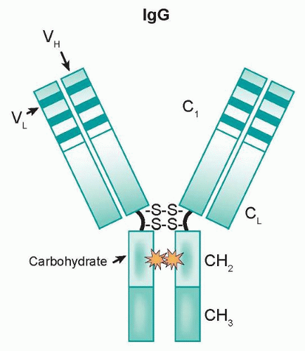
As components of the immune system, effector cells such as natural killer (NK) cells and monocytes/macrophages represent natural lines of defense against oncologically transformed cells. These effector cells express Fcγ receptors (FcγR) on their cell surfaces, which interact with the Fc domain of IgG molecules. This family is comprised of three classes (type I, II, and III) that are further divided into subclasses (IIa/IIb and IIIa/IIIb).36 Recognition of transformed cells by immune effector cells leads to cell-mediated killing through processes such as ADCC and phagocytosis, as shown in Figure 29.2, and can be mediated by FcγRI (CD64), a high affinity receptor capable of binding to monomeric IgG, or FcγRII (CD32) and FcγRIII (CD16), which are low affinity receptors that preferentially bind multimeric complexes of IgG. Signaling through type I, IIa, and IIIa receptors results in the activation of effector cells due to associated immunoreceptor tyrosine-based activation motifs (ITAM), whereas the engagement of type IIb receptors inhibits cell activation through associated immunoreceptor tyrosinebased inhibitory motifs (ITIM).36 Clinical results support the idea that ADCC can play a role in the efficacy of antibody-based therapies. Naturally occurring polymorphisms in FcγRs alter their affinity for human IgG1 and have been linked to clinical response.37,38 A polymorphism in the FCGR3A gene results in either a valine or phenylalanine at position 158 of FcγRIIIa. Human IgG1 binds more strongly to FcγRIIIa-158V than FcγRIIIa-158F, and likewise to NK cells from individuals that are either homozygous for 158F or heterozygous for this polymorphism.39 The FcγRIIIa-158v was a predictor of early response and was associated with improved PFS.
A second polymorphism, FcγRIIa-131H/R, did not predict early response but was an independent predictor of time to progression (TTP).38 Taken together, these data suggest that modulating the affinity of MAbs for FcγRIIIa, FcγRIIa, or both may increase the efficacy of therapeutic MAbs.


Figure 29.2 Antibody-dependent cellular cytotoxicity. The antibody engages the tumor antigen and the Fc domain binds to cellular Fc receptors to bridge effector and target cells. This bridging induces effector cell activation, resulting in natural killer cell cytotoxicity or phagocytosis by neutrophils, monocytes, or macrophages.

Each class of FcγR exhibits a characteristic specificity for IgG subclasses.40 Many groups have focused on modifying the Fc domain of IgGs to optimize the engagement of subclasses of FcγR and the induction of ADCC, based on the findings of Shields et al.,29 who performed a series of mutagenesis experiments to map the residues required for IgG1-FcγR interaction. Antibodies such as ocrelizumab, a humanized version of rituximab, have increased binding to low affinity FcγRIIIa variants and are now in clinical trials.

An alternative to modifying the Fc region of MAbs is to create bispecific antibodies (bsAbs) that recognize both a tumor-associated antigen and a trigger antigen present on the surface of an immune effector cell.43 Simultaneous engagement of both antigens can redirect the cytotoxic potential of the effector cell against the tumor.41,42,43 Such antibodies are capable of eliciting effector function against tumor cell lines in vitro and in animal models. Two HER-2 directed bispecific antibodies, 2B1 and MDX-H210, have been tested in phase I clinical trials.44,45

Bispecific antibodies have a number of distinctive properties, including flexible choices of cytotoxic trigger molecules,46 recruitment of effector function in the presence of excess IgG,42 and custom tailoring of the affinity of the bsAb to match effector cell characteristics. These advantages have been facilitated by improved methods of bsAb production.47 BiTE antibodies represent a novel class of bispecific, single-chain Fv antibodies.48 Promising results have been seen in early phase clinical trials with at least two BiTE antibodies, one of which, blinatumomab, targets CD19/CD3.49 Promising phase I results have also been reported in an interim analysis of an anti-EpCAM/anti-CD3 MT110 BiTE in the setting of advanced lung and gastrointestinal tumors.50


Complement-Dependent Cytotoxicity

In addition to cell-mediated killing (see previous), MAbs can recruit the complement cascade to kill cells via CDC. Although IgM is the most effective isotype for complement activation, it is not widely used in clinical oncology. Similar to ADCC, the human IgG subclass used to construct a therapeutic MAb dictates its ability to elicit CDC; IgG1 is extremely efficient at fixing complement, in contrast to IgG2 and IgG4.51 Antibodies activate complement through the classical pathway, by engaging multiple C1q to trigger activation of a cascade of serum proteases, which kill the antibody-bound cells.52,53 The anti-CD20 MAb rituximab has been found to depend in part on CDC for its in vivo efficacy.54 Antibody engineering approaches have identified residues in the CH2 domain of the Fc region that either suppress or enhance the ability of rituximab to bind C1q and activate CDC.55 The ability to manipulate complement fixation through engineering approaches warrants in vivo testing to determine the impact of these changes on the efficacy and toxicity of MAbs.


ALTERING SIGNAL TRANSDUCTION

Growth factor receptors represent a well-established class of targets for therapeutic intervention. Normal signaling through these receptors often leads to mitogenic and prosurvival responses. Unregulated signaling, as seen in a number of common cancers due to receptor overexpression, promotes tumor cell growth and insensitivity to chemotherapeutic agents. Clinically relevant MAbs can modulate signaling through their target receptors to normalize cell growth rates and sensitize tumor cells to cytotoxic agents. The binding of cetuximab or panitumumab to the epidermal growth factor receptor (EGFR) physically blocks ligand binding56 and prevents the receptor from assuming the extended conformation required for dimerization.57 Pertuzumab binds to the dimerization domain of HER-2, thereby sterically inhibiting subsequent receptor heterodimerization with other ligand-bound family members.58 Alternatively, signaling through growth factor receptors can be indirectly modified by MAbs that bind to activating ligands, as is seen with the anti-vascular endothelial growth factor (VEGF) MAb, bevacizumab.59


IMMUNOCONJUGATES

MAbs that are not capable of directly eliciting antitumor effects, either by altering signal transduction or directing immune system cells, can still be effective against tumors by delivering cytotoxic payloads. MAbs have been employed to deliver a wide variety of agents, including chemotherapy, toxins, radioisotopes, and cytokines (for review see Adams and Weiner60). In theory, the appropriate combination of toxic agents and MAbs could lead to a synergistic effect. For example, delivery of a therapeutic radioisotope by a MAb would be significantly enhanced if, by binding to its target antigen, the MAb also activated a signaling event that increased the target cell’s sensitivity to ionizing radiation.

Catalytic toxins derived from plants catalytic toxins derived from plants (e.g., ricin) and microorganisms (e.g., Pseudomonas) represent two classes of cytotoxic agent that have been investigated for their utility in immunoconjugate strategies.61 Although there are promising preclinical studies,62 few successful clinical trials have been reported using this approach. In a phase I clinical trial in hairy cell leukemia patients who were resistant to cladribine, 11 of 16 patients exhibited complete remissions with minimal side effects with an anti-CD22 immunotoxin with a truncated form of Pseudomonas exotoxin.63 Clinical trials with other immunotoxins have been associated with unacceptable neurotoxicity64 and lifethreatening vascular leak syndrome.65

Only gold members can continue reading. Log In or Register to continue

Stay updated, free articles. Join our Telegram channel

Jun 28, 2016 | Posted by in ONCOLOGY | Comments Off on Monoclonal Antibodies

Full access? Get Clinical Tree

Get Clinical Tree app for offline access