71 Molecular Targeted Therapy
Table 71-1 lists the types of molecular targeted therapies discussed in this chapter. New small-molecule inhibitors, monoclonal antibodies, and viral therapies evolve rapidly and continuously, so the chapter is not intended to delineate a complete list of all agents under development. Instead, we will try to provide an overview of the different strategies available, paying particular attention to the better-known agents likely to affect the clinical approach to cancer.
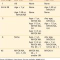
Table 71-1 Categories and Examples of Molecular Targeted Therapies
| Agent | ||
|---|---|---|
| ErbB family inhibitors | Gefitinib | Erlotinib |
| Cetuximab | ||
| Trastuzumab | ||
| Lapatinib | ||
| PDGF receptor inhibitors | Imatinib | |
| VEGF receptor inhibitors | Sorafenib | Sunitinib |
| Bevacizumab | ||
| Farnesyltransferase inhibitors (FTIs) | Tipifarnib | |
| Lonafarnib | ||
| mTOR inhibitors | Sirolimus | |
| Temsirolimus | ||
| Everolimus | ||
| Histone deacetylase inhibitors | SAHA | |
| Nonreplicating viral therapy | Gene replacement | p53 |
| Suicide gene therapy | HSV-tk | |
| Radiation-inducible viral therapy | Egr-TNFα | |
| Replicating viral therapy | Oncolytic adenoviral therapy | Onyx-015 |
| CV707 | ||
| CV787 | ||
| Oncolytic herpesvirus therapy | G207 | |
| G92a |
EGF, Epidermal growth factor; HSV-tk, herpes simplex virus tyrosine kinase; PDGF, platelet-derived growth factor receptor; SAHA, suberoylanilide hydroxamic acid; TNF, tumor-necrosis factor; VEGF, vascular endothelial growth factor.
Molecular Targets
Inhibition of Tyrosine Kinase Receptors
Receptor tyrosine kinases (RTKs) play pleiotropic roles in maintaining homeostasis of individual cells, specific tissues, and entire organisms. The function of RTKs must be tightly regulated, since they mediate fundamental cellular functions including proliferation, survival, adhesion, and differentiation. RTK molecules share a ligand-binding extracellular portion, a transmembrane section, and an intracellular portion that contains the tyrosine kinase catalytic domain.1–3 The family of RTKs includes the epidermal growth factor receptor (EGFR), platelet-derived growth factor receptor (PDGFR), vascular endothelial growth factor receptor (VEGFR), fibroblast growth factor receptor (FGFR), stem-cell factor receptor (SCFR), and nerve growth factor receptor (NGFR). Inactivating mutations in various domains of the protein, including cytoplasmic juxtamembrane and extracellular and kinase domains, confer ligand-independent phosphorylation and kinase activation.4 Activation of RTKs directly contributes to the initiation, progression, and prognosis of several human malignancies.5,6
Monoclonal antibodies (mAbs) can prevent binding of ligands to their receptors, impede the subsequent phosphorylation loop, and thus block signaling through growth factor receptors.7,8 Technical difficulties such as detrimental antibodies patients develop against rodent antibodies have been addressed by advances in antibody construction.9
Small-molecule inhibitors compete with adenosine triphosphate (ATP) for binding sites in the receptor, blocking signaling through RTKs.2 In comparison to mAbs, small-molecular inhibitors against RTKs tend to have less specificity and multiple target receptors. They also have significantly shorter half-lives than mAbs.
Epidermal Growth Factor Receptor Inhibitors
The ErbB family includes four different receptors: ErbB-1 (also known as EGFR), ErbB-2 (also known as HER2/neu), ErbB-3 (also known as HER3), and ErbB-4 (also known as HER4).10–12 EGFR is located on chromosome 7p12, and its activation occurs via several mechanisms including amplification, overexpression, and expression of a truncated constitutively active form. EGFR plays a key role in the pathogenesis of many human malignancies: non–small-cell lung, breast, head and neck, gastric, colorectal, esophageal, prostate, bladder, renal, pancreatic, ovarian, and brain malignancies. EGFR is the most commonly amplified oncogene in glioblastoma (GM), with amplification seen in 40% of tumors.13–15 One-third of GMs in which EGFR is amplified contain a mutant form, most commonly the EGFRvIII mutant, in which deletion in the extracellular domain results in constitutive tyrosine kinase activity.16,17
EGFR offers a promising and exciting target for therapeutic intervention. Several small-molecule inhibitors of EGFR signaling have entered clinical trials for a variety of neoplasms. Gefitinib and erlotinib are two such oral drugs that inhibit EGFR signaling by targeting the ATP-binding site of the receptor.18,19 In a phase III trial of refractory non–small cell large carcinoma (NSCLC) patients, gefitinib did not improve survival in the overall study population, but a subgroup analysis revealed a significant survival benefit in patients of Asian origin or in those who had never smoked.20 Surprisingly, a phase III trial of erlotinib (BR.21) showed an increase in median survival of 2 months in a population of previously treated NSCLC patients.21 Although a survival benefit was found in all subgroups, it was most robust in women, patients with adenocarcinoma, and patients who had never smoked. It is hypothesized that the differences in responses between erlotinib and gefitinib may be the result of the doses used in the trials. In pancreatic cancer patients, a phase III trial of erlotinib in combination with gemcitabine showed impressive improvement in response and overall survival, resulting in approval of this combination as first-line therapy for these patients.22
The efficacies of erlotinib and gefitinib have also been investigated in glioma patients. In unselected GMs, 10% to 20% of patients respond to EGFR inhibitors, and while this proportion is modest, some patients experience dramatic responses.23 Studies have sought to understand the molecular underpinnings that render certain tumors responsive to EGFR inhibitors.24,25 Two complementary studies found that overexpression of wild-type or mutant EGFR promotes sensitivity to EGFR inhibitors, and activation of the phosphoinositide 3-kinase (PI3K)-mediated signaling pathway leads to resistance to such small-molecular inhibitors.26–28
Inhibition of EGFR signaling has been accomplished not only with small molecules but also with mAbs directed against the extracellular domain of EGFR. Human/murine chimeric antibodies offer the advantage of reduced immunogenicity while preserving potency. Cetuximab is such an antibody. Its administration in animal models delays growth of xenograft tumors overexpressing ErbB-1.29 Like the small-molecular inhibitors, the clinical efficacy of Cetuximab has also been investigated in a variety of neoplasms. Phase II trials have shown limited activity for cetuximab in the treatment of NSCLC.30 However, more definitive conclusions must await publication of the results of a recently completed phase III trial (NCT00148798). The benefits of cetuximab for colorectal cancer patients have been thoroughly explored. In phase II trials in metastatic colorectal cancer, response rates to cetuximab alone were between 9% and 12% and in combination with irinotecan rose to 20%.31 A phase III trial of cetuximab plus irinotecan compared to irinotecan alone as second-line therapy showed significant improvements in response rates, progression-free survival, and quality of life.32 More impressively, a phase III trial involving patients who had failed all available therapies showed that cetuximab, compared to best supportive care, improved progression-free survival, overall survival, and quality of life.33 Cetuximab has also proved promising in the treatment of squamous cell carcinoma of the head and neck. When used in combination with radiotherapy, the addition of cetuximab resulted in a significant prolongation of progression-free survival, duration of locoregional control, and overall survival when compared to RT alone.34 Furthermore, the addition of cetuximab to a fluorouracil or platinum-based chemotherapy in recurrent or metastatic disease improved progression-free and overall survival.35
Platelet-Derived Growth Factor Receptor Inhibitors
Whereas some novel signaling inhibitors exhibit specificity toward a given receptor, other drugs can inhibit a number of RTKs. Imatinib is a small-molecule drug that inhibits the tyrosine kinases Abl, PDGFR, and Kit.36 Clinical trials have documented efficacy of imatinib in the treatment of chronic myeloid leukemia, a malignancy driven by constitutive activation of Abl resulting from a chromosomal translocation called Bcr-Abl. Imatinib induces durable responses in the vast majority of these patients, although resistance can develop.37 On the basis of impressive phase II trial results wherein 54% of patients had a partial response to it, imatinib was also approved for the treatment of gastrointestinal stromal tumor (GIST).38
Clinical trials are incorporating treatment with imatinib in the approach to several adult and pediatric malignancies. Studies of gliomas are capitalizing on the inhibitory function of imatinib against PDGFR. The relevance of imatinib for glioma therapy rests in molecular aberrations involving PDGF and PDGFR. PDGFR-α and PDGFR-β are two distinct receptors that bind PDGF ligand, which in turn consists of various dimers of PDGFA and PDGFB chains. Overexpression of the PDGF ligand and receptor are seen in all grades of gliomas, suggesting activation of an autocrine loop that drives glioma proliferation.39,40 In the minority of cases, amplification of the PDGFR-α gene underlies overexpression, but in the majority, the mechanism of overexpression remains unknown.39,41,42 These molecular aberrations in the PDGFR signaling pathway have provided the rationale for treating gliomas of all grades with PDGFR inhibitors. To date, two phase II trials of imatinib in patients with recurrent high-grade gliomas have shown promising results.43,44 Imatinib has also shown some efficacy when combined with docetaxel in treating ovarian neoplasms.45
Vascular Endothelial Growth Factor Receptor Inhibitors
Initiation and maintenance of malignancies rely not only on cell proliferation and survival but also on concomitant development of a blood supply to support the enlarging tumor. Therefore, much effort has focused on developing molecules that block proangiogenic factors and inhibit angiogenesis. The best-described proangiogenic factors are VEGFs that transmit their signals through Flt-1 (also known as VEGF-R1) and Flk-1/KDR (also known as VEGF-R2) receptors.46 Whereas VEGF ligands are secreted by tumor and stromal cells, VEGF receptors are expressed mostly by endothelial cells.
Strategies to block signaling through VEGFR follow the familiar dual paths of small-molecule inhibitors and antibodies directed against receptors or ligands. This theme applies to all RTKs targeted to date. Sorafenib is a small-molecule inhibitor that targets the Faf/MEK/Erk, VEGFR, and PDGFR pathways.47 A second pharmacologic molecule, sunitinib, exhibits wider specificity toward potentially proangiogenic RTKs, including VEGFR2, FLT-3, PDGF, and c-KIT receptors.48 In addition to small molecule inhibitors, antibodies directed against VEGF or its receptors have entered clinical trials.51 Bevacizumab, directed against the receptor ligand VEGF, is the most studied of these recombinant mAbs.
Despite widespread excitement over the positive preclinical results associated with antiangiogenic therapies, early trials proved surprisingly disappointing.52 However, more recent clinical evidence gives reason for optimism. Based upon the encouraging results of a phase III trial of bevacizumab in combination with other chemotherapeutics, the U.S. Food and Drug Administration (FDA) approved its use for treatment of metastatic colorectal cancer. In these patients, bevacizumab increased median survival, progression-free survival, objective response, and duration of response when compared to irinotecan, fluorouracil, and leucovorin treatment alone.53 Bevacizumab has also been approved for treatment of NSCLC in combination with paclitaxel, having demonstrated an ability to increase median and progression-free survival in these patients.54 It is currently undergoing evaluation for possible use in treating kidney, breast, prostate, brain, and ovarian cancers. Besides the mAb therapies, the small-molecule inhibitors sorafenib and sunitinib have also gained approval for the treatment of advanced renal cell carcinoma and GIST.
Inhibition of Downstream Effectors
Farnesyltransferase Inhibitors
Intermediate molecules transmit downstream signals that emanate from engagement of growth factor receptors. The Ras proteins are such intermediaries that help propagate downstream signaling cascades. Ras is a GTPase that cycles between its active guanosine 5-triphosphate (GTP)-bound state and its inactive guanosine 5-biphosphate (GDP)-bound state. Ras regulates many physiologic cellular functions. Biological functions of Ras, including proliferation, survival, cytoskeletal organization, differentiation, and membrane trafficking, rely on its association with the inner surface of the plasma membrane.55–57 Ras proteins are synthesized as cytosolic precursors and converted to membrane-bound forms through posttranslational modifications that begin with the addition of a 15-carbon farnesyl moiety (an isoprene lipid) to a specific motif at the carboxyl-terminus of Ras proteins. Farnesyltransferase catalyzes the transfer of a farnesyl group from farnesyl diphosphate to a cysteine residue within the CAAX box (A is an aliphatic amino acid and X is methionine or serine) of Ras.58
By blocking farnesylation of Ras and inhibiting its function, farnesyltransferase inhibitors (FTIs) can interrupt the effects of tyrosine kinase receptors that signal through Ras. Thus, FTIs may demonstrate antineoplastic activity not only against tumors that contain oncogenic Ras mutations but also against malignancies driven by aberrant signaling through receptor tyrosine kinases. For example, although gliomas rarely possess mutated oncogenic forms of Ras, common genetic aberrations such as EGFR overexpression may be susceptible to therapeutic targeting by FTIs. The precise mode of FTI action remains unclear, since the mutational status of Ras does not consistently correlate with response of cells to FTI treatment. An enlarging body of evidence suggests that FTI activity is mediated in part through inhibition of farnesylation of other Ras family members such as RhoB.59–61 As a result, it remains difficult to use biochemical characteristics of individual tumors to predict which malignancies will prove most susceptible to FTI therapy. These ambiguities notwithstanding, FTI treatment has resulted in objective tumor responses in early clinical trials of various human malignancies.
Tipifarnib and lonafarnib are the two small-molecule FTIs that have been most extensively investigated. Although tipifarnib failed to improve survival and demonstrated minimal activity as a monotherapy in treating metastatic colorectal cancer and NSCLC, it has shown impressive single-agent activity in a variety of hematologic malignancies.62–64 Moreover, tipifarnib, in combination with imatinib, has shown impressive activity in chronic myelogenous leukemia patients with Abl kinase mutations.65
As a monotherapy, lonafarnib has also disappointed, failing to show tumoricidal activity in metastatic colorectal, urothelial, or squamous-cell head and neck cancers.66–68 However, as with tipifarnib, lonafarnib has shown impressive results in several hematologic malignancies.69
Mammalian Target of Rapamycin (mTOR) Inhibitors
The mammalian target of rapamycin (mTOR) is a 289-kD serine/threonine kinase that is critical in regulating cell growth and metabolism during conditions of nutrient abundance. Activation of mTOR itself is mediated by a variety of growth factors and nutrients. Upon activation, mTOR complexes with raptor and mSLT8 to form mTORC1. The mTORC1 complex then, by phosphorylating a variety of downstream effectors, upregulates protein biosynthesis, ribosome biogenesis, and the transcription of genes crucial to cell growth. Because of its intimate involvement in cell growth, and thus cell division, it is not surprising to find mTOR frequently upregulated in certain tumors.70
Because of its importance in tumorigenesis, there has been a recent rash of interest in inhibitors of mTOR. A phase II trial of temsirolimus in advanced refractory renal cell carcinoma demonstrated antitumor activity and encouraging survival, specifically a dose-dependent increase in overall survival.71 In GM, although temsirolimus was well tolerated, it showed only moderate antitumor activity.72,73 In advanced previously treated breast cancer, a phase II trial found a 9.2% objective response rate to treatment with temsirolimus monotherapy. Increasingly, the focus has turned to the benefit of mTOR inhibitors in combination with tyrosine kinase inhibitors, with the two drugs acting synergistically to arrest tumor growth. There are a variety of phase II trials underway to evaluate this novel combination.
Epigenetic Therapies
Although associated with significant toxicities at higher doses, lower doses of the DNA demethylating agents, 5-aza cytidine and 5-aza 2-deoxycytidine, by reactivating tumor-suppressor genes and inducing differentiation, have shown promise in treatment of hematologic malignancies. Recently the FDA approved these two agents for treating myelodysplastic syndrome.74
Histone deacetylase inhibitors (HDACI) represent another promising therapeutic strategy. The acetylation of certain lysine residues on histones results in increased expression of associated genes. In some tumors, the enzyme histone deacetylase is responsible for abrogation of tumor-suppressor expression.75 Suberoylanilide hydroxamic acid (SAHA) is an HDACI that recently has been approved for the treatment of cutaneous T-cell lymphoma. Other HDACIs are under investigation for treatment of a variety of hematologic and solid malignancies.76 Histone methylation is another modification that can, depending upon the residue involved, result in transcriptional repression. Again, certain compounds that result in removal of these methylation marks have shown promise in cancer cell lines.77