Minimally invasive surgery has revolutionized the surgical management of benign foregut disease, as well as pulmonary and other gastrointestinal malignancies. With the potential to reduce operative morbidity and increase patient satisfaction, minimally invasive esophagectomy for the management of esophageal cancer is gaining in popularity. It is unclear, however, whether the minimally invasive approach to esophageal cancer resection has comparable long-term oncologic results. This article discusses the rationale for minimally invasive esophagectomy, describes the surgical technique, and reviews the published results.
Key Points
- •
Esophagectomy remains the best curative treatment of resectable esophageal cancer.
- •
Minimally invasive surgery has revolutionized the surgical management of benign foregut disease, and pulmonary and other gastrointestinal malignancies, and is gaining popularity in the management of esophageal cancer.
- •
The important principles and techniques of minimally invasive esophagectomy are described in this article.
- •
Minimally invasive esophagectomy has short-term results equivalent to those of open resection, with comparable morbidity and mortality, and some reports demonstrate a decreased rate of cardiopulmonary complications.
- •
Long-term oncologic results for minimally invasive esophagectomy are unclear. The number of resected lymph nodes is comparable with that reported for open surgery, but locoregional recurrence rate and long-term survival needs to be studied.
Introduction
Esophagectomy remains the best curative option for the treatment of resectable esophageal cancer, but is a complex operation with significant morbidity and mortality. Over the past decade, minimally invasive esophagectomy (MIE) has been gaining favor as an attractive alternative to open resection with the potential to reduce surgical trauma, decrease morbidity, and shorten the length of hospital stay. While minimally invasive surgery has revolutionized pulmonary surgery, treatment of benign foregut disease, and management of other gastrointestinal malignancies, adoption of MIE has been slow. The aim of this article is to review the technique and results of MIE for esophageal cancer.
History of minimally invasive esophagectomy
Beginning with the first successful esophageal resection by Torek in 1913, esophagectomy has undergone considerable refinement and evolution over the past century. Despite decades of experience, controversy remains as to the optimal open surgical technique for resection of the esophagus and reconstruction of the alimentary tract. Debated issues include the oncologic benefits of a transthoracic versus transhiatal resection and the relative risk of constructing the anastomosis in the neck or chest.
The late 1980s ushered in the modern era of minimally invasive surgery. Techniques and instruments were developed for laparoscopic and video-assisted thoracoscopic surgery (VATS). Over the ensuing 2 decades, minimally invasive surgery for gastroesophageal reflux, paraesophageal hernias, achalasia, obesity, colon cancer, and lung cancer have become standardized and have supplanted open surgery as the standard operation.
As experience in minimally invasive surgery grew, initial attempts at esophageal resection were undertaken. In 1993, Collard and colleagues published their initial experience with thoracoscopic mobilization of the esophagus. In 1995, DePaula and colleagues published an early clinical series of their experience with laparoscopic transhiatal esophagectomy. By the late 1990s initial reports of combined thoracoscopic and laparoscopic esophagectomy and reconstruction were being published, documenting the feasibility of the operation. Despite the great enthusiasm to adopt minimally invasive surgery for other intrathoracic or intra-abdominal diseases, MIE has been slow to be adopted and only accounts for about 15% of all esophagectomies performed.
Rationale for minimally invasive esophagectomy
The advent and refinement of minimally invasive surgery has been a transformative event in surgical science. Worldwide acceptance by clinicians and patients has been driven by improved outcomes and patient satisfaction over open surgery. Classic examples include the tremendous increase in popularity of laparoscopic obesity surgery and laparoscopic cholecystectomy for benign gallbladder disease.
Treatment of benign foregut disease has been revolutionized by minimally invasive surgery. Before laparoscopy, surgical treatment of gastroesophageal reflux disease was reserved for those patients with severe, uncontrollable symptoms and complications from long-standing disease (ie, strictures) due to the morbidity associated with open repair. Beginning in 1991, the technique of laparoscopic Nissen fundoplication was developed, and the operation is now a reasonable alternative to medical therapy even in patients with moderate disease. In addition, laparoscopy has nearly eliminated the need for transthoracic antireflux operations. Laparoscopic antireflux surgery is an extremely safe operation with low mortality and morbidity. A recent analysis of a nationwide database in the United States found the mortality following laparoscopic Nissen fundoplication to be extremely low. Complication rates, length of hospital stay, and patient satisfaction are significantly improved in comparison with open surgery. Similarly, laparoscopic paraesophageal hernia repair and laparoscopic Heller myotomy have become the standard operations for the management of large paraesophageal hernias and achalasia.
With the popularity of minimally invasive surgery for benign disease well established, adoption of a minimally invasive approach for oncologic surgery was initially approached with trepidation out of concern for compromised oncologic outcomes and port-site recurrences. Despite initial concerns, multiple trials for both intrathoracic and intra-abdominal malignancies have reported the early benefits of minimally invasive surgery but have also demonstrated oncologic equivalence. Trials in colon cancer and lung cancer have shown equivalent lymph node harvest, and ability to achieve complete R0 resection and long-term survival. In many centers, laparoscopic colectomy and VATS lobectomy are standard of care for the management of most colon and pulmonary malignancies.
Mortality following open esophagectomy ranges from 2% to 8% in experienced centers and can be as high as 15% to 20% in low-volume centers. Complications following open esophageal resection are common. As a consequence, some centers promote definitive chemoradiotherapy to avoid the risk of resection. Traditionally, pulmonary complications are the major source of morbidity following esophagectomy and are attributed to the thoracotomy and its negative impact on pain, atelectasis, and postoperative pulmonary toilet. Similarly, laparotomy can have a significant negative impact on respiratory function, as pulmonary complications are common even after transhiatal resections.
With the established popularity of minimally invasive surgery, the demonstrated equivalence of minimally invasive surgery for oncologic resection, and the potential to improve the morbidity following open esophagectomy, the minimally invasive approach to esophageal resection and reconstruction may be a prudent alternative to open resection.
Surgical technique
As with open esophagectomy, there are multiple approaches to performing an MIE. Transhiatal, Ivor-Lewis, and Transthoracic (3-hole) esophagectomy are all feasible with minimally invasive techniques. The basic principles of the thoracic and abdominal dissections are similar for each surgical approach, with variations to remove of the specimen, pass the gastric conduit, and create the anastomosis.
The Thoracic Phase
The patient is intubated with a double-lumen endotracheal tube and positioned in the left lateral decubitus position for dissection in the right thoracic cavity. The patient can be rotated more prone to facilitate anterior retraction of the lung and exposure of the esophagus in the posterior mediastinum. A 4-port technique is commonly used to access the right thoracic cavity ( Fig. 1 ). A camera port is positioned in the anterior axillary line at the seventh or eighth intercostal space. A second port is positioned in the posterior axillary line at the eighth or ninth intercostal space. These first 2 ports are placed low on the chest wall to allow dissection of the esophagus starting at the diaphragm and working cephalad. A challenging area to dissect is the costophrenic recess, which is made more difficult if ports are placed too cephalad. An axillary port is placed in the fourth intercostal space just anterior the latissimus dorsi muscle to be used by the assistant for lung retraction, suction, or countertraction of the esophagus or periesophageal tissues as needed. The fourth port is positioned just posterior to the scapular tip. The surgeon typically stands to the patient’s right using primarily the posteriorly placed ports for dissection.
Once access is obtained and the lung collapsed, a fan retractor or atraumatic lung clamp can be used to retract the lung anteriorly to expose the posterior mediastinum. Additional visualization of the lower thoracic esophagus is achieved by retraction of the diaphragm using a heavy suture placed into the central tendon of the diaphragm and brought out through the camera port or a separate stab incision low on the anterolateral chest wall. Dissection of the esophagus proceeds in a caudal to cephalad direction with the aid of ultrasonic dissection. The inferior pulmonary ligament is taken down and the pleura opened in the plane anterior to the esophagus and adjacent to the pericardium. As the dissection is carried cephalad, periesophageal and subcarinal lymph nodes are dissected off the adjacent pericardium and airway en bloc with the esophagus. A plane posterior to esophagus is created just anterior to the cranially directed azygous vein. Dissection is carried down toward the aorta, which serves as the posteromedial limit of dissection. The azygous vein traversing over the esophagus toward the superior vena cava is stapled with an endoscopic linear stapler to facilitate dissection of the esophagus into the thoracic inlet.
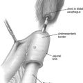
Circumferential dissection is achieved in the mid esophagus, and a Penrose drain can be passed around the esophagus for improved retraction ( Fig. 2 ). Circumferential dissection is frequently easiest just above the level of the inferior pulmonary vein where the esophagus and periesophageal tissue is dissected off the posterior pericardium. The esophagus is fully mobilized from the diaphragm to the thoracic inlet with dissection of paratracheal, subcarinal, paraesophageal, and crural lymph nodes under direct thoracoscopic guidance. Care should be taken to avoid injury to the posterior membranous wall of the trachea, which can be well visualized by the thoracoscopic camera.
Once the esophagus is fully mobilized, the Penrose drain can be loosely tied around the esophagus and positioned into the thoracic inlet, to be retrieved during the cervical phase of the operation. A second Penrose drain can be tied around the esophagus and tucked down near the diaphragm to assist in the hiatal dissection during the abdominal phase of the surgery. A single chest tube is placed and the wounds closed in the standard fashion.
The Abdominal Phase
The setup for the abdominal phase of the operation is similar to a laparoscopic Nissen fundoplication. The patient is positioned in either dorsal lithotomy or supine position with a foot rest to allow for steep reverse Trendelenburg. Typically, 5 ports are placed for a camera, liver retractor, the surgeon’s dissecting ports, and a port for the assistant. With the liver retracted anteriorly, the gastrohepatic ligament is opened up to the level of the crus. If a thoracic mobilization has already been performed, dissection of the phrenoesophageal ligament and crura is delayed until the end of the abdominal phase of the operation to avoid escape of insufflated gas through the thoracic cavity. Along the greater curve of the stomach, the gastrocolic omentum and short gastric arteries are divided. It is crucial to visualize the gastroepiploic arcade. Inability to adequately identify this vessel may necessitate conversion to open laparotomy to avoid injury to the critical blood supply of the gastric conduit. With the short gastrics and gastrocolic ligament mobilized, the lesser sac can be entered. Any residual adhesions to the posterior wall of the stomach and retroperitoneum are divided, and the left gastric artery is identified. After sweeping the lymphatic tissue toward the stomach, the left gastric artery is divided at its origin with a linear endoscopic stapler. The phrenoesophageal ligament and crura are dissected next. The Penrose drain placed during the thoracic phase of the operation can be retrieved, used to retract the gastroesophageal junction, and assist the dissection at the hiatus. It is sometimes necessary to complete the hiatal dissection before dividing the left gastric artery to facilitate passage of the stapler around the vessel with tips of the stapler passing through the hiatus.
The gastric tube is then created with sequential firings of a heavy tissue linear endoscopic stapler ( Fig. 3 ). It can be helpful to perform on-table endoscopy to ensure an adequate distal margin. The gastric tube is fashioned by starting the division of the stomach from the lesser curve and dividing toward the fundus. Strong consideration should be given to insertion of a feeding jejunostomy and pyloric drainage procedure.


