Fig. 19.1
Conceptual schema of central vascular ligation during pancreaticoduodenectomy (quoted from reference [10] with modification). (a) Magnified view of systems around the pancreas head with the components arranged in a single plane. The mesopancreas includes the plPh-1, plPh-2, arteries, and veins. (b) Level 1 dissection, which aims to resect the pancreas head alone without systematic lymph nodes (LNs) dissection. (c) Level 2 dissection, which aims to dissect the pancreas head and the mesopancreas en bloc. (d) Level 3 dissection, which aims to dissect the pancreas head, the mesopancreas, and the hemi-circumferential nerve plexus around the SMA and if needed the celiac axis
19.3 PD-CVL Surgical Technique
19.3.1 Abdominal Exploration and Preparation for PD-CVL
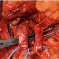
After an upper abdominal midline incision, the peritoneal cavity is explored to confirm the tumor stage and operability. After a wide Kocher maneuver, the para-aortic LNs are explored and resected if necessary. The duodenum is dissected to the left, exposing the superior mesenteric vein (SMV) from the right side. The gastrocolic ligament and greater omentum is then dissected until the pancreas head is well exposed. The superior right colic vein is ligated routinely, followed by further dissection along the same plane to expose the middle colic artery, which is exposed to its root to identify the SMA and to dissect the LNs above the SMA. The SMV is then taped at the level of the transverse portion of the duodenum.
19.3.2 Right Dorsal Dissection of the SMA Using Supracolic Anterior Artery-First Approach
In level 2 dissection usually without SMV co-resection, branches such as Henle’s gastrocolic trunk or the first JV are divided to free the SMV from the pancreas head. A diamond-shaped window is then created by retracting the SMV rightward, the transverse mesocolon caudally, the SMA leftward, and the pancreas neck cranially (Fig. 19.2a). If the middle colic vein obstructs this window, it can be ligated and divided. In this field, the right and dorsal aspects of the SMA are dissected using the supracolic anterior approach while preserving the circumferential plSMA (Fig. 19.2b).
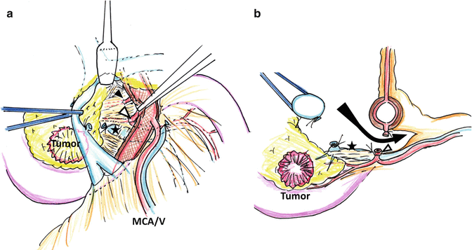

Fig. 19.2
Level 2 supracolic anterior dissection. (a) Frontal view. By retracting the pancreas neck using thin retractor and rotating the SMA at pinpoint, the mesopancreas was detached from plSMA. The IPDA 1 (black arrow) and the common trunk of IPDA 2 and JA 1 (blank arrow) were exposed. The JV (star) was ligated and exposed on the surface of the mesopancreas. (b) Transverse view. The plSMA was entirely preserved. The dissection reaches to the left side of the SMA (black arrow). The common trunk of IPDA 2 and JA was ligated by the central vascular ligation
In level 3 dissection, wherein the cancer has invaded the mesopancreas and SMV, the connective tissue around the SMV is not detached from the SMV. The middle colic vein is routinely ligated and divided. The hemi-circumferential plSMA in the corresponding direction is resected to gain an optimal margin from the area of cancer infiltration (Fig. 19.3a, b).
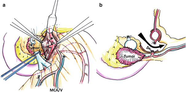

Fig. 19.3
Level 3 supracolic anterior dissection. (a) Frontal view, showing the plSMA dissected from the SMA adventitia hemi-circumferentially under pancreas neck retraction and pinpoint rotation of the SMA. The dissection along the SMA depended on the tumor invasion. The IPDA 1 (black arrow) and the common trunk of IPDA 2 and JA 1 (blank arrow) were exposed. (b) Transverse view. The plSMA was entirely preserved. The dissection reaches to the left side of the SMA (black arrow). The common trunk of IPDA 2 and JA was ligated by the central vascular ligation
In both level 2 and 3 dissection, the JV running behind the SMA should be adequately separated from the SMA, i.e., up to 1 cm left to the SMA (Figs. 19.2b and 19.3b). The root of the IPDA or the common trunk of the IPDA and first JA is exposed, ligated, and cut at this stage. We then convert the procedure to left-sided dissection of the SMA.
19.3.3 Finger-Guided Connection of the Dissection Space of Both Sides of the SMA
At this stage, we can easily identify the root of the second JA to be preserved (Fig. 19.4a). The surgeon inserts the left fingers behind the SMA from the right side at a point just proximal to the second JA (Fig. 19.4b). Under the guidance of the surgeon’s fingers, the serosa of the mesentery is opened, connecting the right and left dissection spaces (Fig. 19.4c, d). A tape for hanging is placed through this hole, encircling the dorsal aspect of the SMA.
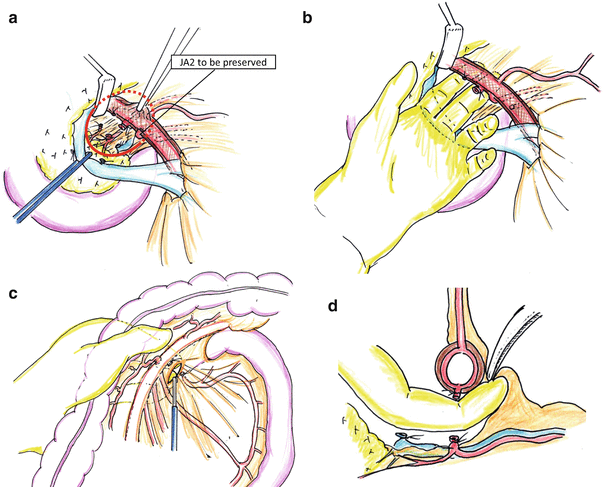

Fig. 19.4
Finger-guided connection of dissection space of the both sides of the SMA. (a) After LV-2 supracolic anterior dissection, the mesopancreas was dissected from the SMA, preserving PL-SMA. The dissection space reached to the left side of the SMA (red circle). The second jejunal artery (JA2) is preserved in this dissection. (b) The surgeon’s finger is inserted into the dissection pocket. (c, d) By the finger guidance, the mesentery is opened by an electric cautery, and the dissection space was opened atraumatically using Kelly clamp, and the SMA was taped
19.3.4 Left-Sided Dissection of the SMA
The transverse colon is then reflected cranially, and the left side of the SMA is dissected so that the previous opening is enlarged toward the root of the SMA, preserving the plSMA (Fig. 19.5a). This procedure can be performed bloodlessly because the root of the first JA has already been ligated and cut, and the JV has been separated from the SMA during the previous supracolic anterior approach. The mesentery of the first JA territory is divided from the remnant, and the corresponding jejunum is cut. The dissection of the left side then progresses, and the ligament of Treitz is identified as a membranous muscular layer that narrows cranially [12, 13]. The ligament is dissected from the SMA, then ligated and cut at the level of the SMA root. The left side of the SMA is thus fully dissected while preserving the plSMA (Fig. 19.5b).
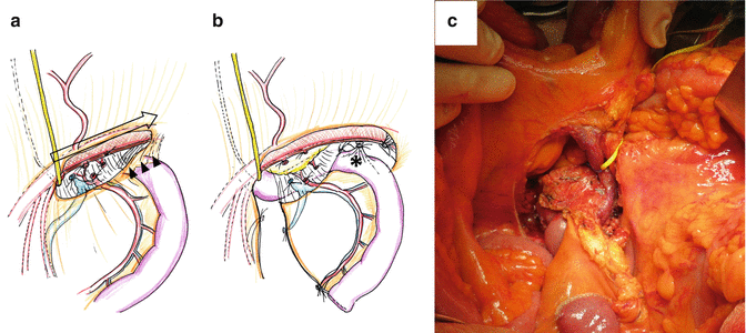

Fig. 19.5
The left-sided dissection around the SMA. (a) Initial part. The dissection begins from the point at which the tape is applied and progresses toward the root of the SMA, preserving the nerve plexus of the superior mesenteric artery. The tape represents the starting point of the longitudinal dissection of the SMA (blank arrow). Left-sided superior mesenteric artery dissection is then promoted further, identifying the ligament of Treitz as a membranous muscular layer (black arrows). (b) Final part. The ligament is detached from the superior mesenteric artery, ligated, and cut beyond the left renal vein (asterisk). At this stage, complete dissection of the left side of the superior mesenteric artery is achieved with preservation of this side of the nerve plexus of the superior mesenteric artery. (c) The intraoperative view after the left-sided dissection of the SMA. The pancreas head and the mesopancreas were detached from the SMA and the SMV
19.3.5 Completion of PD-CVL
After the stump of the jejunum is reflected rightward, the SMV is retracted to the left, and the upper portion of the mesopancreas is dissected (Fig. 19.6a). Once the right-sided dissection reaches the root of the SMA, en bloc dissection around the SMA is completed (Fig. 19.6b). Division of the pancreas neck, common bile duct, stomach, or duodenum is then performed, and PD resection is competed.
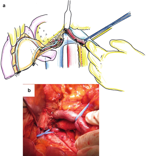

Fig. 19.6
The final stage of the central vascular ligation of the pancreas and the SMA. (a) The proximal jejunal stump is reflected to the right, whereas the superior mesenteric vein is reflected to the left, and the right dorsal aspect of the SMA is well exposed. (b) The intraoperative view of completion of LV-2 dissection around the SMA using central vascular ligation. The mesopancreas is entirely detached from the SMA
19.4 Indication
PD-CVL is indicated for periampullary malignancy requiring LNs dissection and is subclassified according to the extent of dissection: Level 2 dissection should be used for malignancies that need total resection for the mesopancreas. By dividing IPDA and 1st JA at their roots, corresponding LNs and mesojejunum can be resected systematically. Dissection along the SMA is determined according to the site of IPDA branch. The disease suitable for level 2 dissection includes ampullary, distal bile duct, and duodenal cancers or pancreatic cancers with limited invasion. In level 3 dissection, the right half circle of plSMA is resected en bloc with tumor to obtain maximal margin length from potentially invasive tumor, and it should be used for pancreatic ductal cancer or advanced bile duct cancer. Dissection along SMA should be determined based on the extent of tumor invasion. Corresponding mesojejunum is also resected.
19.5 Comment
In this chapter, we described a comprehensive technique for mesopancreas excision during pancreaticoduodenectomy using CVL by a supracolic anterior artery-first approach. Several technical modifications in PD applying so-called artery-first principle have been proposed previously [14–21]. However, few reports have documented the whole outline of the SMA up to its root and the circumference of the SMA. We [22] recently described a technique termed “pancreaticoduodenectomy with systematic mesopancreas dissection (SMD-PD)” with en bloc mesopancreas resection including staged dissection around the SMA using a supracolic anterior artery-first approach. The technique described here is the extension of the concept of SMD-PD, and this allows precise and bloodless SMA dissection as we reported previously [10, 11, 23].
The advantage of level 2 dissection is multifold; first, early ligation of the supplying artery reduces the bleeding during SMA dissection, which is often bloody otherwise. Secondly, complete removal of LNs corresponding to IPDA and JA secures the oncologic clearance. For example, in ampullary cancers, LN metastasis into the proximal jejunal region via mesopancreas is reportedly substantial, and complete dissection of this area has been advocated [24, 25]. Actually, we sometimes encounter LN metastasis in the mesojejunum in patients with ampullary, duodenal, and pancreatic cancers. Likewise, distal bile duct cancer has been reported to be potentially more aggressive compared to ampullary cancers, indicating that the level 2 dissection should be applied. Lastly, PD-CVL using supracolic anterior approach enables straightforward dissection along the SMA without distorting the in situ anatomy, helping the surgeon to grasp the dissection margin clearly. This approach resembles the concept of total mesorectal excision [5], and like rectal cancer, standardization of systematic mesopancreas excision and LN dissection may improve the clearance of cancer spread via the lymphatic pathway.
Stay updated, free articles. Join our Telegram channel

Full access? Get Clinical Tree


