Melanoma of the Head and Neck


Melanoma of the Head and Neck


Cecelia E. Schmalbach

Alison B. Durham

Timothy M. Johnson

Carol R. Bradford


Although melanoma accounts for only 5% of cutaneous cancers diagnosed each year, it is the most lethal form, accounting for over 75% of deaths attributed to skin cancer. Approximately 25% of all cutaneous melanomas arise in the head and neck (HN) region.1 The dramatic increase in melanoma incidence and mortality over the past two decades has led to significant economic burden.2 In 2010, the total direct cost of treating melanoma in the United States exceeded $2.3 billion.3 The leading cause of melanoma remains intense sun exposure; consequently, it is a cancer of young as well as older adults. Melanoma ranks second only to testicular cancer in loss of average adult lifeyears per fatality and a loss of ˜$3.5 billion in productivity.4 These startling melanoma statistics underscore the importance of prevention, accurate staging, and clinical trials.


EPIDEMIOLOGY

The incidence of cutaneous melanoma in the United States continues to rise at epidemic proportions with 137,990 new cases of melanoma estimated in 2014 (61,300 noninvasive; 76,690 invasive).2,5 This rate has consistently increased 2.8% per year since 1981. By 2015, it is estimated that 1 in 50 Americans will develop a melanoma in their lifetime.6 A slight male predominance has been consistently reported throughout the literature.7,8

Although the mortality rate of various cancers has declined, mortality from melanoma has risen by 3% each year since 2004. 9,710 Americans will die from melanoma this year, an estimate that averages to approximately one patient per hour.2 Melanoma typically arises in the fifth and sixth decade.7 However, this statistic is misleading because one in four new melanoma cases will present before the age of 40 years. The increasing incidence among younger patients, especially women, is attributed to sun worshiping tendencies and indoor tanning.9 Melanoma is the most common cancer for young adults aged 25 to 29 years, the second most common cancer for women aged 30 to 34 years, and the second most common cancer in ages 15 to 14.5 Patients in the pediatric age group account for ˜1.66% of the HN melanoma cases, with patients being diagnosed as young as 4 years old.10,11


ANATOMIC DISTRIBUTION

Approximately 25% of all cutaneous melanomas arise in the HN region.1 The majority arise on the cheek, scalp, and neck.7,8,21 Among 857 HN melanoma patients, Fisher et al.7
found that the face and neck regions accounted for over 60% of primary tumors. An additional 26% arise from the scalp, with the ear and nose accounting for only 9% and 4% of primary tumors, respectively. This anatomic distribution was confirmed by O’Brien et al.21 The forehead and cheek regions contain a two- to threefold higher melanocyte density compared to other anatomic sites. This difference, coupled with the increased sun exposure, likely accounts for the distribution of melanomas within the HN region.


Figure 9.1. United States Center for Disease Control and Prevention (CDC) 2010 list of the highest melanoma incidence by state. Statistics include both genders and all races. Melanoma is attributed to intense, intermittent as opposed to cumulative sun exposure, which may account for this high incidence in northern states.


RISK FACTORS

Table 9.1 summarizes the environmental and genetic risk factors implicated in the development of cutaneous melanoma.


Sun Exposure

The leading cause of melanoma is sun exposure. Johnson et al. found that 81% of 1,515 patients with melanoma investigated recalled a history of at least one sunburn.23 Recent epidemiologic trends further support the causal relationship of solar damage and melanoma. A “latitude gradient” has been reported in which the incidence of melanoma increases among similar ethnic populations as distance to the equator decreases.17 A higher rate of melanoma has also been reported among immigrants to areas of increased solar radiation compared to native residents.17,24

Rigel25 analyzed 43 melanoma risk factors among 200 patients. Two of the six key factors associated with increased melanoma risk directly related to sun exposure are three or more blistering sunburns before the age of 20 and three or more outdoor jobs during teenage years. Additional significant risk factors identified included red/blonde hair, family history of melanoma, actinic keratoses, and marked freckling of the upper back. Individuals demonstrating one or two key factors carried a three- to fourfold increased risk for development of melanoma. The risk for melanoma increased 20-fold if a patient was found to have three or more key risk factors.


Table 9.1 Cutaneous Melanoma: Environmental and Genetic Risk Factors


Environmental


Medical/Genetic


Inability to tan


CDKN2A (p16) mutation


Fair skin/freckling


Immunosuppression


Blue/green eyes


Xeroderma pigmentosa


Blonde/red hair


Dysplastic nevus


History of blistering sunburn


Giant congenital melanocytic nevus


Outdoor summer job


History of prior skin cancer


Tanning booth use (UVa radiation)


Family history of melanoma


a UV: ultraviolet-alpha rays (315 to 400 nm).


Adapted from Schmalbach CE, Johnson TM, Bradford CR. The management of head and neck melanoma. Curr Probl Surg. 2006;43:781-835. Ref.22


Genetics

A genetic etiology has also been implicated in the pathogenesis of melanoma.26 Approximately 15% of patients with melanoma report a positive family history.27 The most commonly inherited genomic abnormalities associated with melanoma is the CDKN2A locus, which encodes the p16 tumor suppressor gene.27,28 However, p16 mutation is reported in only 0.2% of the melanoma cases diagnosed.29 The hereditary nature of cutaneous melanoma was first described in the 1970s when Clark et al.30 observed two families in which members
acquired large dysplastic nevi, often in sun-protected regions of the body such as the scalp and trunk. They coined the term “B-K mole syndrome.” During this same time period, Lynch et al.31 independently reported a similar association, which they termed “familial atypical multiple mole-melanoma syndrome” or FAMMM syndrome. Today, the term “atypical mole syndrome” is applied to familial cases of melanoma. The syndrome is inherited in an autosomally dominant fashion. Family members carry a 10-year melanoma risk of 10.7%, which is significantly >0.62% risk reported in control patients. A 56% cumulative risk is estimated in these carriers from age 20 to 59 years. Nearly 100% of patients with atypical mole syndrome develop melanoma by age 76.32

Although a melanoma gene has been postulated, the genetic aspect of this disease is far more complex.33 The first wholegenome melanoma sequence was published in 2010 and identified more than 33,000 mutations compared to the germline control.34 Various tumor suppressors, transcription factors, and oncogene mutations have been associated with melanoma.35 The majority of melanomas are found to harbor one or more mutations related to a kinase signaling pathway.33 A point mutation in BRAF, a serine-threonine protein kinase, has been identified in 65% of melanoma cell lines and 42% of tumors.36,37 Eighty-five percent of the BRAF mutations are associated with a single substitution (V600E).37 Mutations in NRAS, a member of the RAS family of GTPases, have also been implicated in up to 25% of melanoma tumors.33 The c-KIT gene encodes for a tyrosine kinase receptor within the cellular membrane, and mutation of the c-Kit gene has been identified in 19% of melanomas arising in chronically sunexposed regions of the body.38 Although the genetic alterations are promising avenues for targeted therapy (see section below), it is important to realize that an estimated 30% of patients with melanoma lack a detectable genetic abnormality.33

In addition to the familial forms of melanoma described above, there is another syndrome associated with melanoma development termed xeroderma pigmentosum (XP). XP is a rare, autosomally recessive disease associated with skin cancers including melanoma.39 Fibroblasts in XP patients have an impaired ability to repair DNA damaged by UVR,40 which leads to the development of multiple cutaneous malignancies including melanoma, BCC, and SCC. Patients are usually diagnosed with their first cancer before the age of 10. Despite UVR precautions, careful surveillance, and aggressive treatment, the development of skin cancers is relentless, with the majority of XP patients succumbing to cancer during their childhood years.


Immunosuppression

Numerous studies throughout the literature provide supporting evidence for a role of immunosuppression in the development of melanoma. A recent systematic review of the literature identified an association between melanoma and the following immunosuppression settings: solid organ transplant, lymphoproliferative disorders, iatrogenic immunosuppression, and human immunodeficiency virus infection/AIDS.41 Higher rates of premalignant melanocytic nevi in the setting of transplantation, chemotherapy, and childhood leukemia lend further support of this association.42,43 Ultimately, immunosuppressed patients warrant vigilant monitoring for skin cancer, and melanoma care must be coordinated carefully with all medical teams.


Melanotic Nevi

Although melanoma can arise de novo, ˜50% of cases develop from a preexisting pigmented lesion44 (Fig. 9.2). The vast majority of adults have at least one melanotic lesion. Intradermal nevi account for the majority of adult moles. Junctional nevi are common in childhood. This lesion appears as a flat, tanbrown papule, which is smooth and well defined. Overall, junctional nevi are recognized as the most common premalignant nevi.45

Atypical melanocytic nevi (AMN), also known as atypical moles, dysplastic nevi, and Clark nevi, are acquired pigmented lesions with both a clinical and histologic appearance different from that of the common mole.46 AMN are recognized as a marker for increased risk for melanoma risk and as a melanoma precursor. They often display irregular or poorly demarcated borders. They differ from common benign nevi in that they are typically larger in size, measuring between 5 and 12 mm in diameter.

Congenital melanocytic nevi (CMN) are pigmented lesions present at birth or within the first 6 months of infancy.47,48 Up to 6% of children are born with CMN. CMN size ultimately dictates the melanoma risk. Small CMN (<1.5 cm diameter) and medium CMN (1.5 to 1.99 cm diameter) carry the same lifetime melanoma risk as do any other typical nevi. However, large CMN (≥2 cm diameter) carry an increased risk for development of melanoma, with development in an estimated 5% to 20% of individuals.47 These melanomas are usually diagnosed in early childhood, with 70% of cancers being diagnosed before the age of 10.49 For this reason, prophylactic excision is advocated for large CMN if the nevus is in an anatomic location amenable to surgery. Unfortunately, the large size can carry significant cosmetic, as well as psychosocial, implications.48

Lentigo maligna (LM) (see above) is a melanoma subtype in which the cancer is limited to the intraepidermal layer. Classified as melanoma in situ, it is deemed a precursor to invasive melanoma. The exact percentage of LMs that progress to invasive lentigo malignant melanoma (LMM), remains unknown.50 However, the rate of progression is estimated to be between 5% and 33%.


Figure 9.2. Approximately 50% of melanomas develop from a preexisting pigmented lesion.


MELANOMA CLASSIFICATION

Three histologic variants of melanoma are reported within the HN region and are outlined below. It is important to realize that melanoma subtype does not generally influence prognosis once tumor thickness and other prognostic variables such as ulceration are taken into account. For this reason, the melanoma subtype does not impact tumor staging.


Common HN Melanoma Subtypes

The majority of HN cutaneous melanomas are superficial spreading melanoma (SSM), accounting for ˜70% of all cases.19 The characteristic SSM feature is color variation, which is often described as kaleidoscopic with areas of black, dark brown, tan, and blue-gray pigmentation. Areas of pink and white may be present and represent hypopigmentation secondary to tumor regression. Although SSM lesions are well circumscribed, the borders tend to be scalloped and asymmetric. Patients are usually diagnosed within their fourth to fifth decade and often report a preexisting nevus in the region of their newly diagnosed melanoma.

Nodular melanoma (NM) is the second most common melanoma variant, accounting for 15% to 30% of cases.19 The majority of mucosal melanomas are of the nodular variant. The lesion typically appears as a raised, blue-black or blue-red nodule. As hemangioma, blue nevus, pyogenic granuloma, and pigmented BCC can appear similarly, it is important to biopsy lesions with this appearance before treating them to avoid undertreatment.

As mentioned above, LM represents intraepidermal or melanoma in situ. Also known as “Hutchinson melanotic freckle,” it is often diagnosed in the background of chronic solar damage. The invasive counterpart to LM is lentigo malignant melanoma (LMM). The exact percentage of LMs that progress to invasive LMM remains unknown.50 It is speculated that if LM patients live long enough, all will progress to invasive melanoma. LM/LMM is commonly found within the H&N region. The subtype has been associated with older individuals, but the frequency in younger patients is increasing.30 LM/LMM can display subepithelial extension as well as peripheral involvement with atypical junctional melanocytic hyperplasia (AJMH). These findings make achieving adequate surgical margins challenging from both an esthetic and a functional standpoint. Additional challenges associated with LMM are that both amelanotic and desmoplastic melanoma (DM) commonly arise in the setting of LM/LMM.


Desmoplastic Melanoma

DM is a rare subtype of melanoma composed of spindle cells with abundant collagen.51 Although DMs are rare, accounting for only 1% of melanoma cases,52 75% are diagnosed in the HN region. They commonly arise in the setting of LMM. DMs are distinct from other melanoma subtypes in that they present in an older patient population; the median age of diagnosis is 61 years compared to 46 years.
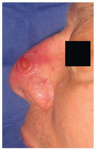
52 Although amelanotic cases account for only 7% of cutaneous melanomas, up to 73% of DM and DNM have been found to be amelanotic.52,53 This atypical appearance (Fig. 9.3), coupled with the spindle cell histology, makes DM somewhat of a diagnostic challenge. Immunohistochemistry (IHC) is helpful because the majority of tumors will stain positive for S100 and vimentin; HMB-45 is less reliable due to the amelanotic appearance. DM is highly infiltrative, has a propensity for neurotropic spread, and is considered locally aggressive. Spread along cranial nerves to the skull base and cavernous sinus is not uncommon. In addition, early local recurrences are reported as high as 49%, and this may be related to undetected perineural spread.53


Figure 9.3. Amelanotic melanoma of the nose mimicking basal cell carcinoma. Note the lack of traditional melanoma ABCD warning signs.


Table 9.2 Indications for Cutaneous SLNB


Localized melanoma ≥1 mm depth of invasion (T1-T4N0)


Localized melanoma <1 mm depth of invasion (T1N0)


Demonstrating a poor prognostic feature include:


Ulceration (T1bN0)


Mitotic rate ≥1/mm2 (T1bN0)


Young age


Angiolymphatic invasion


Positive deep margin


Tumor regression


Primary treatment for DM remains surgical excision, with a minimum of 1 cm margins in order to prevent local recurrence. Pure DMs carry a low (<10%) rate of regional metastasis; therefore, sentinel lymph node biopsy (SLNB) is not recommended in this setting.54 Despite the low rate of regional metastasis, patients with this diagnosis have a similar risk for the development of distant metastases as patients with NM of similar depth of invasion. More commonly, melanomas will be classified as “mixed” DM. These lesions carry the same rate of regional metastasis as do other melanoma subtypes, and SLNB should be offered to patients meeting the criteria outlined in Table 9.2. Evaluation of these lesions by an experienced dermatopathologist is critical in discerning between pure DM and mixed DM lesions.


Mucosal Melanoma

Mucosal melanoma (MM) represents a rare variant of melanoma, accounting for <2% of all cases.55 Review of the Surveillance, Epidemiology, and End Results (SEER) database from 1987 to 2009 identified an increasing incidence of MM in the United States.56 This increase was unique to the nasal cavity subsite, especially for women ages 55 to 84.

MM is regarded as a distinct and separate entity from its cutaneous counterpart. Unlike cutaneous melanoma, etiologic environmental factors have not been linked to the development of MM.57 MM presents on average one decade later than cutaneous lesions.58 In addition, women are diagnosed twice as often as men. Lastly, the BRAF oncogene mutation commonly identified in cutaneous melanoma is rarely found in the mucosal subtype. Instead, a relatively high incidence of KIT mutations has been reported.59

The majority of MMs arise in the nasal cavity. The anterior nasal septum is involved most often (33%), followed by the lateral nasal wall (28%), turbinates (15%), and nasal vestibule (10%).60 The paranasal sinuses are another common site of origin, with the maxillary sinus involved most often. Given these anatomic locations, it is not uncommon for patients with sinonasal MM to present with nasal obstruction and epistaxis. These symptoms often lead to early diagnosis, with 75% of sinonasal patients presenting with localized disease only.
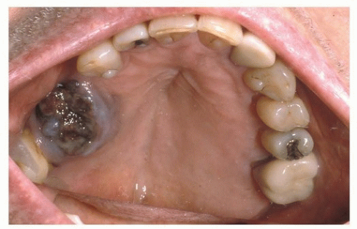
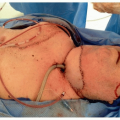
61

Approximately 40% of HN MMs arise in the oral cavity (OC), with the upper alveolus and hard palate (Fig. 9.4) reported as the most common subsite (70%).62 OC MM is often asymptomatic and can go undiagnosed until a neck mass develops from metastasis.63 A review of five major MM series by Batsakis et al.64 found laryngeal primary tumors to account for fewer than 4% of all cases. Within the larynx, the supraglottis was the most common site of origin.

MM arises from respiratory stromal and mucosal melanocytes.
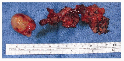
60 The diagnosis can be more challenging than that of cutaneous melanoma due to amelanotic nature of many tumors (Fig. 9.5). For this reason, IHC plays an important role in diagnosis. MM will often stain for S100, HMB-45, and Melan-A (MART1). Olfactory neuroblastoma may stain for S100 and HMB-45; however, the MAP-2, cytokeratin, and epithelial membrane antigen stains will facilitate the correct diagnosis.65 Sinonasal undifferentiated carcinoma (SNUC) will stain for cytokeratin but not S100 or HMB-45. Lastly, plasmacytoma and lymphoma are routinely leukocyte common antigen positive in the absence of S100 staining.


Figure 9.4. Mucosal melanoma of the hard palate. The oral cavity is the second most common site for mucosal melanoma, accounting for 40% of cases.


Figure 9.5. Mucosal melanoma resected from the superior nasal cavity via a subfrontal craniotomy approach. Note the amelanotic nature of MM, which mimics a nasal polyp.

The recent 7th edition of the AJCC cancer staging system now incorporates a dedicated tumor-node-metastases (TNM) staging system for MM.66 The staging system begins with stage III disease because of the overall poor prognosis of MM, even in the setting of limited primary tumor burden. Due to the overall aggressive nature of MM, T1 and T2 categories do not exist. T3 tumors are limited to the mucosa. T4a represents moderately advanced disease with invasion into the deep soft tissue, cartilage, bone, or overlying skin. T4b is reserved for very advanced disease, which includes the brain, dura, skull base, cranial nerve, masticator space, carotid artery, prevertebral space, and mediastinal structures. Regional disease and distant disease also impact patient outcome. Patients with nodal metastasis are classified as N1, which upstages them to stage IVA. Similarly, patients with distant metastasis are designated M1 and are classified as stage IVC.

Wide local excision (WLE) of the primary tumor remains the standard of care, and therapeutic neck dissection is recommended for known nodal metastasis.57 Elective management of the N-zero neck is based upon the site of origin. Sinonasal MM is usually confined to the primary site at presentation.67 For this reason, an elective neck dissection (END) is not typically recommended. However, OC MM carries an increased risk for nodal metastasis and may warrant END.

Adjuvant radiation to the primary MM is recommended, regardless of depth of invasion. Extracapsular spread (ECS), two or more positive nodes, intraparotid nodal metastasis, any node >3 cm in diameter, and tumor recurrence are considered high-risk features warranting adjuvant radiation to the draining nodal basins.57 Radiation planning is based on anatomic subsite and risk. The most common plan for high-risk MMs is conventional fractionation to 60 to 66 Gray (Gy) postoperatively or 70 Gy to gross disease.


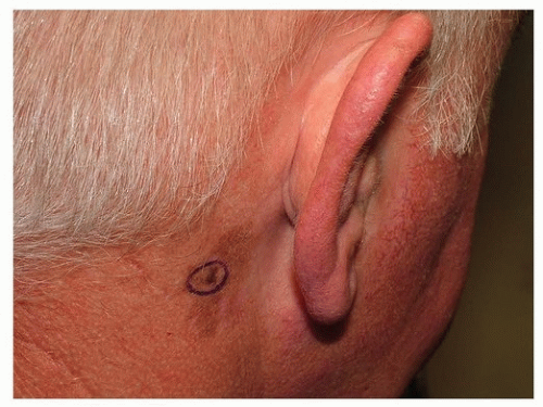
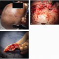
Melanoma of the Auricle

Melanoma of the auricle was originally thought to carry a worse prognosis compared to other HN sites.68 The increased risk was attributed to rich lymphatics, complex anatomic subdivisions of the auricle (6 hillock of His), and a paucity of subcutaneous tissue between the thin skin of the auricle and the underlying perichondrium.69 For these reasons, total auriculectomy was historically considered standard of care. Retrospective reviews failed to demonstrate a difference in
local recurrence based on the extent of auricular excision.69 After accounting for known prognostic features such as tumor thickness, recent studies demonstrate similar survival rates between melanoma of the auricle compared to other anatomic sites.70 It is now recognized that current prognostic indicators and surgical principles can be applied safely to the auricular region. Perichondrium is considered a barrier to the spread of melanoma.71 For this reason, the underlying cartilage requires resection only in the setting of tumor involvement or if previous surgery/biopsy has violated the plane making it impossible for the surgeon to determine if there was direct tumor extension.


EVALUATION OF THE PATIENT


History

Approximately 25% of melanomas are diagnosed during routine office physical examination, whereas the vast majority of these cancers are first detected by the patient or his/her partner.23,72 The earliest signs of melanoma include change in color, size, or shape of an existing lesion. The earliest symptom is persistent pruritus. Bleeding, ulceration, and pain represent later changes concerning for more advanced disease. Patients should be questioned about a previous personal and family history of melanoma. Information elucidated during the history should include previous skin biopsies to include “mole” removal, sun exposure (history of blistering sunburns, tanning booth use, and occupation), and immunosuppression.


Biopsy

Any pigmented lesion that demonstrates the ABCDE warning signs outlined above, has undergone change, or appears different from surrounding nevi warrants a biopsy with histologic evaluation. The differential diagnosis for cutaneous melanoma is quite broad, including seborrheic keratosis, hemangioma, blue nevus, Spitz nevus, pyogenic granuloma, pigmented BCC, and cutaneous SCC. It is important to view the biopsy of a melanotic lesion as a two-staged process: the first step involves histologic diagnosis including microstaging of tumor depth and evaluation of concerning features such as ulceration, mitotic rate, angiolymphatic invasion, and perineural spread. These results then serve as the guide for the second stage, which is definitive treatment with WLE and possible SLNB. Although combining the two steps by excising the lesion at the time of initial biopsy may seem both cost and time effective, clinical accuracy is uncertain. In addition, wide excision of the lesion may compromise the ability to accurately stage the melanoma with SLNB.81

If excisional biopsy is not feasible due to the large size or anatomic location of the concerning lesion, punch biopsy or incisional biopsy through the thickest portion of the neoplasm is recommended. Shave biopsy and fine needle aspiration of a pigmented lesion are discouraged because tumor thickness, which dictates further diagnostic workup as well as treatment, cannot be accurately determined. Both punch and incisional biopsies are subject to sampling error. If a diagnosis of melanoma is not rendered following either procedure, a repeat biopsy is suggested.

The American Academy of Dermatology (AAD)82 and National Cancer Comprehensive Network (NCCN)81 encourage standardization of reporting melanoma pathology. Dermatopathologists are encouraged to report tumor depth of invasion (measured in millimeters and often referred to as Breslow depth), mitotic rate, margin status (deep and peripheral), melanoma subtype to include pure desmoplasia if present, Clark histologic level of invasion for thin (≤1 mm) tumors, vertical growth pattern, tumor-infiltrating lymphocytes, tumor regression, and satellitosis.


Radiographic Imaging

Current NCCN staging guidelines81 are outlined in Table 9.3. The majority of patients with melanoma present with localized lesions. They are usually asymptomatic and lack clinical findings suggestive of regional or distant metastasis. Patients with melanoma in situ and stage IA disease (invasion up to 1 mm depth in the absence of ulceration, involvement beyond Clark level III, and high mitotic rate) are considered early stage, and imaging studies are not indicated.83

The most common site for distant metastasis is the lungs.84 However, the incidence of occult pulmonary metastasis in an asymptomatic stage I and II disease is exceedingly low.85 Routine chest radiograph (CXR) in this low-risk patient population carries a high false-positive rate of 7%,86 necessitates additional evaluation, is not cost-effective, and is not suggested for stage I and II disease.81,83 Evidence supporting the use of other screening modalities such as computed tomography (CT), liver-spleen scans, magnetic resonance imaging (MRI), and bone scans for patients with limited stage I and II disease is lacking.87 Site-specific imaging is only recommended in the event that a patient reports one of the symptoms listed
in Table 9.4.85 Routine blood tests are not recommended for stage I and II melanoma.81 Screening lactate dehydrogenase (LDH) carries a 15% false-positive rate, does not correlate with SLN status, and has not been helpful in detecting occult disease in asymptomatic patients.86 LDH is only recommended for stage I and II disease when the history or physical examination reveals jaundice, abdominal pain, or other specific findings raising concerns for distant metastasis.85


Table 9.3 National Comprehensive Cancer Network Workup Recommendations for Cutaneous Melanoma Based on American Joint Committee on Cancer Staging


Stage66


TNM


Recommendations


Stage 0


In situ melanoma


H&P only


Stage IA


T1aN0: depth ≤1 mm without ulceration or high MR


H&P onlya


Stage IB


T1bN0: depth ≤1 mm with ulceration or high MR


T2aN0: depth 1.01-2.0 mm without ulceration or MR


H&P


SLNB


Stage II


T2bN0: depth 1.01-2.0 mm with ulceration or high MR


T3aN0: depth 2.01-4 mm without ulceration or high MR


T3bN0: depth 2.01-4.0 mm with ulceration or high MR


T4aN0: depth >4.0 mm without ulceration or high MR


T4bN0: depth >4.0 mm with ulceration or high MR


H&P


SLNB


Stage III


Regional disease, satellite lesion, or in-transit metastasis


H&P


FNA


Baseline imaging


Stage IV


Distant metastasis H&P


FNA


CT chest/abdomen/pelvis


Brain MRI and/or PET


a SLNB can be considered in the setting of poor prognostic features outlined in Table 9.2.


TNM, tumor-nodal-metastasis staging description; H&P, history and physical examination; high MR, mitotic rate ≥1/mm2; SLNB, sentinel lymph node biopsy; FNA, fine-needle aspiration of regional or distant disease; LDH, lactate dehydrogenase level; CT, computed tomography scan; MRI, magnetic resonance imaging; PET, positron emission tomography.


From National Cancer Comprehensive Network. NCCN clinical practice guidelines in oncology: melanoma. Available at http://www.nccn.org. Accessed April 14, 2014.

Only gold members can continue reading. Log In or Register to continue

Stay updated, free articles. Join our Telegram channel

Dec 18, 2016 | Posted by in ONCOLOGY | Comments Off on Melanoma of the Head and Neck

Full access? Get Clinical Tree

Get Clinical Tree app for offline access