MDRO Infections: Gram-Positive Organisms (Including Methicillin-Resistant Staphylococcus aureus and Vancomycin-Resistant Enterococcus)


MDRO Infections: Gram-Positive Organisms (Including Methicillin-Resistant Staphylococcus aureus and Vancomycin-Resistant Enterococcus)


Sarah E. Sansom

Mary K. Hayden


PURPOSE AND SCOPE OF CHAPTER

Antibiotic-resistant Gram-positive organisms are common causes of healthcare-associated infections (HAIs). In this chapter, we provide an overview of the epidemiology of antibiotic-resistant Gram-positive bacteria, with a focus on methicillin-resistant Staphylococcus aureus (MRSA) and vancomycin-resistant enterococci (VRE); outline the roles these microbes play in the development of HAIs; and review strategies for prevention of MRSA and VRE transmission and infection in healthcare settings. Less common resistant gram-positive organisms, including coagulase-negative staphylococci, Corynebacterium jeikeium, and Corynebacterium striatum, are also discussed briefly.


METHICILLIN-RESISTANT STAPHYLOCOCCUS AUREUS


Historical Perspective

S. aureus is an important human pathogen that was first described in 1880 by Alexander Ogston when it was isolated from a purulent wound infection. Before the introduction of effective antibiotics, mortality from disseminated S. aureus infections reached nearly 80%. Mortality fell rapidly after the introduction of penicillin. However, penicillin-resistant strains of S. aureus were identified soon after penicillin became widely available in the 1940s. Subsequently, rates of penicillin resistance increased rapidly among hospital and community isolates of S. aureus. Semisynthetic penicillinase-resistant penicillins were then developed; methicillin was the first of these agents to be brought to market in 1959. The first isolates of MRSA were reported in 1961, but the prevalence of resistance to methicillin remained low until the 1980s, when MRSA emerged as a prominent nosocomial pathogen.1,2 Since that time, MRSA has become endemic in most hospitals worldwide, with a few exceptions in countries with strict surveillance and control programs (eg, Denmark, Norway, Sweden, and the Netherlands). MRSA prevalence in Southeast Asian countries is among the highest in the world, in some centers exceeding 80% of detected S. aureus isolates.2

MRSA is a remarkably successful pathogen that can live as a commensal, and it is transmitted in both healthcare and community settings. Despite extensive knowledge gleaned from decades of study, healthcare facilities continue to suffer from its effects and to struggle with its control.1,2


Laboratory Characteristics and Antibiotic Resistance of MRSA

S. aureus is a gram-positive, nonmotile, facultatively anaerobic coccus. In the laboratory, staphylococci tend to grow in grapelike clusters of cells, hence the genus name Staphylococcus, which is derived from the Greek word staphylé or “bunch of grapes.” The species name, aureus, Latin for golden, describes the color of S. aureus colonies growing in culture. Unlike many other bacteria, staphylococci can grow in environments with high concentrations of salt. This feature is commonly used to assist in the laboratory identification of Staphylococcus species. The presence of catalase activity can be used to distinguish staphylococcal species from several other genera of gram-positive cocci, including Streptococcus and Enterococcus. The production of the enzyme coagulase differentiates S. aureus from the other staphylococcal species (ie, the coagulasenegative staphylococci). Similarly, mannitol fermentation can also differentiate S. aureus from most other staphylococcal species.

The antibacterial effect of β-lactam antibiotics is the result of inhibition of penicillin-binding proteins (PBPs), which are bacterial proteins acting as catalysts of cell wall assembly. In MRSA, a single genetic change—acquisition of a gene encoding a variant penicillin-binding protein—confers resistance to nearly all β-lactam antibiotics. In this section, we discuss antibiotic resistance and laboratory characteristics of MRSA. The laboratory detection of MRSA and strain typing methods, both of which are important
components of MRSA surveillance programs and outbreak investigations, are also discussed here briefly.

Laboratory Definition of MRSA and Genetics of Resistance to β-Lactam Antibiotics The antibacterial effect of β-lactam antibiotics is thought to be mediated primarily through the inhibition of penicillin-binding proteins (PBP), which are bacterial proteins that act as catalysts of cell wall assembly. The main inhibitory target for β-lactam antibiotics is PBP2. While simple penicillin resistance in S. aureus is due to a plasmid-encoded penicillinase that cleaves penicillin alone, resistance to methicillin is a result of acquisition by S. aureus of a homologue of PBP2, designated as PBP2a, which has very low affinity for most β-lactams. Although typically referred to as MRSA, these strains are resistant not only to the antistaphylococcal penicillins (such as methicillin, nafcillin, and oxacillin) but also to all other currently available β-lactam antibiotics (with the exception of ceftaroline), including the first- through fourth-generation cephalosporins and the carbapenems.3,4

PBP2a is encoded by the mecA gene, which is located together with other functional genes within mobile staphylococcal cassette chromosomal (SCCmec) elements. The SCCmec has been categorized into multiple types based on genetic diversity, but contains three basic elements: the mec gene complex (including the mec gene and its regulatory elements), the ccr gene complex (recombinase genes), and the joining regions (J regions).2 Divergent mecA genes that also code for low-affinity PBP2, which are designated as mecB and mecC, have been identified in humans, albeit uncommonly. Studies to date indicate that strains that carry mecB and mecC are detected as MRSA by most phenotypic tests but may be missed by molecular assays such as PCR that are specific for the mecA gene.5 The mecB-mediated resistance is particularly concerning, as this gene has been identified on a mobile plasmid that harbors additional genes that encode resistance to aminoglycosides and macrolides.6


TABLE 19-1 Selected Resistance Mechanisms of Staphylococcus aureus


Antibiotic resistance


Mechanism


Gene(s)


Location(s)


Aminoglycoside


Aminoglycoside modifying enzymes (AME)


aacA-aphD, aphA, aadD, ant(4’), ant(9)


Transposon, plasmid, SCCmec


Daptomycin


Alteration of membrane charge


mrpF*


Chromosomal


Fluoroquinolone


DNA gyrase/topoisomerase mutation, efflux pump


gyrA*, norA/B/C*


Chromosomal


Linezolid


Drug alteration or ↓ binding, ribosomal mutation


cfr, optrA, 23S rRNA mutation*


Plasmid, chromosomal


Macrolide


rRNA methylase


ermA, ermC


Transposon, plasmid


Methicillina


Penicillin-binding protein (PBP) 2a


mecA, mecB, mecC


SCCmec


Mupirocin


tRNA synthetase substitution or alteration


mupA (high-level resistance), ileS* (low-level resistance)


Plasmid, chromosomal


Penicillin


β-Lactamase


blaZ


Plasmid, transposon


Rifampicin


Block transcription


rpoB*


Chromosomal


Tetracycline


Efflux, ribosomal protection


tetK, tetM


Plasmid, transposon


Tigecycline


Alteration in efflux pump


mepA*, mepR*


Chromosomal


Trimethoprim


Dihydrofolate reductase (DHFR) alteration


dfrA, dfrB*, dfrK, dfrG


Plasmid, chromosomal


Vancomycin


Change peptidoglycan structure of cell wall


vanA (high-level resistance), multiple mutations (intermediate-level resistance)*


Plasmid (high-level resistance), chromosomal (intermediatelevel resistance)


* Genes marked with asterisks undergo mutation that leads to resistance. Genes not marked with asterisks are acquired resistance genes.

a Confers resistance to all β-lactam antibiotics (except ceftaroline), including methicillin, nafcillin, and oxacillin, first- through fourth-generation cephalosporins, and carbapenems.


Sources: Refs.1,3,4


Resistance to Non-β-Lactam Antibiotics Most strains of healthcare-associated (HA)-MRSA are also resistant to one or more other classes of antimicrobial agents, and multidrug resistance is common. This can be the result of mutations in chromosomal DNA or to acquisition of exogenous antibiotic resistance genes. In many instances, these additional resistance determinants may reside within SCC-mec. Common mechanisms of resistance to non-β-lactam antibiotics are discussed briefly here and summarized in Table 19-1.

Aminoglycoside resistance nullifies synergism between an aminoglycoside and cell wall active agents. It is mediated by acquisition of cytoplasmic aminoglycoside modifying enzymes (AME) that prevent the drug from binding to the ribosome. These genes are carried by mobile genetic elements including transposons (aacA-aphD, aphA) or by plasmids integrated into the SCCmecII (aadD).3,4

Fluoroquinolones target the DNA gyrase and topoisomerase IV in S. aureus. Resistance has been demonstrated through mutations to topoisomerase IV (grIA) or DNA gyrase (gyrA) that reduce drug binding and expression of efflux pumps (NorA, NorB, NorC). Topoisomerase mutants often develop in the setting of subinhibitory concentrations of quinolones.3,4


Tetracycline resistance is typically mediated through efflux pumps or ribosomal protection. Tet efflux pump genes, TetA(K) and TetA(L), are part of the SCCmecIII cassette. TetO and TetM are located on a conjugative transposon and encode a protein with GTPase activity that confers ribosomal protection. Resistance to the related glycylcycline antibiotic, tigecycline, has also been reported rarely.3,4

Trimethoprim, typically used in combination with sulfamethoxazole (TMP-SMX), is a folic acid metabolism inhibitor of the enzyme dihydrofolate reductase (DHFR). This enzyme is essential for bacterial survival. Intermediate resistance has been described by chromosomal mutation in DHFR (dfrB), but high-level resistance is due to horizontal acquisition of genes coding for mutant enzymes (DfrA, DfrK, DfrG).3,4

Daptomycin is a cyclic peptide antibiotic that is used widely to treat serious MRSA infections. It functions by disrupting the bacterial cell wall, resulting in depolarization and cell death. Resistance to daptomycin can occur following prolonged courses of daptomycin, and is accelerated by prior treatment with vancomycin. Mutations associated with vancomycin-intermediate S. aureus (VISA) can also reduce susceptibility to daptomycin. The most common mutation to confer daptomycin resistance results in increased charge of the outer face of the membrane (mrpF), although several other mechanisms have been described.3,4

Linezolid is a synthetic oxazolidinone that is used in difficult-to-treat MRSA infections. A second-generation drug in this class, tedizolid, has also been approved for the treatment of skin and soft tissue infections. Resistance to these drugs has been described rarely. Resistance mechanisms include acquisition of a methyltransferase (Cfr, OptrA) resulting in reduced linezolid binding or alteration of the 23S ribosomal RNA at the linezolid binding site.3,4

Vancomycin and Other Glycopeptide Resistance Vancomycin is a glycopeptide antibiotic that is commonly used to treat infections caused by MRSA in hospitalized patients. Semisynthetic lipoglycopeptides related to vancomycin have also been approved for treatment of MRSA, including oritavancin and telavancin. Vancomycin-resistant S. aureus has been described rarely, usually after prolonged vancomycin treatment.3,4

In S. aureus, intermediate resistance to the glycopeptides is due to mutations in the bacterial chromosome. These mutations cause changes in the structure of the peptidoglycan component of the cell wall, leading to a thicker wall with more un-crosslinked D-alanyl-D-alanine (D-ala-D-ala) terminals. These excess D-ala-D-ala terminals bind to glycopeptide molecules and prevent them from reaching their intended target. Although the terms VISA and glycopeptide-intermediate S. aureus (GISA) are often used interchangeably, some VISA isolates retain in vitro susceptibility to the glycopeptide teicoplanin. Heteroresistant VISA populations (h-VISA) have also been described.3,4

One of the most feared scenarios has been the development of high-level vancomycin resistance in S. aureus (VRSA) due to acquisition of the plasmid-mediated vancomycin resistance gene, vanA, from vancomycin-resistant Enterococcus (VRE). The mechanism of high-level vancomycin resistance involves alteration of the peptidoglycan synthesis pathway; it is discussed in more detail later in this chapter. The first clinical isolate of VRSA was identified in the United States in 2002. Since then, 14 additional cases have been described.7 Commonalities identified among most of the reported cases include prior history of VRE colonization or infection, prior history of MRSA colonization or infection, and prior receipt of vancomycin therapy.8 The failure of VRSA strains to expand and disseminate remains poorly understood, but there is some evidence that carriage of vanA confers a fitness cost. It has been shown that in the absence of vancomycin, VRSA growth rates are similar to MRSA strains. However, induction of vanA by exposure to vancomycin results in significant growth reduction of VRSA.9

MRSA Virulence Factors MRSA has the potential to carry a variety of virulence factors. Many of these factors are acquired by horizontal transfer of mobile genetic elements. A number of toxins have been described including hemolysins, leukocidins, enterotoxins, toxic shock syndrome toxin 1 (TSST-1), and exfoliative toxins. Enzymes to promote tissue invasion (eg, hyaluronidase) and factors to evade immune response (eg, protein A) also contribute to the pathogenicity of MRSA. Some of the most studied factors include arginine catabolic mobile element (ACME) and Panton-Valentine leukocidin (PVL); genes that encode these factors are carried on mobile genetic elements found mostly in USA300 MRSA strains. Additionally, genomic islands can carry a wide variety of mobile virulence factors that remain stable following horizontal gene transfer.1 An in-depth discussion of virulence factors is outside of the scope of this chapter but can be found in recent reviews.1,2

Laboratory Methods for Detection and Identification of MRSA Contemporary clinical microbiology laboratories can choose from among a wide range of accurate, simple, commercially available detection and identification methods for MRSA. Culture-based direct detection methods include mannitol salt agar plus oxacillin and with or without other components, and various proprietary selective and differential chromogenic agars. Overnight broth enrichment has often been demonstrated to improve the yield of surveillance cultures. Methods to identify MRSA from isolated colonies include the oxacillin screen agar test, cefoxitin disk test, and the rapid PBP2a antigen test.10 Commercial tests for the rapid molecular detection of genes specific for MRSA are also available; these tests can be used directly on clinical specimens, such as anterior nares swabs; on broth cultures; and on isolated colonies. Characteristics of some common tests are described below and summarized in Table 19-2.

Phenotypic detection of MRSA Chromogenic agars allow for simple and relatively rapid identification of methicillinresistant strains of S. aureus. Clinical or surveillance samples are plated onto selective agars that inhibit the growth of methicillin-susceptible strains of S. aureus and produce specific color changes in colonies of MRSA. Results can be obtained in as little as 24 hours. Multiple formulations of chromogenic agar are available commercially with some differences in performance reported.11,12

The cefoxitin disk diffusion screening test is a highly sensitive and specific phenotypic means of detecting mecA-mediated methicillin resistance in S. aureus culture. This test has mostly replaced the oxacillin screen agar test because cefoxitin is a more potent inducer of the mecA
gene and resistance is less likely to be expressed heterogeneously than with oxacillin.11,12 Both the cefoxitin disk test and the oxacillin screen agar test require overnight incubation of pure cultures of S. aureus.

Latex agglutination and immunochromatographic assays are available that directly detect PBP2a in pure cultures of MRSA. These assays allow for rapid detection of resistance (<1 hour) and are considered the “gold standard” of phenotypic methods for identification of MRSA.11,12


TABLE 19-2 MRSA Laboratory Detection and Identification Methods


Method


Description


Test type


Specimen type


Time


Mannitol salt agar + oxacillina


Specimens plated directly to agar containing oxacillin or upon which an oxacillin disk has been placed, followed by variable length incubation at 35°C.


Selective and/or differential culture


Clinical or surveillance specimen, for example, anterior nares swab


24-48 h


Chromogenic agara,b


Specimens plated onto chromogenic agar and incubated for 24-48 hours at 35°C. MRSA produces specific color change.


Selective and differential culture


Clinical or surveillance specimen, for example, anterior nares swab


24-48 h


Oxacillin screen agar testc


Mueller-Hinton agar with 4% sodium chloride and 6 µg/mL oxacillin incubated at 35°C


Selective agar culture


Isolated bacterial colony


24 h


Cefoxitin disk diffusion test


Mueller-Hinton agar with a 30-µg cefoxitin disk, incubated at 33°C to 35°C


Antibiotic susceptibility


Isolated bacterial colony


12-16 h


Detection of PBP2a


Latex agglutination assays and immunochromatographic assays


Rapid antigen detection


Isolated bacterial colony


<1 h


Nucleic acid amplification, including polymerase chain reaction (PCR)


Nucleic acid amplification and amplicon detection targeted at SCCmec


Rapid molecular identification


Clinical specimen, for example, anterior nares swab; isolated bacterial colony; broth bacterial growth, for example, positive blood culture


<1-3 h


a May be preceded by a period of broth enrichment.

b Multiple formulations of chromogenic agar are available commercially with some differences in performance reported.

c Oxacillin screen agar test has been replaced by cefoxitin disk testing in many laboratories.


Molecular detection of MRSA Nucleic acid amplification tests (NAAT) such as PCR are available for the detection of MRSA are available for screening of both clinical and surveillance specimens, including nasal and skin swabs. PCR-based assays are also approved for detection of MRSA from colony or broth culture specimens. As compared with culture-based methods, PCR is highly sensitive and specific for detection of mecA-mediated methicillin resistance and is usually considered to be the “gold standard” molecular test for MRSA detection. Performance of these assays is dependent in part on the specific genetic sequence that is targeted within the SCCmec cassette. Many assays have targeted the SCCmecorfX junction that may also be present in methicillin-resistant coagulase-negative staphylococci, leading to false-positive results. Newer assays detect a combination of multiple genetic sequences to improve performance. As noted above, mecB and mecC may not be detected by PCR methods that are specific for mecA. Advantages to PCR are primarily speed: definitive results are available within <1-3 hours of receipt of the specimen in the laboratory. The cost of commercial PCR tests for MRSA detection are typically higher than the cost of culture-based testing methods.11,12

Rapid identification of MRSA with the use of PCR-based assays has implications for antimicrobial stewardship and may allow more rapid implementation of infection control measures designed to reduce the risk of MRSA transmission.6,13 However, in order to achieve these outcomes, preanalytic and postanalytic factors must be optimized, such as rapid delivery of specimens to the laboratory for testing, and rapid release of test results to a healthcare provider who can change antibiotic orders or initiate infection control measures.14 Utility of rapid MRSA identification to reduce transmission remains controversial and will be discussed later in this chapter.11,12

MRSA Strain Typing Strain characterization and typing of MRSA isolates is important to help delineate the epidemiology of transmission, and detection of virulence and antibiotic resistance markers can have clinical implications. Several historical strain typing methods and MRSA designations are listed in Tables 19-3 and 19-4. Whole genome sequencing (WGS) has largely replaced these older typing methods, because of its greater discriminatory power and ability to identify chains of transmission as well as infection clusters. Analysis by WGS was cost prohibitive in the past, but now benchtop instruments and lower cost systems have made it more accessible to many laboratories. The biggest challenge for implementation of WGS remains the postsequencing workflow, which requires computing and bioinformatics knowledge that may not be available readily in clinical laboratories. WGS
is predicted to become increasingly more widely used, and has already proved to be a powerful tool in MRSA transmission and outbreak investigation.2,15


TABLE 19-3 Selected Strain Typing Methods for MRSA


Technique


Description


Comments


MLST


Comparison of partial sequences of housekeeping genes, strains determined by combinations of alleles


Reproducible, good for long-term global epidemiology, expensive


PFGE


DNA fragmented by restriction enzymes and then separated by size on a pulsed electric field gel


Declining in popularity. Labor intensive. Interlaboratory variability


Spa typing


Analysis of sequence in variable tandem repeats or repeat region of spa gene


Rapid, good for outbreak investigations. Moderately expensive


SCCmec typing


PCR-based identification of SCCmec types, assigned to allotype of ccr and mecA genes


Moderately expensive


WGS


Analyze core or entire genome for single-nucleotide variants. WGS has largely replaced older typing methods.


Highly discriminatory, need data analysis and bioinformatics expertise, moderately expensive


Modified from Turner NA, Sharma-Kuinkel BK, Maskarinec SA, et al. Methicillin-resistant Staphylococcus aureus: an overview of basic and clinical research. Nat Rev Microbiol. 2019;17(4):203-218.


Epidemiology

Prevalence of MRSA in Healthcare Settings MRSA is endemic in the majority of healthcare centers worldwide. In the United States, ˜50% of S. aureus HAIs are caused by MRSA, with MRSA reported as an important cause of ventilator-associated pneumonia (VAP), surgical site infections (SSI), and central line-associated bloodstream infections (CLABSI).16 A similar distribution of MRSA infection types is reported for intensive care unit (ICU)-acquired HAIs in Europe, with MRSA representing ˜30% of S. aureus isolates, although there is substantial variability in prevalence between European countries.17


TABLE 19-4 Typing Designations of Commonly Identified MRSA Strains


Historical geographic distribution


MLST


SCCmec


PFGE (CDC, USA)


PFGE (Canada)


spa (Ridom)


spa (Kreiswirth)


1


IVa


USA400


CMRSA-7


t128


UJJFKBPE


New York, Japan (pediatric)


5


II


USA100


CMRSA-2


t002


TJMBMDMGMK


IV


USA800


8


IVa


USA300


CMRSA-10


t008


YHGFMBQBLO


8


II, IV


USA500


CMRSA-5


t064


YHGCMBQBLO


8


VIII


CMRSA-9


t008


YHGFMBQBLO


EMRSA-15


22


IV


CMRSA-8


t022


TJEJNF2MF2 MOMOKR


30


IV


USA1100


t019


XKAKAOMQ


EMRSA-16


36


II


USA200


CMRSA-4


t018


WGKAKAOMQQQ


Berlin


45


II


USA600


CMRSA-1


t004


A2AKEEMBKB


59


IV


USA1000


t216


ZDMDMNKB


72


IVa


USA700


t126


UJGFMGGM


Brazil, Hungary


239


III


CMRSA-3/6


t037


WGKAOMQ


The Netherlands (Pig Strain)


398


V


nontypeable


nontypeable


t034


XKAOAOBQO


Strain typing designations for some of the widely used typing systems as applied to prevalent strains of MRSA clones. Reprinted from: CLSI. Surveillance for Methicillin-Resistant Staphylococcus aureus: principles, practices, and challenges; a report. CLSI document X07-R. Wayne, PA: Clinical and Laboratory Standards Institute; 2010.


In a retrospective review of S. aureus among all hospital isolates reported to the U.S. National Nosocomial Infections Surveillance (NNIS) System, the percentage of MRSA increased from 2.4% in 1975 to 29% in 1991.18 MRSA infections were initially reported to occur among those who frequented healthcare facilities (eg, hemodialysis units) or among those admitted into acute or long-term care facilities. However, MRSA became an increasingly common cause of community-onset and invasive staphylococcal infections during the next two decades, mostly attributed to clonal expansion of pulsed-field gel electrophoresis-defined USA300. MRSA is now a leading cause of both HAIs and community skin and soft tissue infections (SSTI).16,19

Recent reports reveal a decreasing overall prevalence of MRSA infections since the early to mid-2000s. This trend is demonstrated among multiple large geographic
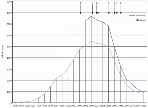
cohorts in the United States, in Europe, and among both adult and pediatric populations.20,21 A decrease in the overall number of hospitalizations related to all S. aureus infections (including both MSSA and MRSA) was appreciated from 2010 to 2014, with a decrease of ˜15% in MRSA infections.22 Total hospital-associated community-onset and hospital-onset MRSA infections decreased by ˜25%-50% from 2005 to 2011, with decreased prevalence of invasive MRSA infections, including CLABSI, also reported during this period.20,23,24,25 Duerden et al. (2011) demonstrated an 80% reduction in MRSA bloodstream infections (BSI) in England following implementation of mandatory reporting and targeted infection control and prevention initiatives (Fig. 19-1).21 The extent to which observed reductions in MRSA are due to targeted MRSA control efforts, global infection prevention efforts, and/or natural changes in MRSA prevalence over time is unclear.26 Although the observed reductions in MRSA HAI are encouraging, it is important to realize that MRSA remains prevalent in most healthcare facilities.


FIGURE 19-1 Decrease in numbers of methicillin-resistant S. aureus bloodstream infections (MRSA BSI) reported in England from 1990 to 2012. Vertical arrows indicate interventions designed to reduce MRSA BSI including mandatory reporting, hand hygiene campaign, mandatory targets to decrease MRSA BSI, legislation requiring quarterly reporting on HAI, reductions in cephalosporin and fluoroquinolone prescribing, and implementation of MRSA screening. (Reprinted with permission from Duerden B, Fry C, Johnson AP, Wilcox MH. The control of methicillin-resistant Staphylococcus aureus blood stream infections in England. Open Forum Infect Dis. 2015;2(2):ofw035.)


TABLE 19-5 Genetic and Epidemiologic Differences Between HA-MRSA and CA-MRSA


Characteristic


HA-MRSA


CA-MRSA


SCCmec types


I, II, III


IV (V, VI)


PFGE types


USA100, USA200


USA300, USA400


MLST


ST5, ST8, ST22, ST36, ST45


ST8, ST30, ST1, ST80


PVL gene


Rare


Common


Additional resistance


Resistance to multiple classes of antimicrobial agents is common


Often resistant to ≤2 additional classes of antimicrobial agents


Epidemiologic risk factors


Exposure to healthcare: hospitalization, residence in LCTF, surgery, dialysis


Crowded living conditions; skin-to-skin contact; cuts, abrasions, exposure to contaminated surfaces and items; poor hygiene


Common infections


BSI, SSI, pneumonia, UTI


Skin and soft tissue infection, pneumonia


HA-MRSA, healthcare-associated methicillin-resistant Staphylococcus aureus; CA-MRSA, community-associated methicillin-resistant Staphylococcus aureus; BSI, bloodstream infection; SSI, surgical site infection; UTI, urinary tract infection.


Evolving Epidemiology of Community-Associated and Hospital-Associated MRSA Over the past two decades, the epidemiology of MRSA has changed as a result of clonal dissemination of novel MRSA strains that are genetically and epidemiologically distinct from typical HA-MRSA strains (Table 19-5). Community-associated
MRSA (CA-MRSA) appears to have arisen as the result of migration of SCCmec type IV and V into MSSA, with USA300 (ST8) as the dominant strain in the United States. CA-MRSA strains are often resistant only to β-lactam antibiotics and perhaps one or two additional antibiotic classes.2 However, resistance to additional classes of antibiotics is being reported with increasing frequency.

Historically, the major determinant in characterizing an MRSA infection as either “healthcare associated” or “community associated” was the time of onset, or the time to identification of the infection after admission to the hospital. A recent epidemiologic classification scheme that acknowledges the ongoing risk of healthcare exposure even after discharge from a healthcare facility is shown in Table 19-6. As noted earlier, though, molecular strain typing has also been used to attempt to distinguish healthcare- and community-associated risks of MRSA infections. The ability to reliably distinguish the likely sources of MRSA strains has become more difficult as strains of MRSA that were traditionally considered to be CA-MRSA, such as USA300, have become established in hospitals and HA-MRSA strains have entered the community.27

Nosocomial outbreaks of CA-MRSA have been reported since 2003.2 In some institutions, USA300 is now the major nosocomial clone of invasive disease, replacing USA100 as the predominant isolate.28 Complex local community and hospital transmission networks of USA300 have been identified, with the community as the primary driver of MRSA BSI.29 From 2005 to 2013, the incidence of BSI caused by USA100 declined by >60%, with >80% decline in hospital-onset USA100 BSI. USA100 continues to be associated with classical healthcare exposures (ie, central venous catheters, prior hospitalization) despite the expansion of USA300.30 A full discussion of the evolving epidemiology of MRSA clones is beyond the scope of this chapter, but excellent reviews can be found in the literature.2


TABLE 19-6 Epidemiologic Classification of MRSA Isolates and Infections


Classification strategy


Classification


Definition


Temporal


Healthcare facility-onset (HO) MRSAa


The specimen from which MRSA was isolated was obtained more than 3 calendar days after the patient was admitted to the healthcare facility. (Note: The day of admission is the first calendar day.)


Community-onset (CO) MRSA


The specimen from which MRSA was isolated was obtained 3 or fewer calendar days after admission to the hospital (or other healthcare facility).


Clinical


Healthcare-associated (HA) MRSA


MRSA that is isolated from a patient with documented healthcare-associated risk factors for MRSA acquisition such as: current or recent admission to a hospital, long-term care, or rehabilitation facility; indwelling vascular catheter; recent surgery; or outpatient hemodialysis.


Nosocomial MRSA


A subset of HA-MRSA, this classification refers to MRSA that is likely to have been acquired during the patient’s admission to the hospital (or other healthcare facility).


Community-associated (CA) MRSA


MRSA that is isolated from a patient without documented healthcareassociated risk factors for MRSA acquisition.


aAlthough the term is “hospital onset,” it is meant to indicate that the onset of MRSA occurred within the healthcare facility in which surveillance is being performed. Thus, a better term may be “facility onset” in order to allow wider applicability, such as in long-term care and other types of healthcare facilities. Adapted with permission from Cohen AL, Calfee D, Fridkin SK, et al. Recommendations for metrics for multidrugresistant organisms in healthcare settings: SHEA/HICPAC Position paper. Infect Control Hosp Epidemiol. 2008;29(10):901-913.


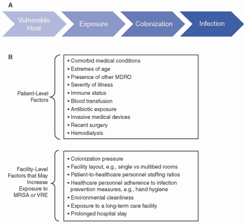
Risk factors for MRSA MRSA infection is the result of a complex interplay of multiple factors, including those specific to the host (eg, comorbid conditions, sex, age, immune defenses, exposure to antibiotics or invasive medical devices), the institution (eg, colonization pressure, adherence of healthcare providers with hand hygiene and other prevention measures). Many of the patient-level and facility-level risk factors for development of nosocomial MRSA infection are common to other multidrug-resistant organisms (MDROs). As is generally true for other MDROs, MRSA colonization typically precedes infection (Fig. 19-2). MRSA colonization itself is a major risk factor for MRSA infection. Here, we discuss risk factors for MRSA at each step from acquisition of colonization to development of MRSA infection.

Patient-level risk factors for MRSA A number of host factors have been identified that increase the risk of MRSA acquisition, establishment of colonization, and progression to MRSA infection (Fig. 19-2). Due to overlap, it is difficult to discern which factors contribute the most to each step in the pathway toward MRSA infection. In addition, studies of MRSA infection may not have surveilled for preceding colonization, thus making it difficult to distinguish risk factors specific for colonization versus infection.

Commonly reported risk factors for MRSA colonization in the host include certain underlying comorbidities such as diabetes, extremes of age, open skin lesions, chronic renal insufficiency, neurologic disorders (prior stroke, dementia, hemiplegia), human immunodeficiency virus (HIV) infection, and cystic fibrosis. Receipt of antibiotics, the presence of an invasive medical device (such as indwelling urinary, vascular catheters, tracheostomy tubes, and feeding tubes) recent surgery, or colonization with another MDRO are also associated with increased risk of MRSA. Facility-associated risk factors for MRSA colonization include high MRSA
colonization pressure, admission from a long-term care facility, and prolonged hospitalization.1,31,32,33,34,35,36 These and other risk factors are shown in Figure 19-2.


FIGURE 19-2 Pathway to nosocomial multidrug-resistant organism infection and associated risk factors. A. The pathway to healthcare-associated infection with multidrug-resistant organisms (MDROs) such as or VRE (vancomycin-resistant enterococcus) methicillin-resistant Staphylococcus aureus (MRSA) or VRE (vancomycin-resistant enterococcus) involves multiple steps including host susceptibility and exposure to the MDRO, which may lead to colonization, which may subsequently proceed to infection. B. Multiple patient- and facility-level risk factors have been identified that are associated with colonization and/or infection with MDROs such as MRSA or VRE.

MRSA Reservoirs in Healthcare Facilities A variety of factors within the healthcare system and the healthcare delivery process have been implicated in the acquisition or transmission of MRSA. The most important MRSA reservoir in the acute care setting is patients who are colonized or infected with MRSA. The risk of acquisition of MRSA during hospitalization increases as the prevalence of MRSA among hospital patients increases (designated as “colonization pressure.”).37 In one study, it was observed that when the weekly colonization pressure exceeded 30%, the risk of MRSA acquisition was five times higher than when colonization pressure was <10%.38 It should be noted, however, that because MRSA can asymptomatically colonize patients, the true prevalence of MRSA in most healthcare facilities is often not known unless MRSA screening is performed. MRSA prevalence or colonization pressure in healthcare facilities varies substantially. In one study, a range of 3.7%-20% was observed in MRSA prevalence across multiple ICUs.33

Contamination of environmental surfaces and healthcare equipment in the rooms of MRSA colonized or infected patients is relatively common. Once in the environment, S. aureus can persist for extended periods.39 In one study, an average environmental contamination of >20% was found on numerous high-touch hospital surfaces.40 HA-MRSA strains were more often identified on surfaces in contact with hospital healthcare personnel (HCP) (ie, medicine room, chart holders, access doors, medicine cart), while public surfaces (ie, hand rails, coffee machine, elevator) had equal detection of both HA- and CA-MRSA strains (ie, USA100 and USA300).40 Numerous environmental foci have been implicated in MRSA contamination, including the air, clinical tools (ie, stethoscopes, blood pressure cuffs), bedding equipment (ie, mattress, bed wheels), furniture, sinks, toilets, HCP and patient clothing, and stationary utensils (ie, pens, keyboards).39 Thus, thorough environmental cleaning of both high-touch and common area surfaces is considered an important component in the control of the spread of MRSA in the healthcare setting.

HCP may transmit MRSA indirectly from patient to patient, or indirectly from an environmental source to a patient, through transient contamination of their hands or clothing. Less commonly, HCP may transmit MRSA directly from a colonized site on their own body to a patient. Rarely, airborne transmission of S. aureus from nasally colonized HCP, so-called cloud disseminators, has also been epidemiologically linked to hospital outbreaks. The risk of S. aureus airborne transmission has been associated with viral upper respiratory infection.41 In a meta-analysis of
127 published studies, the average MRSA prevalence worldwide among HCP was found to be 4.6%.42 A separate meta-analysis of 31 studies from the United States and Europe found a lower overall prevalence of 1.8%, although when one study from the Netherlands with very low rates of colonization was excluded the rate increased to 4.4%.43 Risk factors for MRSA carriage by HCP include cutaneous lesions or skin conditions, sinusitis/rhinitis, recent antibiotic use, employment in areas with high patient MRSA prevalence, close contact with patients, and poor attention to infection control. In one study, the risk of colonization in nurses was nearly twofold higher than in other healthcare staff; the study’s authors hypothesized that this finding was explained by nurses’ more frequent and close contact with patients.43 Routine MRSA screening and decolonization of HCP is not recommended currently. However, treatment of colonized HCP implicated in MRSA outbreaks has been an important step in outbreak control.44

MRSA colonization in acute care hospital patients S. aureus is a common component of the indigenous microbiota of humans and many animals. Population-based studies suggest that approximately one-third of the population is asymptomatically colonized with S. aureus, with about 1.3%-1.5% colonized with MRSA.45,46 The overall prevalence of U.S. S. aureus nasal colonization decreased from 2001 to 2004, but the proportion with MRSA increased during that interval from 0.8% to 1.5%. Persistent or transient carriage of MRSA is most commonly detected in the anterior nares, but carriage on other mucous membranes and skin is also detected. Common sites of cutaneous carriage include the axilla, groin, perianal and perineal areas, wounds and sites of chronic skin disease, as well as foreign body exit sites.47,48 Inguinal or perirectal colonization is more common in men than in women, with men who have sex with men disproportionately represented.48 If screening is not performed, it is estimated that >50% of asymptomatically colonized patients would be missed.33 Approximately 20% of patients remain persistently colonized with MRSA, while 60% are only intermittently colonized. Both intermittent and persistent colonization increase subsequent risk of invasive MRSA infection.47

The duration of MRSA colonization is not well defined, and may be prolonged in certain patient populations. Persistent carriage is associated with ongoing residence in a healthcare institution, immunosuppressive therapy, hemodialysis status, skin disease, and presence of decubitus ulcers. In one prospective study, nearly 50% of hospitalized patients remained colonized at 1 year and ˜20% at 4 years.49 The estimated half-life of MRSA colonization was 40 months in one earlier study,50 with 50% clearance appreciated at 88 weeks in a separate recent analysis.51

MRSA colonization and reservoirs in long-term care facilities MRSA has emerged as an important microorganism in postacute long-term care facilities (LTCF). Epidemiologic descriptions of MRSA in these facilities are heterogeneous, likely reflecting the heterogeneity of the patient populations. Residents of these facilities are important reservoirs for MRSA transmission within the healthcare system.52 Transmission of MRSA within the healthcare network is complex, with bidirectional movement of MRSA between acute and long-term care facilities.52,53

A wide range of MRSA prevalence rates in long-term care facilities have been reported. In one study of MRSA nares colonization in elderly patients admitted to LTCF in the United States, the overall prevalence was 20%, but there was significant variation between facilities.54 Some cohorts have even described prevalence rates exceeding 50%.55

Risk factors for MRSA carriage in LTCF include antibiotic exposure, bed-bound status, recent hospitalization, severe comorbidities, presence of wound or chronic skin disease, and presence of medical devices.56 Decreased risk for MRSA carriage has been associated with MRSA decolonization following hospital discharge, use of infection prevention interventions within the facility, and increased HCP-to-patient ratio.57,58

MRSA colonization and reservoirs in the community CA-MRSA now serves as an important reservoir for importation of MRSA into the hospital. Skin and soft tissue infections are the most common type of infection caused by CA-MRSA, but other manifestations include pneumonia, BSI, ear infections, and joint infections.59 Risk factors for CA-MRSA infection include extremes of age (<2 years or >65 years), HIV infection, athletic activity, injection drug use, male sexual activity, active military service, incarceration, residence in a homeless shelter, and close contact with a person colonized or infected with MRSA.60,61

MRSA Transmission In healthcare settings, MRSA is generally spread through contact with contaminated HCP hands, fomites, or medical equipment. Use of WGS within an English ICU revealed that ˜20% of MRSA acquisitions could be explained by transmission between patients who were coresident in the ICU. The authors also demonstrated related isolates in patients who were not admitted to the ICU concurrently, suggesting that some acquisitions may have occurred outside the ICU or from other nosocomial sources.62 In a separate large integrated epidemiologic analysis, Coll et al.63 found that more than half of cases of hospital MRSA transmission with epidemiologic and bacterial genomic links were part of transmission clusters with hospital contacts, most frequently on the same ward. Patterns of transmissions within certain hospital wards, as well as movement of specific MRSA-infected individuals through multiple hospital units, were both observed.63

Transmission dynamics of MRSA in the community are complex and appear to have to have important geographic connotations.63 Infection prevention of MRSA within the community is an area of ongoing research and has important implications for the spread of MRSA isolates within both the community and hospital setting.


MRSA Infection

Risk of MRSA infection in patients with MRSA colonization MRSA colonization is the most important risk factor for invasive MRSA infection, with the majority of infections caused by the same colonizing MRSA strain.47,64 Patients who remain hospitalized or who are discharged home are more likely to maintain identical MRSA strains; patients who are discharged to nonhospital residential facilities are more likely to become infected with new MRSA strains.65 Nares carriage of MRSA appears to be an important factor in the development of subsequent MRSA infection and may
be associated with increased risk of infection in patients following surgery or receiving dialysis.66

With shortened hospitalizations, it is increasingly recognized that patients who acquire MRSA colonization during a hospital stay may not develop signs of infection until after discharge. Risk factors associated with postdischarge invasive MRSA infection include MRSA colonization, transfer from or discharge to a nursing home, discharge with a central venous catheter or other invasive device, respiratory failure, receipt of a transfusion, and presence of a chronic wound.64,67 In high-risk tertiary care patients, the risk of subsequent invasive MRSA disease within 1 year of newly identified MRSA colonization exceeds 30%, with 14% of patients developing infection during the index hospitalization.68,69 In one Veterans Affairs (VA) study, 13% of MRSA carriers were subsequently readmitted with MRSA infection.70 Over 2 years, risk of invasive infection in one cohort was 21% in persistent carriers and 13% in intermittent carriers.47 This risk is likely to vary substantially among individuals and populations depending on the setting and individual risk factors.

Effect of MRSA infection on patient outcomes and cost In observational studies, MRSA infection has been associated with increased morbidity, mortality, and hospital costs. In studies of hospitalized patients with S. aureus infections, those infected with MRSA had an increased risk of hospital death and increased length of stay.71 Meta-analyses and observational studies of invasive MRSA infections suggest higher in-hospital mortality and bacteremia recurrence in patients with MRSA BSI.72 MRSA infections in the European Union have resulted in an estimated increase of 1 million extra hospitalization days and overall annual cost of 380 million Euros.73,74 In a large Canadian cohort, the estimated excess cost per infected MRSA patient averaged ˜$12000.75 Significant drivers of increased cost include personnel, diagnostics, laboratory investigations, closed beds, barrier precautions, and antimicrobial therapy.76 These findings should be interpreted carefully, as they are from observational studies and unmeasured confounding is likely.


Clinical Manifestations of Healthcare-Associated Infections Caused by MRSA

Bloodstream Infection (Including CLABSI) and Infective Endocarditis S. aureus is a leading cause of BSI worldwide and was the second most common cause of CLABSI in the United States from 2011 to 2014. Of these isolates, >50% were MRSA.16,77 The prevalence of MRSA bacteremia is highly variable and dependent on geographic and individual patient population factors, with estimates ranging from 1% to >50%.78 For patients with hospital-onset MRSA infection, bloodstream infection was the most common presentation.20

Complications of S. aureus BSI are common, estimated at 11%-53%. Predictors of complications include persistent positive blood cultures at 48-96 hours, community acquisition, skin findings suggesting acute systemic infection, and persistent fever. Patients with immunocompromise, such as HIV infection or malignancy, are at higher risk of complications. Nearly any body site can be seeded during bacteremia, with infective endocarditis as a common complication. Patients with prosthetic valves are at higher risk of MRSA infective endocarditis compared to patients with native valves.79

Healthcare-Associated Pneumonia MRSA is an important cause of nosocomial pneumonia, including hospital-acquired pneumonia (HAP) and ventilator-associated pneumonia (VAP).16 Current guidelines recommend utilizing hospital antibiogram data to reduce the unnecessary use of empiric anti-MRSA antibiotic treatment for nosocomial pneumonia. An active agent against MRSA for the empiric treatment of suspected nosocomial pneumonia is only recommended in patients with risk factors for antimicrobial resistance, high risk of mortality, or in patients in units with high prevalence (>10%-20%) of MRSA.80 An important clinical predictor of MRSA nosocomial pneumonia is receipt of intravenous antibiotics within the previous 90 days. A positive MRSA screen from nasal or respiratory samples and residence in units where >20% of S. aureus isolates are MRSA have also been suggested as a predictors of MRSA nosocomial pneumonia.80 Diagnosis of MRSA nosocomial pneumonia can be difficult due to variability of symptoms and difficulty of obtaining a microbiologic diagnosis. However, negative nasal MRSA PCR screening has excellent negative predictive value for MRSA pneumonia and has been useful in guiding de-escalation of antibiotic therapy.13

Skin and soft tissue infections (SSTI) and SSI S. aureus is the most common cause of SSTI and SSI and causes a wide variety of clinical manifestations, ranging from erysipelas to deep tissue infections and abscesses. Approximately 30% of breast, cardiac, neurological, prostate, and vascular HAI SSI and >40% of orthopedic HAI SSI were attributed to S. aureus in the United States from 2011 to 2014. MRSA prevalence in these isolates was ˜40%.16 Nasal and skin colonization are the major risk factors for development of complicated SSTI due to MRSA in hospitalized patients. Other risk factors include previous MRSA infection, recent antibiotic treatment, age >65 years, chronic illness (ie, diabetes, cardiovascular disease, chronic renal failure), HIV infection, or immunocompromised state.81


MRSA Infections in Special Populations

MRSA in hospitalized neonates MRSA outbreaks in the neonatal ICU (NICU) are among the most common outbreaks in academic hospitals.82 MRSA can cause a variety of neonatal infections including BSI, surgical site infection, skin and soft tissue infection, conjunctivitis, bone and joint infections, endocarditis, meningitis, and pneumonia.83 Neonates commonly become colonized with S. aureus shortly after birth, and the prevalence of MRSA within neonatal ICUs varies widely between institutions.84 Common sites of colonization include the umbilical cord, skin, nasopharynx, and gastrointestinal (GI) tract. Risk factors for invasive MRSA infection in neonates include lower birth weight, preterm birth (gestational age <32 weeks), parenteral nutrition, surgery, indwelling central venous catheter, or intubation.84,85 As in adults and older children, preceding MRSA colonization is an important predictor of MRSA infection in neonates. In one study, infants colonized with MRSA had a >10 times increased rate of MRSA infection compared to uncolonized infants.83

MRSA in hemodialysis patients Patients receiving hemodialysis are at increased risk of MRSA colonization and infection due to frequent healthcare exposure, presence
of indwelling devices, and impaired immunity.86 The incidence of invasive MRSA infection, generally presenting as BSI, is significantly higher in patients receiving hemodialysis than the general population (45 per 1000 vs 0.4 per 1000 population).87 Dialysis-associated infections are often related to vascular access; the rate of BSI is highest among patients with a central venous catheter, and much lower in patients with an arteriovenous fistula or graft.88 In hemodialysis patients identified with staphylococcal colonization and treated with decolonization, those who remained persistently colonized were at increased risk of subsequent bacteremia.86


ENTEROCOCCUS SPECIES, INCLUDING VANCOMYCIN-RESISTANT ENTEROCOCCI


Historical Perspective

Enterococcus was first isolated from the human gastrointestinal tract by Thiercelin, followed soon after by the earliest clinical description of a fatal enterococcal endocarditis syndrome by MacCallum and Hastings in the late 1800s. Originally designated as Micrococcus and then subsequently updated to group D Streptococcus, this group of organisms was finally designated as a separate genus in 1984 based on DNA and rRNA hybridization experiments. Since that time, enterococci have been recognized as indigenous microbiota of both humans and animals, as well as routinely identified in the natural environment. While early on enterococci were viewed as fairly innocuous organisms, they have emerged as important nosocomial pathogens due to intrinsic and acquired antibiotic resistance and because of their ability to cause a wide variety of clinical infections.89,90 VRE were first reported in the 1980s and have now become endemic in the healthcare setting.91


Clinically Significant Enterococcal Species

The number of identified Enterococcus species continues to increase as methods for differentiation have advanced. A total of 35 species have been identified, and more are likely to be added.92 Enterococcus faecalis remains the major human pathogen, accounting for approximately half of clinical isolates of enterococci. Enterococcus faecium is the second most commonly isolated species, now accounting for about one-third of enterococcal clinical isolates.16,93 Of the remaining species, only E. gallinarum and E. raffinosus have been identified as a cause of outbreaks and nosocomial acquisition, and then rarely (Table 19-7). Vancomycin-resistant isolates continue to increase in prevalence worldwide, with E. faecium as the predominant species implicated93,95


Laboratory Features and Genetics of Antibiotic Resistance in Enterococcus

Enterococci are gram-positive facultatively anaerobic, catalase-negative, oval cocci, generally appearing in pairs or variable-length chains on Gram staining. They are able to survive harsh conditions; characteristic features include the ability to grow in the presence of up to 6.5% sodium chloride or 40% bile, at extremes of temperature (range of 10-45°C) and pH (up to 9.6), and they are resistant to desiccation. These features may contribute to the ability of Enterococcus species to survive in the dry, inanimate healthcare environment.96,97,98


TABLE 19-7 Prevalence of Selected Clinically Significant Enterococcus Species


Species


Prevalence (%)


VRE identified


Reference


E. faecalisa


64.0-74.1


Well-described. Estimated prevalence 1.9%


93


E. faeciuma


18.4-32.6


Common. Estimated prevalence 43%


93


E. gallinarum


0.8-1.4


Low-level intrinsic resistance


E. raffinosus


<0.1-0.7


Rare


94


a E. faecalis and E. faecium cause the majority of clinical Enterococcal infections. Adapted from Pfaller MA, Cormican M, Flamm RK, Mendes RE, Jones RN. Temporal and geographic variation in antimicrobial susceptibility and resistance patterns of Enterococci: results from the SENTRY Antimicrobial Surveillance Program, 1997-2016. Open Forum Infect Dis. 2019;6(suppl 1):S54-S62.


Antimicrobial Resistance Enterococcal infections are a therapeutic challenge because of both intrinsic and acquired resistance to many common antibiotics. Intrinsic resistance to cephalosporins, aminoglycosides, clindamycin, and trimethoprim-sulfamethoxazole (TMP-SMX) limits initial antibiotic selection. Additionally, enterococci have a highly adaptable genome that readily allows acquisition of further resistance elements. High-level resistance to ampicillin, aminoglycosides, and vancomycin have all been well described, and enterococci with high-level resistance to multiple antimicrobials have been recognized world-wide.96 Evolving enterococcal resistance portends infections that are more difficult to treat, thus increasing the urgency to prevent dissemination of these organisms within and between healthcare institutions.

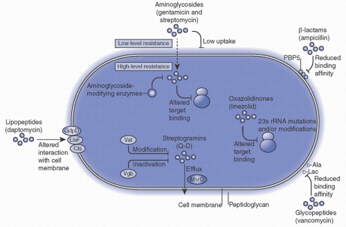
Intrinsic resistance Most enterococci are inherently resistant to many antimicrobials, as shown in Table 19-8 and Figure 19-3. The genes coding for intrinsic resistance reside on the chromosome and confer resistance to cephalosporins, penicillinase-resistant penicillins, clindamycin, low levels of aminoglycosides, and TMP-SMX. The relative resistance to β-lactam antimicrobials is due to low affinity of enterococci for the penicillin-binding proteins.96,99 The MICs of E. faecalis to penicillin are ˜10-100 times greater than those for most streptococci, with E. faecium strains even more resistant. In addition, all enterococci exhibit resistance to low concentrations of aminoglycosides due to limited cell wall permeability. Even in the presence of low-level aminoglycoside resistance, aminoglycosides may be used in combination with a cell-wall active agent (ie, a penicillin or vancomycin) to achieve synergistic killing.100


Acquired resistance

β-Lactam resistance In the absence of acquired resistance mechanisms, enterococci are susceptible to only a few β-lactam antibiotics including penicillin, ampicillin, and piperacillin. Fifth-generation cephalosporins, such as
ceftaroline, also have activity against Enterococcus; however, resistance has emerged with use. High-level resistance to ampicillin was first seen in E. faecium in the 1970s in the United States. Resistance to ampicillin is usually acquired through mutations in penicillin-binding proteins or, rarely, due to production of a β-lactamase. High-level ampicillin resistance remains rare in E. faecalis but occurs in ˜90% of hospital-associated E. faecium isolates. E. faecium isolates with resistance to both ampicillin and vancomycin continue to increase worldwide and result in significant treatment challenges.91


TABLE 19-8 Selected Resistance Mechanisms of Enterococci


Antibiotic resistance


Mechanism


Gene(s)


Location(s)


Intrinsic Resistance


Aminoglycosides


Low cell-wall permeability


β-lactams


Low affinity of antibiotic for penicillin-binding protein (PBP) target, altered PBP target


pbp4/5 production* L,D-transpeptidase*


Chromosomal


Clindamycin


Putative efflux


lsa(A)*


Chromosomal


Trimethoprim/sulfamethoxazole


Utilize environmental folate to bypass antibiotic effect


Acquired Resistance


Aminoglycosides


High-level resistance. Aminoglycoside modifying enzymes (AME) or inactivate antibiotic


AME, ribosomal mutation*


Transposon, plasmid, chromosomal


β-Lactams


High-level ampicillin and imipenem resistance, ↓ affinity for antibiotic or inactivate antibiotic


pbp4/5 mutation*, β-lactamases


Chromosomal, transposon


Daptomycin


Change membrane phospholipids


liaFSR*, gdpD*, variety of mutations dependent on species


Chromosomal


Vancomycin


Modified peptidoglycan precursors


van genes


Transposon


Lincosamides


Efflux, ribosomal protection


ermA/B, vat genes


Transposon


Linezolid


Ribosomal protection


rRNA point mutation*, cfr


Chromosomal, plasmid


Quinolones


↓ affinity for antibiotic


gyrA*, parC*


Chromosomal


Tetracycline


Ribosomal protection or efflux


tet(M), tet(L)


Transposon


Tigecycline


Ribosomal protection or efflux


tet(M), tet(L)


Plasmid


* Genes marked with asterisks undergo mutation that leads to resistance. Genes not marked with asterisks are acquired resistance genes.
Sources: Refs.96,99


High-level aminoglycoside resistance As discussed above, intrinsic mechanisms confer low-level aminoglycoside resistance in enterococci. Acquisition of mobile genetic elements encoding genes for aminoglycoside-modifying enzymes, usually found on a transferable plasmid, is the cause of high-level resistance in both E. faecium and E. faecalis.99 A second mechanism of high-level streptomycin resistance involves modification of the ribosomal target.96 Since first identified in the 1970s, high-level aminoglycoside resistance has increased dramatically worldwide.101 High-level resistance to gentamicin in most clinical isolates is mediated by a bifunctional aminoglycosidemodifying enzyme with 6′-acetyltransferase and 2″-phosphotransferase activity that is capable of inactivating all aminoglycosides other than streptomycin. The gene encoding this enzyme has DNA sequence homology to the gene-conferring gentamicin resistance in S. aureus, and has been localized to transposons found on conjugative plasmids and chromosomes. High-level resistance abolishes the synergistic effect normally observed with combinations of cell-wall active agents and an aminoglycoside.102

Vancomycin resistance VRE were first described in the late 1980s and have increased in prevalence dramatically worldwide, with these organisms now globally endemic.90 The mechanism of resistance to vancomycin and other glycopeptide antibiotics involves alteration of the peptidoglycan synthesis pathway, mostly due to substitution of the glycopeptide target D-alanine-D-alanine (D-Ala-D-Ala) by D-alanine-D-lactate (D-Ala-D-Lac) or D-alanine-D-serine (D-Ala-D-Ser). In general, D-Ala-D-Lac type operons (ie, vanA, vanB, vanD, vanM) confer high levels of resistance to both vancomycin and teicoplanin. VanA and VanB are the predominant phenotypes in clinical isolates of VRE. D-Ala-D-Ser type operons (ie, vanC, vanE, vanG, vanL, vanN) confer lower levels of resistance to vancomycin and maintain susceptibility to teicoplanin (Table 19-9). This latter group includes the intrinsic VanC-type resistance observed in Enterococcus gallinarum, Enterococcus casseliflavus, and Enterococcus flavescens.96

VanA resistance is of the greatest clinical and epidemiological significance. The vanA gene is carried by a gene cluster located within a transposon, Tn1546. The transposon is usually found on a plasmid, which is transferable to other gram-positive species.103 Although vanA is found mainly in E. faecium and E. faecalis, it has been identified
in other enterococcal species and rarely in other grampositive genera.104 VanB strains express variable levels of resistance to vancomycin (MICs 16 to >1000 µg/mL) but in general remain susceptible to teicoplanin. The genes that code for VanB trait are very similar to vanA and are usually found within large mobile elements that are located on the chromosome and that can be transferred to other enterococci.105


FIGURE 19-3 Illustrated common enterococcal resistance mechanisms. The most common mechanisms of antibiotic resistance are illustrated here, including high-level aminoglycoside, β-lactams, oxazolidinones, lipopeptides, and glycopeptides. (Reprinted with permission from Arias CA, Murray BE. The rise of the Enterococcus: beyond vancomycin resistance. Nat Rev Microbiol. 2012;10(4):266-278.)


TABLE 19-9 Phenotypes of Vancomycin-Resistant Enterococci


Characteristic


Phenotype


vanA


vanB


vanC


vanD


vanE


vanG


Vancomycin MIC (µg/mL)


64 to >1000


4 to >1000


2-32


16-64


16


12-16


Ligase activity


D-ala-D-lac


D-ala-D-lac


D-ala-D-ser


D-ala-D-lac


D-ala-D-ser


ND


Genetic


Acquired


Acquired


Intrinsic, chromosomal


Acquired


Acquired


ND


Major species


E. faecium


E. faecalis


E. durans


E. mundtii


E. avium


E. faecalis


E. faecium


E. casseliflavus


E. gallinarum


E. faecium


E. faecalis


E. faecalis


MIC, minimum inhibitory concentration; ND, not done.


Resistance to newer antimicrobial agents Daptomycin is a cyclic lipopeptide that has activity against most enterococci, including VRE. Resistance to daptomycin is sporadic, is associated with multiple different chromosomal mutations, and the mechanism of resistance remains incompletely understood.96 Daptomycin exposure within the previous 90 days is associated with acquisition of daptomycin-resistant Enterococcus,106 although acquisition has also been described in patients without daptomycin exposure. As the prevalence of VRE increases worldwide, use of daptomycin has similarly escalated. Rates of reduced susceptibility to daptomycin have been described in up to 55% of VRE isolates in some studies.107

Linezolid is an oxazolidinone that also has activity against most enterococci, including VRE. Resistance of VRE to linezolid is uncommon and is mediated mostly through mutations in ribosomal proteins.108 Susceptibility of VRE
to linezolid has decreased significantly in some cohorts. Individual centers have reported susceptibility to linezolid in VRE falling from >95% to 45% of enterococcal isolates over an 11-year period, although the worldwide prevalence of linezolid resistance remains low.109 VRE with combined resistance to both daptomycin and linezolid have also been described.110

Tigecycline is a newer broad-spectrum glycylcycline antimicrobial that is active against most enterococci, including VRE. Tigecycline is a derivative of the tetracycline class of antimicrobials but overcomes common resistance mechanisms associated with this class. Enterococcal resistance to tigecycline was first reported in 2015 but remains very rare (<1%).111 Resistance is conferred by increased expression of tetracycline-resistance mechanisms [conferred by tet(L) and tet(M)genes].96 As resistance to other second-line treatment options continues to increase, it is expected that tigecycline will play a progressively larger role in the treatment of multidrug-resistant Enterococcus as a last-line option.

Virulence Factors of Enterococcus Although enterococci are not generally considered to be highly virulent organisms, they have accumulated a number of virulence factors that contribute to their success in the hospital environment.96 Epidemic strains often carry virulence genes associated with more severe disease. This may in part explain the difference in severity of infection between VRE isolates from nosocomial and nonnosocomial acquisition.90 Virulence factors remain incompletely understood and are a subject of ongoing research. Identified virulence factors include secreted toxins (GelE, SagA, Cyl, Hylefm), cell surface molecules (LTA, WTA, capsule), stress response (PTS), transport systems, and gene regulators. Cyl (hemolysin) was one of the earliest identified virulence factors that functions by secretion of a cytolysin that damages the host cell membrane, thus promoting infection.96,112 A number of factors have been identified with biofilm formation and surface adhesion (ie, Esp, Ace, Acm, SgrA, EchA, Pili, BepA). These surface proteins contribute to cell adhesion and pathogenesis. For example, Esp has been demonstrated to be involved in the pathogenesis of urethral colonization, endocarditis, and enhancement of biofilm formation.113,114,115 Many of these virulence factors are present in both E. faecium and E. faecalis, although some virulence factors have been identified only in single species.112

Laboratory Methods for Detection and Identification of Enterococcus Selective culture media, often containing bile salts, sodium azide, antibiotics, and/or esculin, are available for the isolation of clinically relevant Enterococcus species.96 Chromogenic agars that detect VRE are commercially available, and some formulations may also differentiate between E. faecium and E. faecalis. Although these media have excellent sensitivity, they may result in false-positive results due to breakthrough growth by other microbes.89 Many clinical laboratories do not identify enterococcal isolates to the species level if they are isolated in a normally nonsterile source such as urine, which can hinder microbiologic surveillance efforts.96

Identification to the species level can have important clinical implications for the pathogenicity and the likelihood of antibiotic resistance. Molecular techniques are now widely used for species identification of putative enterococcal isolates in broth or agar cultures. Common methodologies include matrix-assisted laser desorption ionization time of flight mass spectrometry (MALDI-TOF MS), peptide nucleic acid fluorescent in situ hybridization (PNA-FISH), and (NAAT). Some of these methods can also be utilized for enterococcal strain typing.96 These rapid identification methods are advantageous due to decreased turnaround times compared to more traditional biochemical methods89 and thus may have important implications for antimicrobial stewardship programs and infection prevention.

Only gold members can continue reading. Log In or Register to continue

Stay updated, free articles. Join our Telegram channel

Jun 8, 2021 | Posted by in INFECTIOUS DISEASE | Comments Off on MDRO Infections: Gram-Positive Organisms (Including Methicillin-Resistant Staphylococcus aureus and Vancomycin-Resistant Enterococcus)

Full access? Get Clinical Tree

Get Clinical Tree app for offline access