Fig. 1
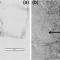
Morphological evaluation of two differently processed parts of a bone marrow trephine biopsy (H&E). a, c Fixation in 10 % neutral buffered formalin (NBF) at room temperature (RT) for at least 6 h followed by decalcification for 18 h at 65 °C in a solution containing formaldehyde and EDTA at pH 7.0 leads to sufficient bone decalcification and preservation of cellular details. b, d Fixation in 10 % NBF for 1 h at RT and 1 h at 37 °C, followed by decalcification in MOLDecal for 4 h at 37 °C leads to incomplete decalcification of the bone with fragmentation of trabecules during cutting of the block. The resulting section is thick hampering evaluation of cellular details
Decalcification for 18 h at 37 °C
These specimens showed a sufficient decalcification of bony trabecula, resulting in thin sections with good preservation of cellular details. There was also a slight shrinkage of the cells detectable (Fig. 2).
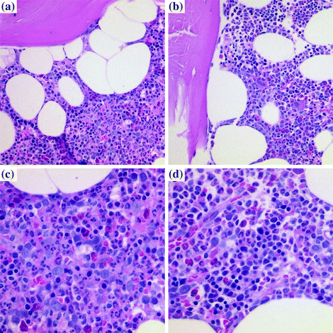

Fig. 2
Morphological evaluation of two differently processed parts of a bone marrow trephine biopsy (H&E). a, c Fixation in 10 % neutral buffered formalin (NBF) at room temperature (RT) for at least 6 h followed by decalcification for 18 h at 65 °C in a solution containing formaldehyde and EDTA at pH 7.0 leading to sufficient bone decalcification and preservation of cellular details. b, d Fixation in 10 % NBF for 1 h at RT and 1 h at 37 °C, followed by decalcification in MOLDecal also for 18 h at 37 °C. There is only a slight shrinkage of the cells detectable, while bone trabecules are sufficiently decalcified and the cellular details easily evaluable
3.2 Immunohistochemistry
Decalcification for 4 h at 37 °C: The nuclear antigen detected by Ki67 was equally good preserved in sections from both protocols. Also the expression patterns of CD3 and CD138 were identical independent of fixation/decalcification protocol. Only the immunostains for CD20 and CD117 were weaker after decalcification with MOLDecal and the reaction product was also more granular. This weaker intensity resulted also in an incorrect estimation of the number of positive cells.
Decalcification for 18 h at 37 °C: Both specimens showed weaker and more granular immunostains for CD20 and CD138 (Fig. 3) after decalcification with MOLDecal, while the results for CD3, CD117, and Ki67 (Fig. 3) were largely identical in both protocols.
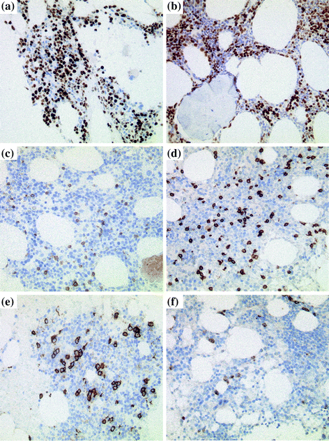
Fig. 3
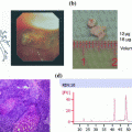
Immunohistological evaluation of two differently processed parts of a bone marrow trephine biopsy. Following antigens have been detected: Ki67 (a, b), CD3 (c, d) and CD138 (e, f). Our current protocol (fixation at RT for at least 6 h followed by decalcification for 18 h at 65 °C in a solution containing formaldehyde and EDTA at pH 7.0) leads to good preservation of the various antigens (a, c, e). The novel protocol with shorter fixation (1 h at RT and 1 h at 37 °C) and decalcification in MOLDecal for 18 h at 37 °C leads also to similar results with the exception of CD138 detection where the labelling is weaker and more granular (b, d, f)
3.3 DNA Quality Assessment
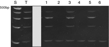
The multiplex control PCR amplification according to the Biomed-2 protocol resulted in a ladder of four fragments (100, 200, 300 and 400 bp) only in the specimens decalcified in the MOLDecal solution after acrylamide gel electrophoresis. In contrast, all samples decalcified with our current protocol showed only 100 bp fragments (Fig. 4).
