, Hans Guski2 and Glen Kristiansen3
(1)
Carl-Thiem-Klinikum, Institut für Pathologie, Cottbus, Germany
(2)
Vivantes Klinikum Neukölln, Institut für Pathologie, Berlin, Germany
(3)
Universität Bonn, UKB, Institut für Pathologie, Bonn, Germany
7.1 Gastrointestinal Epithelial Tumors
7.1.1 Diagnostic Antibody Panel for Gastrointestinal Carcinoma
Cytokeratin profile, CDX-2, SATB-2, CDH-17, CEA, and villin
7.1.2 Diagnostic Antibody Panel for Gastrointestinal Neuroendocrine Carcinoma
Cytokeratin profile, CDX-2, SATB-2, synaptophysin, chromogranin, somatostatin, and Ki-67
CDX-2 | ||
|---|---|---|
Expression pattern: nuclear | ||
Main diagnostic use | Expression in other tumors | Expression in normal cells |
Colorectal adenocarcinoma | Gastric adenocarcinoma, carcinoids of gastrointestinal tract, islet pancreas tumors, sinonasal carcinoma, adenocarcinomas of urinary bladder, ovarian mucinous adenocarcinoma, adenocarcinoma of uterine cervix | Intestinal epithelium and intestinal metaplasia, pancreatic epithelial cell |
Positive control: appendix | ||
Diagnostic Approach
Caudal-related homeobox 2 (CDX-2 ) is an intestine specific transcription factor protein regulating the differentiation and proliferation of intestinal epithelial cells. The expression of CDX-2 begins normally in the post-gastric mucosa in the late stages of embryogenesis of the gastrointestinal tract and is characteristic for different types of adult intestinal mucosa including absorptive, goblet, and Paneth cells in addition to neuroendocrine cells.
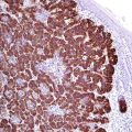
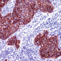
The expression of CDX-2 protein is found in esophageal and gastrointestinal adenocarcinomas in addition to gastrointestinal neuroendocrine tumors in different intensities, whereas the highest frequency and intensity is characteristic for the colorectal adenocarcinomas (Fig. 7.1) [1]. CDX-2 is also an early marker for esophageal Barrett’s metaplasia as the expression of CDX-2 initiates the transformation of squamous epithelium into columnar epithelium with goblet cells.
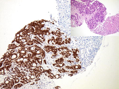

Fig. 7.1
Strong nuclear CDX-2 expression in metastatic colonic adenocarcinoma
The expression of CDX-2 is usually associated with the expression of cytokeratin 20. CDX-1 is a further transcription factor and a marker for gastrointestinal tumors analogous to CDX-2.
Diagnostic Pitfalls
The expression of CDX-2 is reported in many non-gastrointestinal adenocarcinomas. High expression level of CDX-2 is found in bladder adenocarcinoma derived from intestinal urachus, pancreatic adenocarcinoma, biliary adenocarcinoma, and mucinous ovarian carcinoma. CDX-2 expression is also reported in rare cases of prostatic cancer. Pulmonary adenocarcinoma with mucinous differentiation can also be positive for CDX-2; this type of pulmonary adenocarcinoma is also positive for cytokeratin 20 and lacks the expression of TTF-1 [2, 3]. Some neuroendocrine tumors outside the GIT are also reported to be positive for CDX-2 [4]. The loss of CDX-2 expression has been noted in anaplastic high-grade gastrointestinal adenocarcinomas and in medullary adenocarcinomas.
SATB-2 | ||
|---|---|---|
Expression pattern: nuclear | ||
Main diagnostic use | Expression in other tumors | Expression in normal cells |
Colorectal adenocarcinoma and medullary carcinoma, osteosarcoma | Hepatocellular carcinoma, laryngeal squamous cell carcinoma, neuroendocrine tumors of the colon and rectum | Colorectal epithelium, neuronal cells of the central nervous system, hepatocytes, kidney, epithelial cells of the epididymis and seminiferous ducts |
Positive control: appendix | ||
Diagnostic Approach
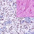
Special AT-rich sequence-binding protein 2 (SATB-2 ) is a nuclear matrix-associated transcription factor and DNA-binding protein involved in the differentiation of osteoblasts. In the gastrointestinal tract, SATB-2 is selectively expressed in colorectal epithelium, while gastric and small intestinal mucosa and pancreatic epithelium lack the expression of SATB-2. SATB-2 is a specific marker for colorectal adenocarcinomas including medullary carcinoma (Fig. 7.2). In routine histopathology, SATB-2 is usually used in combination with cytokeratin 20. SATB-2 is also selectively expressed in neuroendocrine tumors of the left colon and rectum whereas other neuroendocrine tumors reported to be negative or weak positive for this marker [5]. Low expression level of SATB-2 is reported in a subset of pulmonary adenocarcinomas in addition to ovarian carcinomas. Adenocarcinomas of the upper gastrointestinal tract and pancreas typically lack the expression of SATB-2. SATB-2 is also an important diagnostic marker for osteosarcoma [6, 7].
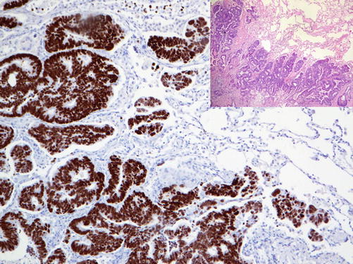

Fig. 7.2
Nuclear SATB-2 expression in metastatic rectal adenocarcinoma (lung metastases)
Cadherin-17 (CDH17) | ||
Expression pattern: membranous and cytoplasmic | ||
Main diagnostic use | Expression in other tumors | Expression in normal cells |
Esophageal and gastrointestinal adenocarcinoma | Pancreatic ductal carcinoma, gastrointestinal and pancreatic neuroendocrine tumors, cholangiocellular carcinoma, osteosarcoma | Gastrointestinal epithelium, pancreas, gall bladder mucosa, adrenal cortex, pituitary gland |
Positive control: appendix | ||
Diagnostic Approach
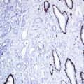
Calcium-dependent adhesion molecule 17 (CDH17) also known as liver-intestine cadherin (LI-cadherin) is a member of the cadherin family regulated by CDX-2. CDH17 is normally expressed in gastrointestinal and pancreatic epithelium and related adenocarcinomas (Fig. 7.3) [8, 9].
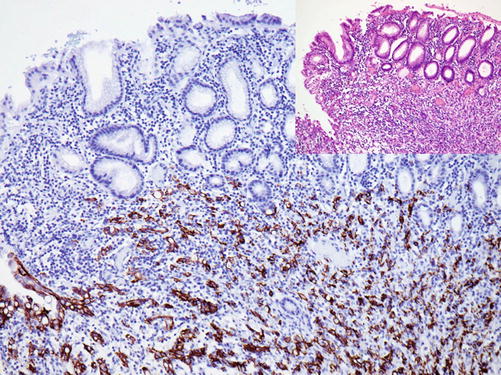

Fig. 7.3
CDH17 expression in cells of gastric adenocarcinoma
CDH17 is generally negative in pulmonary adenocarcinoma, breast carcinoma, papillary thyroid carcinoma, transitional cell carcinoma, renal cell carcinoma, hepatocellular carcinoma, and mesothelioma.
Villin:
Villin is an actin-binding protein and a component of brush border of different epithelial types including cells of intestinal mucosa, mucosa of fallopian tubes, and seminiferous ducts and cells lining proximal renal tubules. Villin is a marker for gastrointestinal adenocarcinomas. Ovarian, endometrioid, and renal cell carcinomas may also be positive for villin. Villin expression is also reported in well-differentiated neuroendocrine tumors of different origin.
Immunoprofile of gastrointestinal tumors | ||||
|---|---|---|---|---|
Tumor type | + in >90% (+) | + in 50–90% (±) | + in 10–50% (∓) | + in <10% (−) |
A. Esophageal and gastric tumors | ||||
Squamous cell carcinoma of the esophagus | CK5/6, CK8, CK14, CK18, CK19, p63, p40 | β-Catenin, cyclin D1 | CK7, CK20 | |
Adenocarcinoma of the esophagus | CK7, CK8, CK18, CK19 | E-Cadherin, CDX-2, cyclin D1, villin | CK20 | CK5/6, p40 |
Adenocarcinoma of the stomach | CK8, CK18, CK19, villin, EMA, CDH-17 | CK7, CEA, CDX-2, glicentin | CK20 | CK5/6, CK14, CK17, CA125, SATB-2 |
B. Intestinal tumors | ||||
Adenocarcinoma of the duodenum and small bowel | CK8, CK18, CK19, CDX-2 a, villin | CK7, CK20, PDX-1, AMACR | Hep Par-1 | SATB-2 |
Adenocarcinoma of the ampullary region | CK8, CK18, CK19, CK7, PDX-1 | CK20,CDX-2 | ||
Colorectal adenocarcinoma | CK8, CK18, CK19, CK20, CDX-2, SATB-2, CEA, villin, MUC-2 | β-Cateninb, CD10 | CK7
Stay updated, free articles. Join our Telegram channel
Full access? Get Clinical Tree
 Get Clinical Tree app for offline access

| |
