, Hans Guski2 and Glen Kristiansen3
(1)
Carl-Thiem-Klinikum, Institut für Pathologie, Cottbus, Germany
(2)
Vivantes Klinikum Neukölln, Institut für Pathologie, Berlin, Germany
(3)
Universität Bonn, UKB, Institut für Pathologie, Bonn, Germany
12.1 Renal Tumors
Diagnostic Antibody Panel for Renal Tumors
PAX-8 | ||
|---|---|---|
Expression pattern: nuclear | ||
Main diagnostic use | Expression in other tumors | Expression in normal cells |
Renal cell carcinoma (clear cell, papillary, chromophobe, and collecting duct carcinomas), nephroblastoma, thyroid carcinoma (papillary, follicular, and anaplastic), ovarian carcinoma (serous, clear cell, and endometrioid carcinoma), adenocarcinoma of seminal vesicle and rete testis | Pancreatic neuroendocrine tumors and other NET of the gastrointestinal tract, parathyroid adenoma and carcinoma, endocervical adenocarcinoma, thymoma (types A and B, thymic carcinoma), seminoma, yolk sac tumor, Merkel cell carcinoma, B-cell lymphomas, medulloblastoma | Thyroid follicular cells, parathyroid cells, thymic cells, of renal tubules, endocervical and endometrial epithelial cells, non-ciliated epithelium of fallopian tubes, epididymal cells, seminal vesicles, a subset of B-lymphocytes |
Positive control: thyroid tissue | ||
Diagnostic Approach
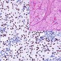
PAX-8 is a transcriptional factor and a member of the paired box (PAX) family consisting of nine members (PAX-1–9). PAX-8 is involved in the fetal development of the central nervous system, eye, inner ear, thyroid gland, kidney, and upper urinary system as well as the Müllerian organs and organs derived from the mesonephric duct [3]. In normal tissue, PAX-8 is highly expressed in thyroid follicular cells, parathyroid cells, and non-ciliated cells of fallopian tubes, mucosa, and renal tubules; consequently, tumors developed from these tissue types are generally positive for PAX-8. Follicular and papillary thyroid carcinomas show high expression level of PAX-8 but not medullary thyroid carcinoma. Clear cell, papillary, and chromophobe renal cell carcinomas besides nephroblastoma are also positive for PAX-8 in addition to the majority of collecting duct carcinoma and oncocytomas and about 50% of sarcomatoid renal cell carcinoma. PAX-8 is also highly expressed in serous, endometrioid, and clear cell ovarian carcinomas, while mucinous carcinoma is usually negative. The expression of PAX-8 is also reported in different percentage of well-differentiated neuroendocrine tumors of pancreatic, gastroduodenal, appendicular, and rectal origin. [4]
Diagnostic Pitfalls
As mentioned, PAX-8 is expressed of a wide range of tumors and must be used as a part of diagnostic panel. A diagnostic panel of PAX-8, WT-1, and two cytokeratins is necessary to confirm the diagnosis of ovarian carcinoma. The PAX-8 expression was noted in about 23% of transitional cell carcinoma of the renal pelvis (Fig. 12.1), which is important to consider in the differential diagnosis of primary renal tumors [5]. PAX-8 is useful to exclude pulmonary adenocarcinoma and breast carcinomas, which usually lack the expression of PAX-8 but express TTF-1 and GATA-3, respectively. The expression of PAX-8 in B-lymphocytes must be also considered in the interpretation of the PAX-8 stain, which is also a useful positive internal control.
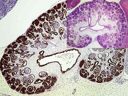

Fig. 12.1
The kidney of a 12-week-old embryo; PAX-8 highlighting the urothelium of the renal collecting system and renal pelvis
PAX-2:
PAX-2 is a further member of the paired box family of transcription factors analogous to PAX-8, is also involved in the renal development, and appears slightly later than PAX-8. PAX-2 has a wide expression range and is found in most renal cell carcinomas with the exception of chromophobe renal cell carcinoma and in tumors of Müllerian origin including ovarian, endometrioid, and endocervical carcinomas in addition to lobular breast carcinoma, hepatocellular carcinoma, epididymal tumor, and Merkel cell carcinoma. PAX-2 is a useful marker to differentiate between benign cervical glandular proliferation positive for PAX-2 and endocervical adenocarcinoma usually lacking the PAX-2 expression. PAX-2 is also expressed in parathyroid cells and parathyroid tumors but constantly negative in thyroid tissue and thyroid carcinomas. Similar to PAX-8, PAX-2 is also positive in B-lymphocytes and related lymphoma types.
Renal cell carcinoma marker (RCC; gp200) | ||
|---|---|---|
Expression pattern: cytoplasmic/membranous | ||
Main diagnostic use | Expression in other tumors | Expression in normal cells |
Renal cell carcinoma (clear cell, chromophobe, and papillary renal cell carcinoma) | Parathyroid adenoma, breast carcinoma, embryonal carcinoma | Renal proximal tubular brush border, epididymal tubular epithelium, breast parenchyma, thyroid follicles |
Positive control: renal tissue or renal cell carcinoma | ||
Diagnostic Approach
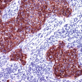
Renal cell carcinoma marker (RCC) is a glycoprotein expressed on the brush border of proximal renal tubules but absent in other renal areas. RCC is detected in about 90% of primary but less frequently in metastatic renal cell carcinoma, including clear cell, chromophobe, and papillary renal cell carcinoma, whereas the highest expression intensity is noted in clear cell carcinoma [6, 7]. Collecting duct carcinoma, sarcomatoid (spindle cell) carcinoma, oncocytoma, mesoblastic nephroma, nephroblastoma, and transitional cell carcinoma are negative for RCC.
Diagnostic Pitfalls
RCC is occasionally detected in rare tumors other than renal cell carcinoma such as primary and metastatic breast carcinoma, embryonal carcinoma, and parathyroid adenoma, which are to be considered in the differential diagnosis.
CD10:
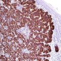
CD10 is listed in detail with the lymphoma markers. CD10 it is also a helpful marker in the differential diagnosis of renal cell tumors. CD10 is positive in the majority of clear cell and papillary renal cell carcinomas in addition to collecting duct carcinoma demonstrating a typical apical expression but negative in chromophobe renal cell carcinoma, which is usually positive for CD117 (Fig. 12.2) [7, 8].
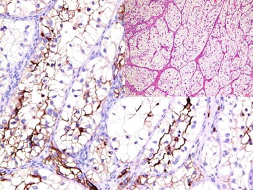

Fig. 12.2
Clear cell renal cell carcinoma stained by CD10 with expression accentuated on the apical side of the cell membrane
Diagnostic Pitfalls
CD10 is also expressed in tumors with similar morphology such as tumors of the adrenal cortex and hepatocellular carcinoma and later lacks the expression of PAX-8 that can be used to discriminate between both tumors.
Paxillin:
Paxillin is a cytoskeletal protein involved in the formation of focal adhesion complexes between F-actin and integrin and widely expressed in epithelial, neuronal, and mesenchymal cells. Paxillin is a helpful marker to differentiate between chromophobe renal cell carcinoma and renal oncocytoma both positive for paxillin and clear cell and papillary renal cell carcinoma negative for this marker [9]. Paxillin is not a specific renal cell carcinoma marker and can be expressed in different carcinoma types of the breast, lung, and liver.
Carbonic anhydrase IX (CA IX) | ||
|---|---|---|
Expression pattern: membranous/cytoplasmic | ||
Main diagnostic use | Expression in other tumors | Expression in normal cells |
Renal cell carcinoma (clear cell and papillary renal cell carcinoma) | Cervical and endometrial carcinoma, transitional cell carcinoma, breast carcinoma, alveolar soft part sarcoma | Gastric and gall bladder mucosa |
Positive control: renal cell carcinoma | ||
Diagnostic Approach
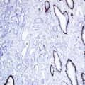
Carbonic anhydrase IX (CA IX) is member of the carbonic anhydrases family zinc metalloenzymes catalyzing the hydration of carbon dioxide. CA IX is a transmembrane isoenzyme taking part in the cell proliferation and cell adhesion as well as the regulation of intra- and extracellular pH. Normally, the expression of CA IX is suppressed by the wild type of von Hippel-Lindau protein and is negative in normal renal tissue. The expression of CA IX is activated during the malignant transformation, and CA IX is markedly expressed in clear cell and a part of papillary renal cell carcinomas. CA IX is helpful in the interpretation of small renal biopsies. It is also a useful marker to discriminate between benign renal cysts generally negative for CA IX and cystic renal cell neoplasm (Fig. 12.3) [10]. Chromophobe cell carcinoma and renal oncocytoma usually lack the expression of CA IX.
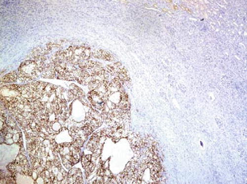

Fig. 12.3
CA IX expression in clear cell carcinoma. The expression is restricted to tumor areas; normal renal tissue is negative
Diagnostic Pitfalls
Carbonic anhydrase IX is not a specific marker for renal cell tumors, and different expression levels are found in various tumors of different origin including pulmonary carcinoma, esophageal carcinoma, renal transitional cell carcinoma, breast carcinoma, neuroendocrine tumors, cervical squamous cell carcinoma and high-grade intraepithelial neoplasia, endometrial carcinoma, embryonal carcinoma, mesothelioma, Sertoli cell tumor, and adrenocortical carcinoma [11].
Human Kidney Injury Molecule-1:
KIM-1 (also known as hepatitis A virus cellular receptor 1) is a type I transmembrane glycoprotein usually not detectable in normal renal tissue but expressed in the epithelial cells of proximal tubules after acute or chronic toxic or ischemic injury. KIM-1 is expressed in different renal cell carcinoma types [12]. In extrarenal tumors, KIM-1 is positive ovarian and uterine clear cell carcinoma, hepatocellular carcinoma, and a subset of colorectal carcinomas in addition to germ cell tumors, which may have a similar morphology to clear cell renal cell carcinoma [13].
Transcription Factor-E3:
TFE-3 a transcription factor encoded by a gene located on Xp11.2. TFE-3 reacts with other transcription factors regulating macrophage and osteoclast differentiation and cell proliferation in addition to activation of B- lymphocytes. The t(X;17) translocation associated with one of the rare types of renal cell carcinoma causes the overexpression of this transcriptional factor, which is considered as a specific immunohistochemical marker for the Xp11.2 translocation-associated renal cell carcinoma [14]. The expression of TFE-3 is also characteristic for the alveolar soft part sarcoma due to an equivalent translocation.
Immunoprofile of renal tumors | ||||
|---|---|---|---|---|
Tumor type | + in >90% (+) | + in 50–90% (+/−) | + in 10–50% (−/+) | + in <10% (−) |
Oncocytoma | CK8, CK14, CK18, E-cadherin, claudin-8, MSH-2a | CK20, PAX-2, PAX-8, EMA, CD15, CD117, DOG-1, cyclin D1, S100A1 | CK7 | Vimentin, RCC38, PAX-2, CD10, KIM-1, CA IX |
Papillary adenoma | CK5, CK8, CK14, CK18, EMA | P504S (AMACR) | ||
Metanephric adenoma | CK8, CK18, vimentin, CD57, S100, PAX-2, PAX-8 | WT-1 | CK7, CK19, EMA, p504S (AMACR) | |
Multilocular cystic renal neoplasm of low malignant potential | PAX-8, CA IX | |||
Clear cell renal cell carcinoma | CK8, CK18, PAX-2, PAX-8, KIM-1, MOC31, α B-crystallin | Vimentin, RCC (gp200), CD10, CA IX, CK19, EMA | CD9 | p504S (AMACR), CK7, CK13, CK20, CD117, inhibin, MSH-2 |
Chromophobe renal cell carcinoma | CK7, CK8, CK18, EMA, CD117, paxillin | CK19, CD9, MSH-2b, DOG-1 E-cadherin, Ki-67 (MIB-1 clone) b, parvalbumin | RCC (gp200), PAX-8, PAX-2, CD10c | Vimentin, CD15, p504S (AMACR), cyclin D1, CA IX, KIM-1, claudin-8, CK13, CK20 |
Papillary (chromophile) renal cell carcinoma | CK8, CK18, PAX-8 | EMA, p504S (AMACR), PAX-2, RCC (pg200), CK19, CD9, CD10, CK7, CK19, CAIX, vimentin
Stay updated, free articles. Join our Telegram channel
Full access? Get Clinical Tree
 Get Clinical Tree app for offline access

| ||
