, Hans Guski2 and Glen Kristiansen3
(1)
Carl-Thiem-Klinikum, Institut für Pathologie, Cottbus, Germany
(2)
Vivantes Klinikum Neukölln, Institut für Pathologie, Berlin, Germany
(3)
Universität Bonn, UKB, Institut für Pathologie, Bonn, Germany
23.1 Diagnostic Antibody Panel for Skeletal Muscle Tumors
Desmin, myoglobin, myogenin, myosin, MyoD1, EGFR, fibrillin-2, and p-cadherin [1, 2].
Desmin | ||
|---|---|---|
Expression pattern: cytoplasmic | ||
Main diagnostic use | Expression in other tumors | Expression in normal cells |
Rhabdomyosarcoma and rhabdomyoma, smooth muscle tumors | Desmoplastic small round cell tumor, alveolar soft part sarcoma, malignant rhabdoid tumor, myofibroblastoma, tenosynovial giant cell tumor | Smooth and striated muscle, myoblasts and myofibroblasts, mesothelial cells, endometrium |
Positive control: appendix | ||
Diagnostic Approach
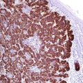
Desmin is a type III intermediate filament protein present in intercalated disks and Z-lines of cardiac muscle and Z-line of skeletal muscle. Desmin stains cardiac, skeletal, and smooth muscle cells and tumors derived from these cells. The intensity of desmin expression correlates with the differentiation grade of muscle or muscle tumor. Desmin is an important diagnostic marker for all myogenic tumors and tumors with myogenic differentiation, whereas myoepithelial cells are negative (Fig. 23.1).
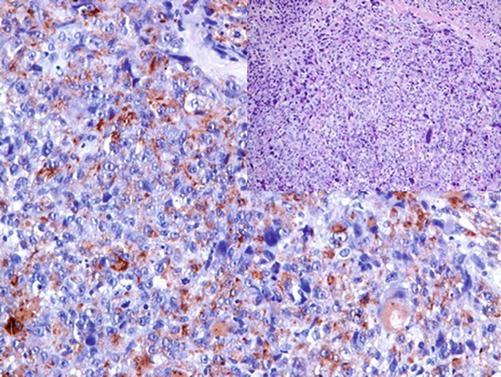

Fig. 23.1
Cells of pleomorphic rhabdomyosarcoma exhibiting marked cytoplasmic expression of desmin
Diagnostic Pitfalls
Desmin is found in other tumors with similar morphology to rhabdomyosarcoma such as desmoplastic small round cell tumor and alveolar soft part sarcoma; hence, the diagnostic panel for rhabdomyosarcoma must include at least one of the antibodies to myogenic transcriptional regulatory proteins (myogenin, Myo D-1, or Myf-3). Markers for smooth muscle differentiation can be also included. It is noteworthy that mesotheliomas (mainly sarcomatous type) and very rarely carcinomas can show focal positivity to desmin; this makes it necessary to determine the cytokeratin profile in doubtful cases.
Myoglobin | ||
|---|---|---|
Expression pattern: cytoplasmic | ||
Main diagnostic use | Expression in other tumors | Expression in normal cells |
Tumors with skeletal muscle differentiation/rhabdomyosarcoma | Various carcinomas, e.g., breast, prostate, colorectal, head, and neck (see below) | Striated muscle, secretory epithelium, goblet cells |
Positive control: skeletal muscle | ||
Diagnostic Approach
Myoglobin is an iron- and oxygen-binding single chain polypeptide that appears in the early stages of muscle differentiation. Myoglobin is expressed in skeletal muscle, cardiac muscle, rhabdomyoblasts and adult-type skeletal muscle tumors. Embryonal muscle tumors and smooth muscle tumors as well as other sarcoma types lack the expression of myoglobin.
Diagnostic Pitfalls
Weak to moderate expression is reported in various carcinomas (e.g., breast, prostate, colon, head and neck) associated with hypoxia and steroid hormone receptor positivity.
Myogenin and MyoD1 | ||
|---|---|---|
Expression pattern: nuclear | ||
Main diagnostic use | Expression in other tumors | Expression in normal cells |
Rhabdomyosarcoma | Wilms’ tumor | Fetal muscle, myoblasts |
Positive control: rhabdomyosarcoma/fetal muscle | ||
Diagnostic Approach
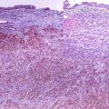
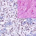
The Myo D family of myogenic transcriptional regulatory factors includes MyoD1 (Myf-3), myogenin (Myf-4) myf-5, and MRF-4 (Myf-6). These transcriptional factors participate in the activation of muscle stem cells and take a part in the regulation of skeletal muscle differentiation in early embryonal stages, maintenance of myogenic program, and repair. The expression of MyoD1 and myogenin is downregulated in mature skeletal muscle, and the expression of both markers is specific for all rhabdomyosarcoma types (Figs. 23.2 and 23.3) [3, 4].
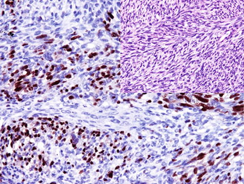
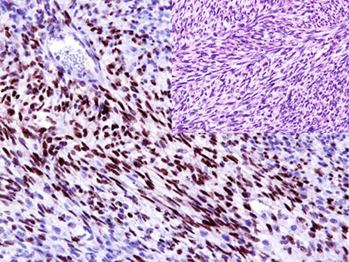

Fig. 23.2
Strong nuclear myogenin expression in rhabdomyosarcoma

Fig. 23.3
Strong MyoD1 nuclear expression in cells of rhabdomyosarcoma
Diagnostic Pitfalls
Both myogenic transcriptional factors can be positive in nonneoplastic myoblasts found within regenerative and atrophic muscle lesions [5]. The expression myogenin and MyoD1 is also reported in some cases of desmoid tumors, infantile fibrosarcoma, mesenchymoma, and Wilms’ tumor. In the interpretation of myogenin and MyoD1 stains, only nuclear stain can be considered as positive; other stain types (cytoplasmic or membranous) are nondiagnostic artifacts.
PAX-5:
PAX-5 is a member of the PAX family of transcription factors was mentioned as a B-lymphocyte marker and a marker for some neuroendocrine carcinomas. In non-lymphoid neoplasms, PAX-5 stains alveolar rhabdomyosarcoma, but it is constantly negative in embryonal-type rhabdomyosarcoma [6].
Epidermal Growth Factor Receptor-1:
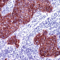
EGFR is a member of type 1 receptor tyrosine kinase family described in a previous chapter (see Chap. 2). EGFR is a transmembrane glycoprotein normally expressed on the membrane of various types of normal epithelial and non-epithelial cells. The expression of EGFR is a characteristic marker for many epithelial and non-epithelial tumors and is diagnostic marker for embryonal rhabdomyosarcoma discriminating it from other rhabdomyosarcoma types (Fig. 23.4) [7].
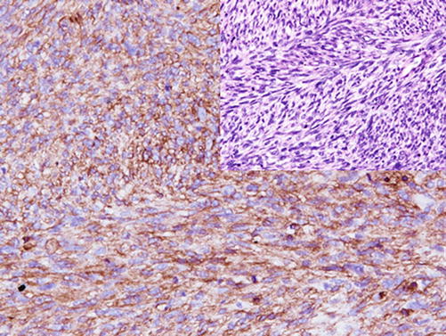

Fig. 23.4
Strong EGFR expression in embryonal rhabdomyosarcoma
Immunoprofile of skeletal muscle tumors | ||||
|---|---|---|---|---|
Tumor type | + in >90% (+) | + in 50–90% (+/−) | + in 10–50% (−/+) | + in <10% (−) |
Fetal rhabdomyoma : | Desmin, sr-actin, myosin, myoglobin | MyoD1, vimentin | GFAP | Pan-CK |
Adult rhabdomyoma: | Desmin, sr-actin, myoglobin
Stay updated, free articles. Join our Telegram channel
Full access? Get Clinical Tree
 Get Clinical Tree app for offline access

| |||
