, Hans Guski2 and Glen Kristiansen3
(1)
Carl-Thiem-Klinikum, Institut für Pathologie, Cottbus, Germany
(2)
Vivantes Klinikum Neukölln, Institut für Pathologie, Berlin, Germany
(3)
Universität Bonn, UKB, Institut für Pathologie, Bonn, Germany
Melanoma is a high malignant tumor with exceptionally variable morphologic appearance that can mimic different epithelioid and sarcomatoid tumors. Generally, the diagnosis of malignant melanoma must be based on the morphology, immunoprofile, and clinical data. In metastatic tumors with ambiguous morphology, it is always advisable to rule out melanoma.
Diagnostic Antibody Panel for Malignant Melanoma
HMB45, MART-1, tyrosinase, Sox-10, microphthalmia transcription factor (MITF), WT-1, S100, CD63 (NK-C3), PHH3, and Ki-67.
HMB-45 | ||
|---|---|---|
Expression pattern: cytoplasmic | ||
Main diagnostic use | Expression in other tumors | Expression in normal cells |
Malignant melanoma, Spitz and cellular blue nevi, clear cell sarcoma | PEComa (angiomyolipoma, sugar tumor of lung), lymphangioleiomyomatosis, pheochromocytoma, hepatoblastoma, ependymoma | Retinal pigmented cells, junctional-activated melanocytes and melanocytes of fetal skin, mononuclear cells |
Diagnostic approach: melanoma | ||
Diagnostic Approach
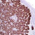
HMB45 (human melanoma black 45) also known as gp100 is a melanosomal glycoprotein involved in the maturation of melanosomes from stage I to II. In normal tissue, HMB45 is found in retinal pigment epithelium and fetal melanocytes but absent in mature melanocytes and intradermal nevi. HMB45 is a marker for melanocytic tumors and tumors with melanocytic differentiation including different types of malignant melanoma, dysplastic nevi, Spitz and blue nevi, as well as clear cell sarcoma (Fig. 21.1).
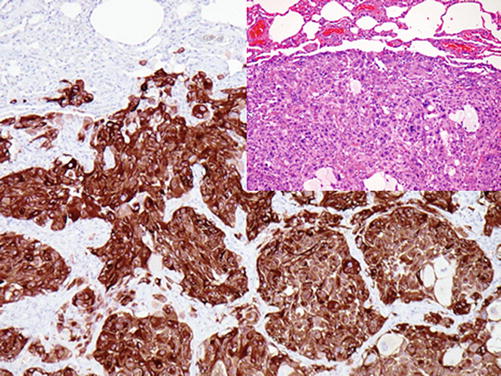

Fig. 21.1
Metastatic melanoma positive for HMB45
Diagnostic Pitfalls
About 10% of malignant melanoma (more frequently amelanotic melanoma, desmoplastic and spindle cell melanomas) lacks the HMB45 expression. The use of an antibody cocktail containing different anti-melanoma markers (usually HMB45, MART-1, and tyrosinase) will markedly increase the sensitivity. Additionally, tumors with similar morphology such as pheochromocytoma and clear cell tumor of the lung (sugar tumor) may be positive for HMB45, but these are usually negative for tyrosinase or Sox-10.
MART-1 (Melan A) | ||
|---|---|---|
Expression pattern: cytoplasmic | ||
Main diagnostic use | Expression in other tumors | Expression in normal cells |
Melanoma, adrenal cortical tumors, sex cord-stromal tumors | Angiomyolipoma, osteosarcoma | Adrenal cortex, melanocytes, brain tissue, granulosa and theca cells, Leydig cells |
Positive control: adrenal cortex | ||
Diagnostic Approach
MART-1 (also known as Melan A) is melanocyte antigen and member of the MAGE family involved in melanosomal maturation and regulation of pigmentation expressed in the endoplasmic reticulum of normal skin melanocytes and retinal cells and tumors derived from these cell types. The MART-1 antigen is recognized by cytotoxic T-lymphocytes.
Stay updated, free articles. Join our Telegram channel

Full access? Get Clinical Tree


